Clinical significance of serum laminin and type‑IV collagen levels in cutaneous melanoma patients
- Authors:
- Faruk Tas
- Elif Bilgin
- Senem Karabulut
- Derya Duranyildiz
View Affiliations
Affiliations: Department of Medical Oncology, Institute of Oncology, University of Istanbul, Istanbul 34390, Turkey
- Published online on: April 25, 2016 https://doi.org/10.3892/mco.2016.873
-
Pages:
195-200
Metrics: Total
Views: 0 (Spandidos Publications: | PMC Statistics: )
Metrics: Total PDF Downloads: 0 (Spandidos Publications: | PMC Statistics: )
This article is mentioned in:
Abstract
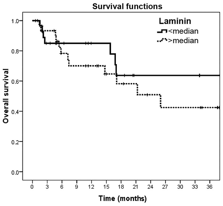
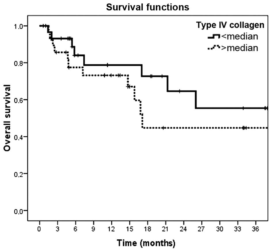
Laminin and type-IV collagen constitute a significant portion of the extracellular matrix. The objective of the present study was to evaluate whether the serum concentrations of laminin and type-IV collagen may serve as biomarkers for cutaneous melanoma. Sixty pathologically confirmed melanoma patients were enrolled in the study. Serum laminin and type‑IV collagen levels were assessed using an ELISA. Thirty healthy controls were also examined. No significant differences in the baseline serum levels of laminin were identified between melanoma patients and healthy controls (P=0.45). However, the baseline serum levels of type‑IV collagen were significantly elevated in melanoma patients compared with those in the control group (P<0.001). Clinical parameters, including patient age, gender, localization of lesion, histopathology, stage of disease, serum lactate dehydrogenase concentrations and responsiveness to chemotherapy were found not to be associated with the serum levels of laminin and type‑IV collagen (P>0.05). Furthermore, the serum levels of laminin and type‑IV collagen had no prognostic value regarding the outcome for melanoma patients (P=0.36 and P=0.26, respectively). While laminin levels showed no diagnostic value, the serum concentrations of type‑IV collagen were indicated to serve as a diagnostic marker in patients with cutaneous melanoma. In conclusion, type‑IV collagen levels may be used as a diagnostic marker for cutaneous melanoma, while being void of any prognostic value.
View References
1
|
Chen HB, Chen L, Zhung JK, Chow VW, Wu BQ,
Wang ZH, Cheng SB and Chew EC: Expression of laminin in metastatic
melanoma cell lines with different metastatic potential. Anticancer
Res. 21:505–508. 2001.PubMed/NCBI
|
2
|
Mortarini R, Gismondi A, Maggioni A,
Santoni A, Herlyn M and Anichini A: Mitogenic activity of laminin
on human melanoma and melanocytes: Different signal requirements
and role of beta 1 integrins. Cancer Res. 55:4702–4710.
1995.PubMed/NCBI
|
3
|
Oikawa Y, Hansson J, Sasaki T, Rousselle
P, Domogatskaya A, Rodin S, Tryggvason K and Patarroyo M: Melanoma
cells produce multiple isoforms and strongly migrate on α5
laminin(s) via several integrin receptors. Exp Cell Res.
317:1119–1133. 2011. View Article : Google Scholar : PubMed/NCBI
|
4
|
Lugassy C, Torres-Muñoz JE, Kleinman HK,
Ghanem G, Vernon S and Barnhill RL: Overexpression of
malignancy-associated laminins and laminin receptors by angiotropic
human melanoma cells in a chick chorioallontoic membrane model. J
Cutan Pathol. 36:1237–1243. 2009. View Article : Google Scholar : PubMed/NCBI
|
5
|
Gradilone A, Gazzaniga P, Cigna E, et al:
Fibronectin and laminin expression in sentinel lymph nodes of
patients with malignant melanoma. Br J Dermatol. 157:398–401. 2007.
View Article : Google Scholar : PubMed/NCBI
|
6
|
Ramos DM, Berston ED and Kramer RH:
Analysis of integrin receptors for laminin and type IV collagen on
metastatic B16 melanoma cells. Cancer Res. 50:728–734.
1990.PubMed/NCBI
|
7
|
Mackie RM, Clelland DB and Skerrow CJ:
Type IV collagen and laminin staining patterns in benign and
malignant cutaneous lesions. J Clin Pathol. 42:1173–1177. 1989.
View Article : Google Scholar : PubMed/NCBI
|
8
|
Burchardt ER, Hein R and Bosserhoff AK:
Laminin, hyaluronan, tenascin-C and type VI collagen levels in sera
from patients with malignant melanoma. Clin Exp Dermatol.
28:515–520. 2003. View Article : Google Scholar : PubMed/NCBI
|
9
|
Hodgson L, Henderson AJ and Dong C:
Melanoma cell migration to type IV collagen requires activation of
NF-kappaB. Oncogene. 22:98–108. 2003. View Article : Google Scholar : PubMed/NCBI
|
10
|
Pasco S, Brassart B, Ramont L, et al:
Control of melanoma cell invasion by type IV collagen. Cancer
Detect Prev. 29:260–266. 2005. View Article : Google Scholar : PubMed/NCBI
|
11
|
Daniels KJ, Boldt HC, Martin JA, et al:
Expression of type IV collagen in uveal melanoma: Its role in
pattern formation and tumor progression. Lab Invest. 75:55–66.
1996.PubMed/NCBI
|