Solitary primary intracranial leptomeningeal glioblastoma invading the normal cortex: Case report

  • Authors:
    • Takamichi Katsuhara
    • Nobuhiro Moro
    • Takashi Ohta
    • Taku Homma
    • Atsuo Yoshino
  • View Affiliations

  • Published online on: January 25, 2018     https://doi.org/10.3892/mco.2018.1561
  • Pages: 466-470
Metrics: Total Views: 0 (Spandidos Publications: | PMC Statistics: )
Total PDF Downloads: 0 (Spandidos Publications: | PMC Statistics: )


Abstract

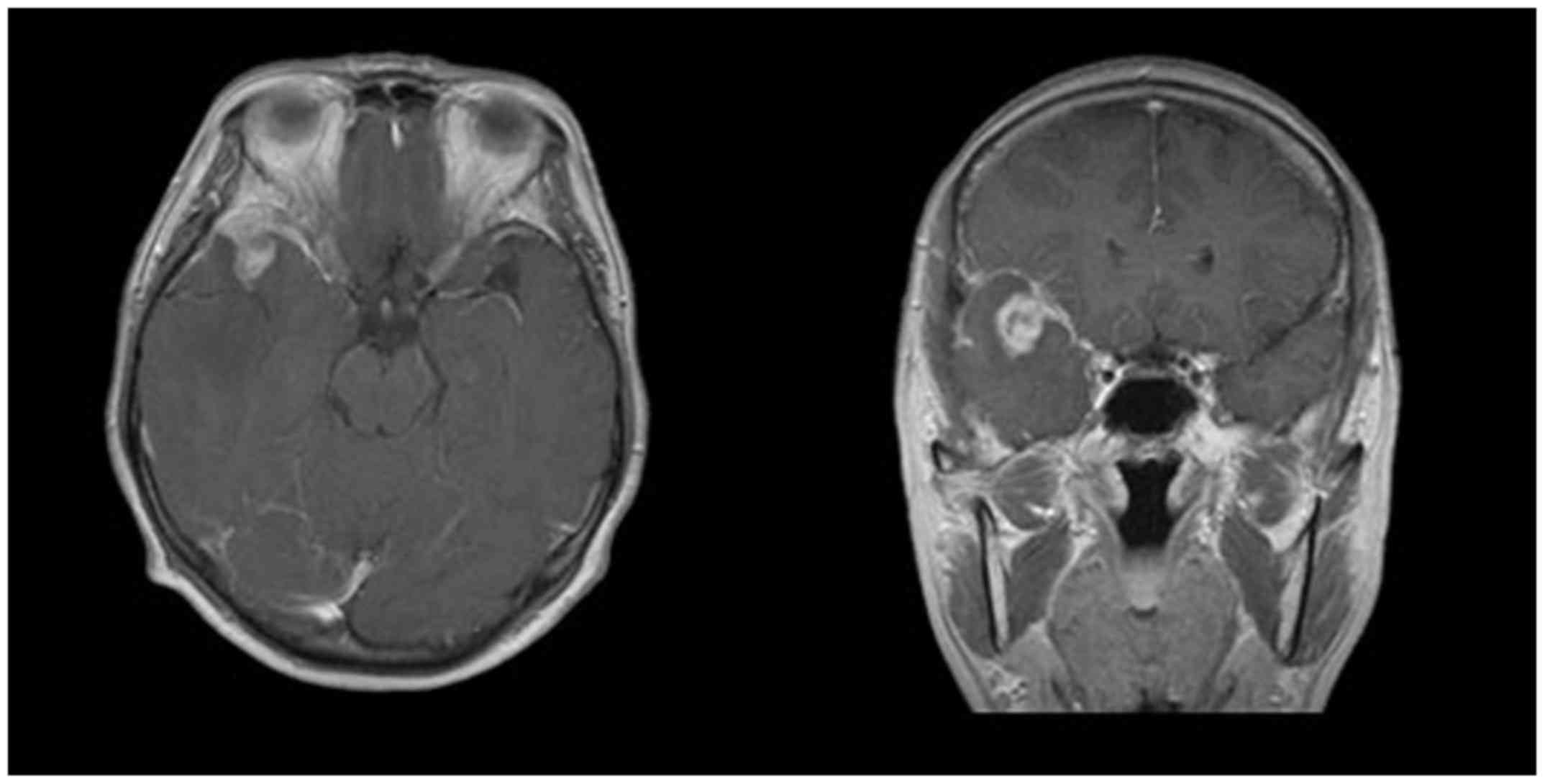
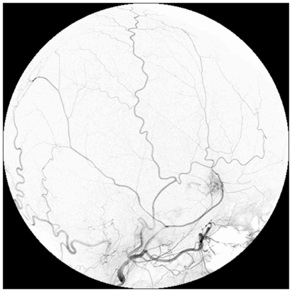
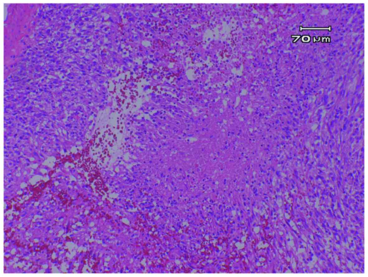
Solitary primary intracranial leptomeningeal glioma (PLG) is a rare entity of glioma. PLG arises from the heterotopic glial tissue in the subarachnoid space and usually grows there without parenchymal invasion. The present study reported a case of solitary PLG, pathologically diagnosed as glioblastoma, that invaded the temporal cortex and finally disseminated to the spinal cord. A 55-year-old woman had headaches and visited Nihon University, Itabashi Hospital. Head magnetic resonance imaging showed a solid mass mainly located in the right middle fossa extending to the frontal base with strong enhancement effect after contrast medium injection. A conventional angiogram showed a tumor arising from the middle meningeal artery. Fronto-temporal craniotomy was performed to remove the tumor. During reflection of the dura matter, there were numerous small vessels connecting the dura matter and the cortical surface. The tumor was located in the Sylvian fissure and extended around the middle cerebral artery. The border between the tumor and the normal temporal lobe was unclear. Temporal lobectomy was done, but the tumor was left around the perforators of the middle cerebral artery. Hematoxylin and eosin staining showed typical glioblastoma with high cellularity, mitosis, pseudopallisading and vascular proliferation. The tumor cells were immunohistochemically negative for isocitrate dehydrogenase (IDH)1-R132H indicating glioblastoma, IDH-wild type. The patient received chemotherapy and radiation therapy, and was discharged from the hospital. Six months later, local regrowth and spinal dissemination were found. Despite additional chemotherapy and radiation therapy, the tumor became uncontrollable and the patient succumbed. Only 15 cases of solitary PLGs have been reported previously. The IDH status of these tumors have not been investigated in most cases; however, pathological grading varies from lower to higher grade glioma. Together with the pathological difference of astrocytic or oligodendrocytic tumors, solitary PLGs may develop due to various gene alterations similar to intra-axial gliomas.
View Figures
View References

Related Articles

Journal Cover

March-2018
Volume 8 Issue 3

Print ISSN: 2049-9450
Online ISSN:2049-9469

Sign up for eToc alerts

Recommend to Library

Copy and paste a formatted citation
x
Spandidos Publications style
Katsuhara T, Moro N, Ohta T, Homma T and Yoshino A: Solitary primary intracranial leptomeningeal glioblastoma invading the normal cortex: Case report. Mol Clin Oncol 8: 466-470, 2018.
APA
Katsuhara, T., Moro, N., Ohta, T., Homma, T., & Yoshino, A. (2018). Solitary primary intracranial leptomeningeal glioblastoma invading the normal cortex: Case report. Molecular and Clinical Oncology, 8, 466-470. https://doi.org/10.3892/mco.2018.1561
MLA
Katsuhara, T., Moro, N., Ohta, T., Homma, T., Yoshino, A."Solitary primary intracranial leptomeningeal glioblastoma invading the normal cortex: Case report". Molecular and Clinical Oncology 8.3 (2018): 466-470.
Chicago
Katsuhara, T., Moro, N., Ohta, T., Homma, T., Yoshino, A."Solitary primary intracranial leptomeningeal glioblastoma invading the normal cortex: Case report". Molecular and Clinical Oncology 8, no. 3 (2018): 466-470. https://doi.org/10.3892/mco.2018.1561