Introduction
Solitary fibrous tumor (SFT) is a relatively rare
mesenchymal tumor. SFT and hemangiopericytoma (HPC) were previously
considered to be distinct disease entities; however, the discovery
of NAB2-STAT6 gene fusion in the majority of SFT and HPC
cases resulted in the recognition of these two entities as a single
clinicopathological disease entity referred to as SFT (1,2). SFT can
affect virtually any site in the body; however, it occurs
preferentially in the pleural cavity. Meningeal SFT is rare,
accounting for <1% of all central nervous system tumors
(3). This type of tumor occasionally
develops extracranial metastases, such as to the lung and liver
(3). To the best of our knowledge,
this is the first cytological report of primary meningeal SFT
metastatic to the lung with an immunocytochemical analysis of
signal transducer and activator of transcription 6 (STAT6), with a
comparison of the cytological characteristics to those of
pleuropulmonary SFT.
Case report
On February 2017, a 58-year-old Japanese male
patient was found to have multiple nodules in the bilateral lungs
by thoracic computed tomography at Kansai Medical University
Hospital 6 years following surgery for right meningeal SFT. Partial
resection of the right lung nodules (2 lesions from the upper lobe,
and 1 lesion each from the middle and lower lobes) was performed
following a clinical diagnosis of metastatic SFT. Touch smears of
the lung nodules were performed. Specimens of the tumor touch
smears were stained with Papanicolaou stain.
Formalin-fixed and paraffin-embedded specimens of
the resected tumors were processed for routine histological
examination and immunohistochemical analyses.
In this report, immunohistochemical and
immunocytochemical analyses were performed using an autostainer (XT
System Benchmark, Roche Diagnostics, Basel, Switzerland, and
Autostainer link 48; DAKO Cytomation, Glostrup, Denmark). The
primary antibodies used in this report were mouse monoclonal
antibody against B-cell lymphoma 2 (Bcl-2; 124), mouse monoclonal
antibody against CD34 (QBEnd10), mouse monoclonal antibody against
Ki-67 (MIB1) (all from DAKO) and mouse monoclonal antibody against
STAT6 (D-1, Santa Cruz Biotechnology, Dallas, TX, USA).
For detection of NAB2-STAT6 fusion, reverse
transcription-polymerase chain reaction (RT-PCR) analysis was
performed.
Cytological findings of the tumor
touch smears
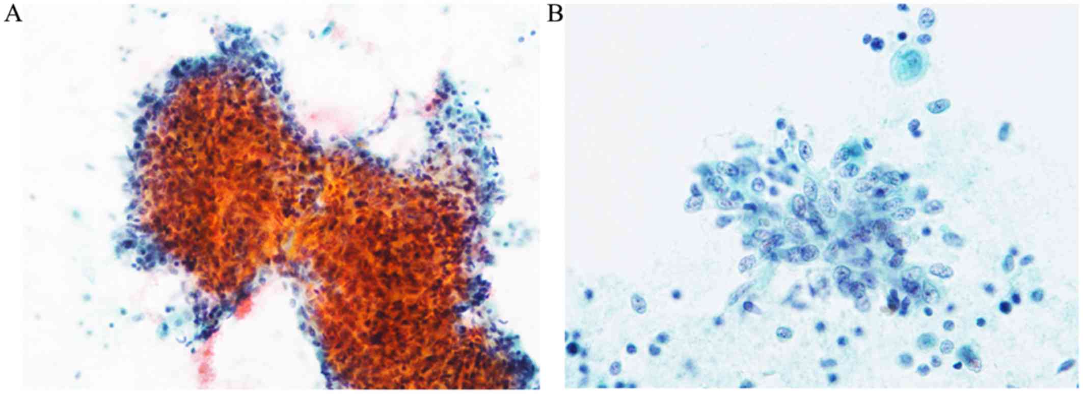
The Papanicolaou smear revealed cohesive
hypercellular clusters or sheets of polygonal to elongated
neoplastic cells in an inflammatory background (Fig. 1A). These neoplastic cells had scant
cytoplasm and oval to short spindle-shaped nuclei with small
nucleoli containing coarse chromatin (Fig. 1B). Mild-to-moderate nuclear
pleomorphism was observed (Fig. 1B).
Neither collagenous stroma nor dilated vascular structures were
noted. Mitotic figures were not found.
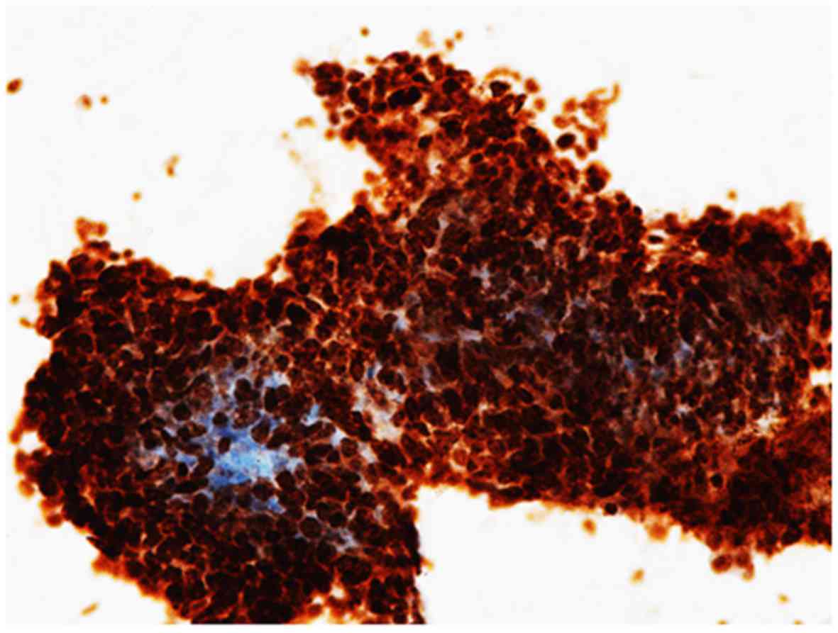
Immunocytochemical findings
STAT6 was diffusely expressed in the nuclei of the
neoplastic cells (Fig. 2).
Accordingly, metastatic SFT was suspected.
Histopathological findings
The four resected pulmonary lesions exhibited
fundamentally the same characteristics. The tumors were composed of
sheet-like proliferations of polygonal to short spindle-shaped
neoplastic cells (Fig. 3A). These
neoplastic cells had scant cytoplasm and oval to short
spindle-shaped nuclei with small nucleoli containing coarse
chromatin (Fig. 3B). Slit-like
vessels were focally observed; however, fibrous stroma was
absent.
A review of the slides revealed that the
histopathological characteristics of the meningeal tumor were
identical to those of the lung lesions.
Immunohistochemical findings
The neoplastic cells in the lung diffusely expressed
STAT6, Bcl-2 and CD34 (Fig. 4). The
meningeal tumor displayed the same immunohistochemical
characteristics, and the Ki-67 labeling index was 1%. Accordingly,
the diagnosis of meningeal SFT metastatic to the lung was
confirmed.
RT-PCR
The lung tumor exhibited NAB2ex6-STAT6ex16
fusion.
Discussion
In this article, we described the first cytological
report of meningeal SFT metastatic to the lung. The characteristic
cytological features of extracranial SFT are as follows: i)
Presence of hypercellular clusters, ii) oval, elongated, round, or
stellate neoplastic cells with limited nuclear pleomorphism, iii)
neoplastic cells with wispy cytoplasm or naked nuclei and iv)
presence of pink collagenous intercellular stroma and occasional
vessel-like structures (4). In
particular, the presence of collagenous intercellular stroma is
characteristic for this type of tumor, and this finding is observed
in almost all cases of benign SFTs (99.5%) (4). Moreover, it has been reported that
cytological characteristics indicative of malignant SFT are the
presence of moderate to extensive nuclear pleomorphism, mitotic
figures and necrosis (4). In the
present case, the presence of hypercellular clusters composed of
polygonal to elongated neoplastic cells with scant cytoplasm and
oval to short spindle-shaped nuclei corresponded to the above
mentioned cytological characteristics. The striking cytological
finding in the present case was the absence of collagenous stroma,
which is noted in 14% of malignant SFTs, and this makes it
difficult to diagnose SFT. Thus, immunocytochemical analysis for
STAT6 is required to confirm the diagnosis of SFT (described
below).
SFT displays a spectrum of histopathological
characteristics. Conventional (typical) SFT exhibits proliferation
of spindle cells with scant cytoplasm and uniform oval to spindled
nuclei within a prominent fibrous stroma and staghorn vessels. This
paucicellular type is classified as fibrous SFT. Tumors with higher
cellularity and more rounded cells, but with prominent perivascular
fibrosis, are referred to as cellular SFT, whereas highly cellular
tumors with well-developed thin-walled or dilated vessels without
perivascular fibrosis are classified as HPC (5). Interestingly, Barthelmess et al
reported that there is a clinicopathological difference among
NAB2-STAT6 fusion variants (5). NAB2ex4-STAT6ex2/3 corresponds to
the classical thoracic SFT with fibrous histology and better
prognosis. By contrast, NAB2ex6-STAT6ex16/17 represents the
cellular SFT and HPC histology, with more aggressive behavior and
extrathoracic occurrence (5).
Moreover, it has been documented that over half of the cases of
meningeal SFT harbor NAB2ex6-STAT6ex16/17 fusion, and the
histopathological characteristics also reflect the fusion variant
in the meningeal SFT, as well as non-meningeal SFT (6). In the present case, the metastatic
tumor harbored NAB2ex6-STAT6ex16 fusion, consistently with
the histopathological and cytological characteristics of HPC-type
SFT.
STAT6 has been recognized as a useful
immunohistochemical marker for both meningeal and non-meningeal
SFTs, without regard for fusion variants (5–8).
Moreover, the usefulness of the immunocytochemical analysis for
STAT6 in formalin-fixed fine-needle aspiration specimens has also
been reported (4). In the present
case, immunocytochemical staining for STAT6 led to cytodiagnosis of
metastatic SFT, although the cytological characteristics were not
typical of conventional SFT, including the absence of collagenous
stroma. Therefore, immunocytochemical analysis for STAT6 is
recommended when SFT is suspected clinically and cytologically.
Acknowledgements
Not applicable.
Funding
No funding was received.
Availability of data and materials
Not applicable.
Authors' contributions
KS, MI and KO contributed to cytological diagnosis
and manuscript preparation. YE performed immunocytochemical and
immunohistochemical staining. MI and KT performed histopathological
diagnosis. KF, TS and TM contributed to patient data collection.
TN, KH and CO performed RT-PCR. The final version of the manuscript
has been read and approved by all authors.
Ethics approval and consent to
participate
Not applicable.
Consent for publication
The authors obtained the consent of the patient to
the publication of the case details.
Competing interests
The authors declare that they have no competing
interests.
References
|
1
|
Chmielecki J, Crago AM, Rosenberg M,
O'Connor R, Walker SR, Ambrogio L, Auclair D, McKenna A, Heinrich
MC, Frank DA and Meyerson M: Whole-exome sequencing identifies a
recurrent NAB2-STAT6 fusion in solitary fibrous tumors. Nat Genet.
45:131–132. 2013. View
Article : Google Scholar : PubMed/NCBI
|
|
2
|
Robinson DR, Wu YM, Kalyana-Sundaram S,
Cao X, Lonigro RJ, Sung YS, Chen CL, Zhang L, Wang R, Su F, et al:
Identification of recurrent NAB2-STAT6 gene fusions in solitary
fibrous tumor by integrative sequencing. Nat Genet. 45:180–185.
2013. View
Article : Google Scholar : PubMed/NCBI
|
|
3
|
Giannini C, Rushing EJ, Hainfellner JA,
Bouvier C, Figarella-Branger D, von Deimling A, Wesseling P and
Antonescu CR: Solitary fibrous tumour/haemangiopericytomaWHO
Classification of Tumours of the Central Nervous System. Revised
4th edition. Louis D, Ohgaki H, Wiestler OD and Cavenee WK: IARC;
Lyon: pp. 249–254. 2016
|
|
4
|
Tani E, Wejde J, Åström K, Wingmo IL,
Larsson O and Haglund F: FNA cytology of solitary fibrous tumors
and the diagnostic value of STAT6 immunocytochemistry. Cancer
Cytopathol. 126:36–43. 2018. View Article : Google Scholar : PubMed/NCBI
|
|
5
|
Barthelmess S, Geddert H, Boltze C,
Moskalev EA, Bieg M, Sirbu H, Brors B, Wiemann S, Hartmann A,
Agaimy A and Haller F: Solitary fibrous tumors/hemangiopericytomas
with different variants of the NAB2-STAT6 gene fusion are
characterized by specific histomorphology and distinct
clinicopathological features. Am J Pathol. 184:1209–1218. 2014.
View Article : Google Scholar : PubMed/NCBI
|
|
6
|
Yuzawa S, Nishihara H, Wang L, Tsuda M,
Kimura T, Tanino M and Tanaka S: Analysis of NAB2-STAT6 gene fusion
in 17 cases of meningeal solitary fibrous tumor/hemangiopericytoma:
Review of the literature. Am J Surg Pathol. 40:1031–1040. 2016.
View Article : Google Scholar : PubMed/NCBI
|
|
7
|
Schweizer L, Koelsche C, Sahm F, Piro RM,
Capper D, Reuss DE, Pusch S, Habel A, Meyer J, Göck T, et al:
Meningeal hemangiopericytoma and solitary fibrous tumors carry the
NAB2-STAT6 fusion and can be diagnosed by nuclear expression of
STAT6 protein. Acta Neuropathol. 125:651–658. 2013. View Article : Google Scholar : PubMed/NCBI
|
|
8
|
Koelsche C, Schweizer L, Renner M, Warth
A, Jones DT, Sahm F, Reuss DE, Capper D, Knösel T, Schulz B, et al:
Nuclear relocation of STAT6 reliably predicts NAB2-STAT6 fusion for
the diagnosis of solitary fibrous tumour. Histopathology.
65:613–622. 2014. View Article : Google Scholar : PubMed/NCBI
|