Fine‑needle aspiration cytology of Warthin‑like mucoepidermoid carcinoma: A case report with cytological review
- Authors:
- Published online on: November 8, 2021 https://doi.org/10.3892/mco.2021.2438
- Article Number: 5
-
Copyright: © Noda et al. This is an open access article distributed under the terms of Creative Commons Attribution License.
Metrics: Total
Views: 0 (Spandidos Publications: | PMC Statistics: )
Total PDF Downloads: 0 (Spandidos Publications: | PMC Statistics: )
Abstract
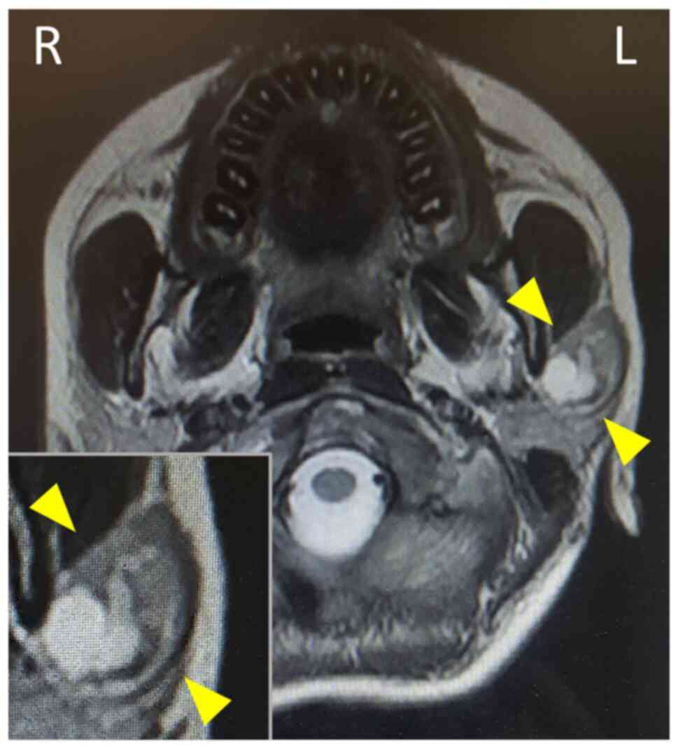
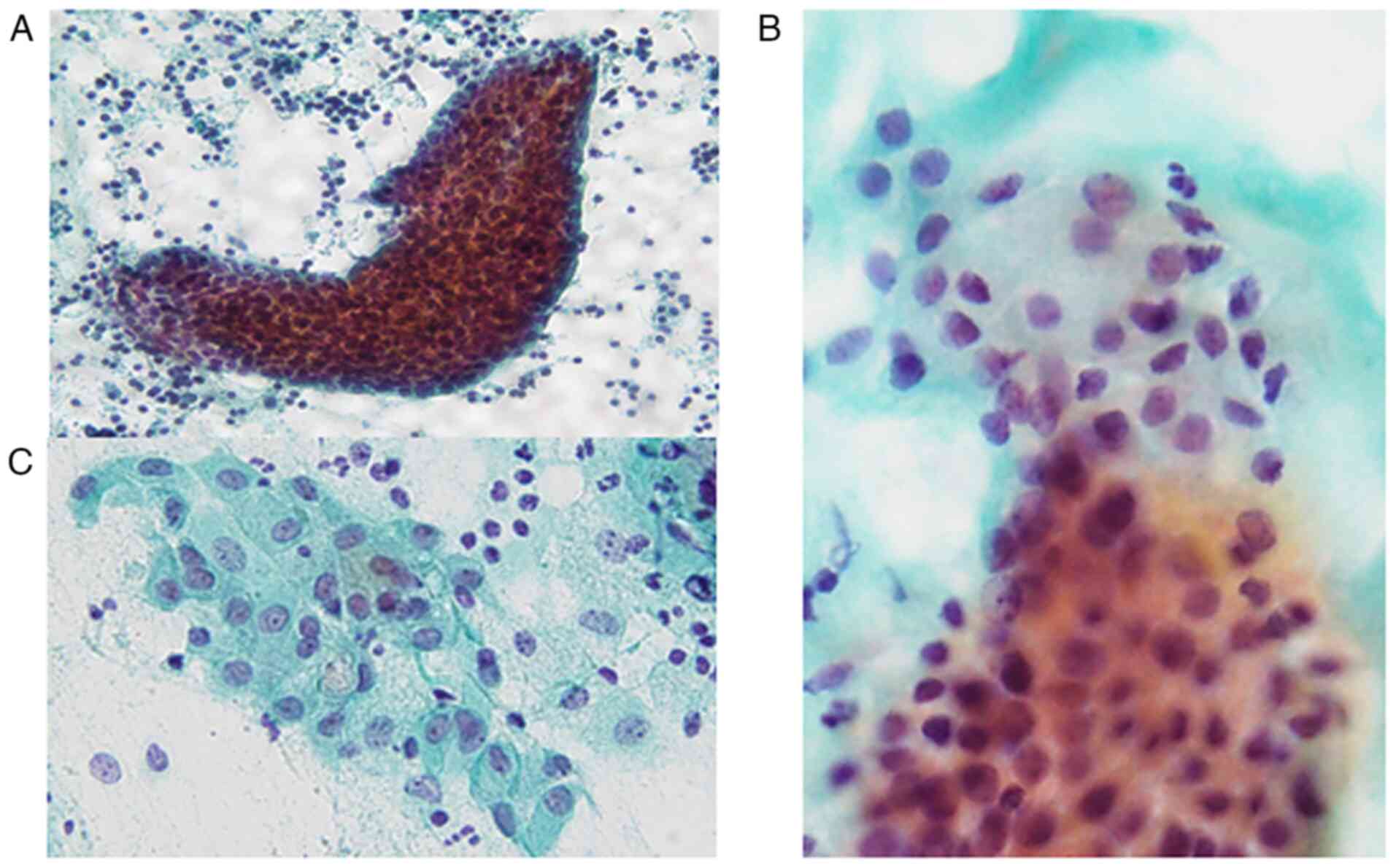
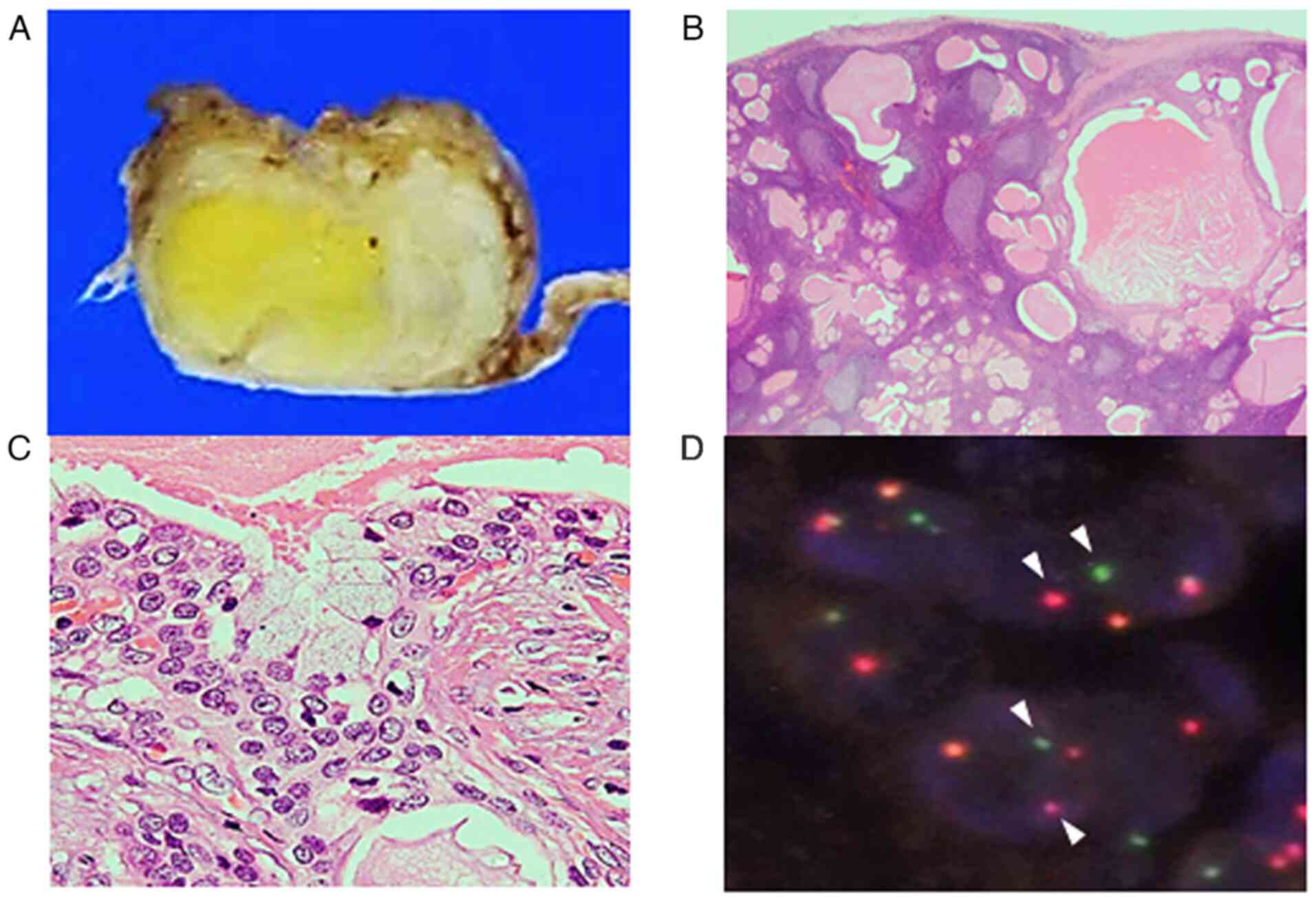
Warthin‑like mucoepidermoid carcinoma (MEC) is a novel and rare subtype of MEC and is characterized histopathologically by the presence of abundant lymphocytic infiltration and cystic changes. A small number of cytological reports of this MEC variant is currently available. The present study reported on the sixth cytological case of Warthin‑like MEC, reviewed the cytological features of the tumour and discussed the cytological differential diagnosis. A 16‑year‑old Japanese female presented with a painful mass in the left parotid gland. Fine‑needle aspiration for cytological examination of the parotid gland tumour was performed, followed by partial parotidectomy. Cytological examination revealed sheet‑like and folded epithelial cell clusters in a mucinous background accompanying abundant lymphocytic infiltration. Epithelial clusters comprised round cells with mildly enlarged round to oval nuclei, polygonal cells with relatively rich cytoplasm and slightly enlarged round to oval nuclei. Certain polygonal cells contained intracytoplasmic mucin. Histopathological examination of the resected parotid gland tumour indicated multiple cystic lesions with abundant lymphocytic infiltration accompanying lymphoid follicle formation. The cysts were lined by intermediate cells with occasional mucinous cells. Fluorescence in situ hybridization using the surgically resected specimen indicated mastermind‑like transcriptional coactivator 2 (MAML2) rearrangement, a characteristic of Warthin‑like MEC. Consequently, the patient was diagnosed with Warthin‑like MEC. The literature review revealed that the characteristic cytological features of Warthin‑like MEC are the presence of intermediate cells and lack of oncocytic cells in the mucinous material under an abundant lymphocytic background. Clinicopathological features may help with a differential diagnosis, particularly from Warthin's tumour, and detection of MAML2 rearrangement is able to improve the accuracy of diagnosis.