Sann-Joong-Kuey-Jian-Tang inhibits hepatocellular carcinoma Hep-G2 cell proliferation by increasing TNF-α, Caspase-8, Caspase- 3 and Bax but by decreasing TCTP and Mcl-1 expression in vitro
- Authors:
- Yao-Li Chen
- Meng-Yi Yan
- Su-Yu Chien
- Shou-Jen Kuo
- Dar‑Ren Chen
- Chun-Yuan Cheng
- Chin-Cheng Su
View Affiliations
Affiliations: Department of Surgery, Changhua Christian Hospital, Changhua County 500-06, Taiwan, R.O.C., Department of Pharmacy, Changhua Christian Hospital, Changhua County 500-06, Taiwan, R.O.C.
- Published online on: March 20, 2013 https://doi.org/10.3892/mmr.2013.1381
-
Pages:
1487-1493
Metrics: Total
Views: 0 (Spandidos Publications: | PMC Statistics: )
Metrics: Total PDF Downloads: 0 (Spandidos Publications: | PMC Statistics: )
This article is mentioned in:
Abstract
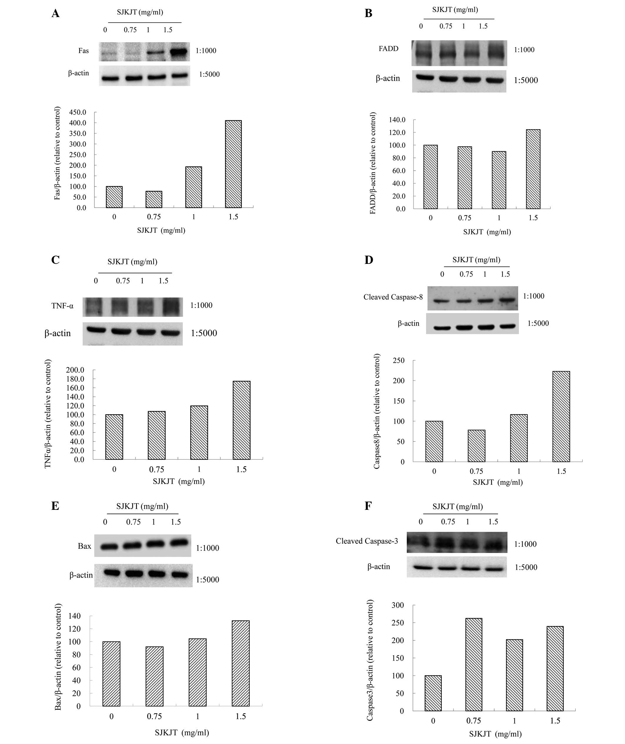
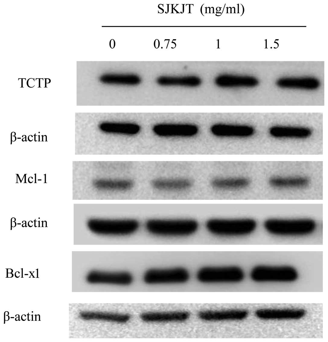
Hepatic cancer remains a challenging disease and there is a need to identify new treatments. Sann-Joong-Kuey-Jian-Tang (SJKJT), a traditional medicinal prescription, has been used to treat lymphadenopathy and exhibits cytotoxic activity in many types of human cancer cells. Our previous studies revealed that SJKJT is capable of inhibiting colon cancer colo 205 cells by inducing autophagy and apoptosis. However, the effects and molecular mechanisms of SJKJT in human hepatocellular carcinoma have not been clearly elucidated. In the present study we evaluated the effects of SJKJT in human hepatic cellular carcinoma Hep-G2 cells. The cytotoxicity of SJKJT in Hep-G2 cells was measured by MTT assay. The cell cycles were analyzed by fluorescence‑activated cell sorting (FACS). The protein expression of translationally controlled tumor protein (TCTP), Mcl-1, Fas, TNF-α, Caspase-8, Caspase-3 and Bax in Hep-G2 cells treated with SJKJT was evaluated by western blotting. The protein expression of Caspase-3 was also detected by immunofluorescence staining. The results showed that SJKJT inhibits Hep-G2 cells in a time- and dose‑dependent manner. During SJKJT treatment for 48 and 72 h, the half-maximum inhibitory concentration (IC50) was 1.48 and 0.94 mg/ml, respectively. The FACS results revealed that increased doses of SJKJT were capable of increasing the percentage of cells in the sub-G1 phase. Immunofluorescence staining showed that Hep-G2 treated with SJKJT had increased expression of Caspase-3. The western blot results showed that the protein expression of Fas, TNF-α, Caspase-8, Caspase- 3 and Bax was upregulated, but that of TCTP and Mcl-1 was downregulated in Hep-G2 cells treated with SJKJT. In conclusion, these findings indicated that SJKJT inhibits Hep-G2 cells. One of the molecular mechanisms responsible for this may be the increased Fas, TNF-α, Caspase-8, Caspase- 3 and Bax expression; another mechanism may be via decreasing TCTP and Mcl-1 expression in order to induce apoptosis.
View Figures |
Figure 1
|
|
Figure 2
|
|
Figure 3
|
|
Figure 4
|
|
Figure 5
|
|
Figure 6
|
|
Figure 7
|
|
Figure 8
|
View References
1
|
Jemal A, Bray F, Center MM, Ferlay J, Ward
E and Forman D: Global cancer statistics. CA Cancer J Clin.
61:69–90. 2011. View Article : Google Scholar
|
2
|
Statistics of Causes of Death, 2010.
Department of Health, Executive Yuan; Taipei, Taiwan, R.O.C: 153.
2012
|
3
|
Leung TW and Johnson PJ: Systemic therapy
for hepatocellular carcinoma. Semin Oncol. 28:514–520. 2001.
View Article : Google Scholar : PubMed/NCBI
|
4
|
Hsu YL, Yen MH, Kuo PL, et al:
San-Zhong-Kui-Jian-Tang, a traditional Chinese medicine
prescription, inhibits the proliferation of human breast cancer
cells by blocking cell cycle progression and inducing apoptosis.
Biol Pharm Bull. 29:2388–2394. 2006. View Article : Google Scholar
|
5
|
Cheng CY, Lin YH and Su CC:
Sann-Joong-Kuey-Jian-Tang increases the protein expression of
microtubule-associated protein II light chain 3 in human colon
cancer colo 205 cells. Mol Med Rep. 2:707–711. 2009.
|
6
|
Cheng CY, Lin YH and Su CC:
Sann-Joong-Kuey-Jian-Tang up-regulates the protein expression of
Fas and TNF-α in colo 205 cells in vivo and in vitro.
Mol Med Rep. 3:63–67. 2010.PubMed/NCBI
|
7
|
Cohen CGM: Caspases, the executioners of
apoptosis. Biochem J. 326:1–16. 1997.
|
8
|
Lowe LSW, Cepero E and Evan G: Intrinsic
tumour suppression. Nature. 432:307–315. 2004. View Article : Google Scholar : PubMed/NCBI
|
9
|
Carswell EA, Old LJ, Kassel RL, et al: An
endotoxin-induced serum factor that causes necrosis of tumors. Proc
Natl Acad Sci USA. 72:3666–3670. 1975. View Article : Google Scholar : PubMed/NCBI
|
10
|
Gaur U and Aggarwal BB: Regulation of
proliferation, survival and apoptosis by members of the TNF
superfamily. Biochem Pharmacol. 66:1403–1408. 2003. View Article : Google Scholar : PubMed/NCBI
|
11
|
Graidist P, Phongdara A and Fujise K:
Antiapoptotic protein partners fortilin and MCL1 independently
protect cells from 5-fluorouracil-induced cytotoxicity. J Biol
Chem. 279:40868–40875. 2004. View Article : Google Scholar : PubMed/NCBI
|
12
|
Liu H, Peng HW, Cheng YS, et al:
Stabilization and enhancement of the antiapoptotic activity of
mcl-1 by TCTP. Mol Cell Biol. 25:3117–3126. 2005. View Article : Google Scholar : PubMed/NCBI
|
13
|
Zhang D, Li F, Weidner D, et al: Physical
and functional interaction between myeloid cell leukemia 1 protein
(MCL1) and Fortilin. The potential role of MCL1 as a fortilin
chaperone. J Biol Chem. 277:37430–37438. 2002. View Article : Google Scholar : PubMed/NCBI
|
14
|
Susini L, Besse S, Duflaut D, et al: TCTP
protects from apoptotic cell death by antagonizing bax function.
Cell Death Differ. 15:1211–1220. 2008. View Article : Google Scholar : PubMed/NCBI
|