Chondrogenic differentiation of human umbilical cord blood‑derived mesenchymal stem cells by co‑culture with rabbit chondrocytes
- Authors:
- Published online on: August 16, 2013 https://doi.org/10.3892/mmr.2013.1637
- Pages: 1169-1182
Metrics: Total
Views: 0 (Spandidos Publications: | PMC Statistics: )
Total PDF Downloads: 0 (Spandidos Publications: | PMC Statistics: )
Abstract
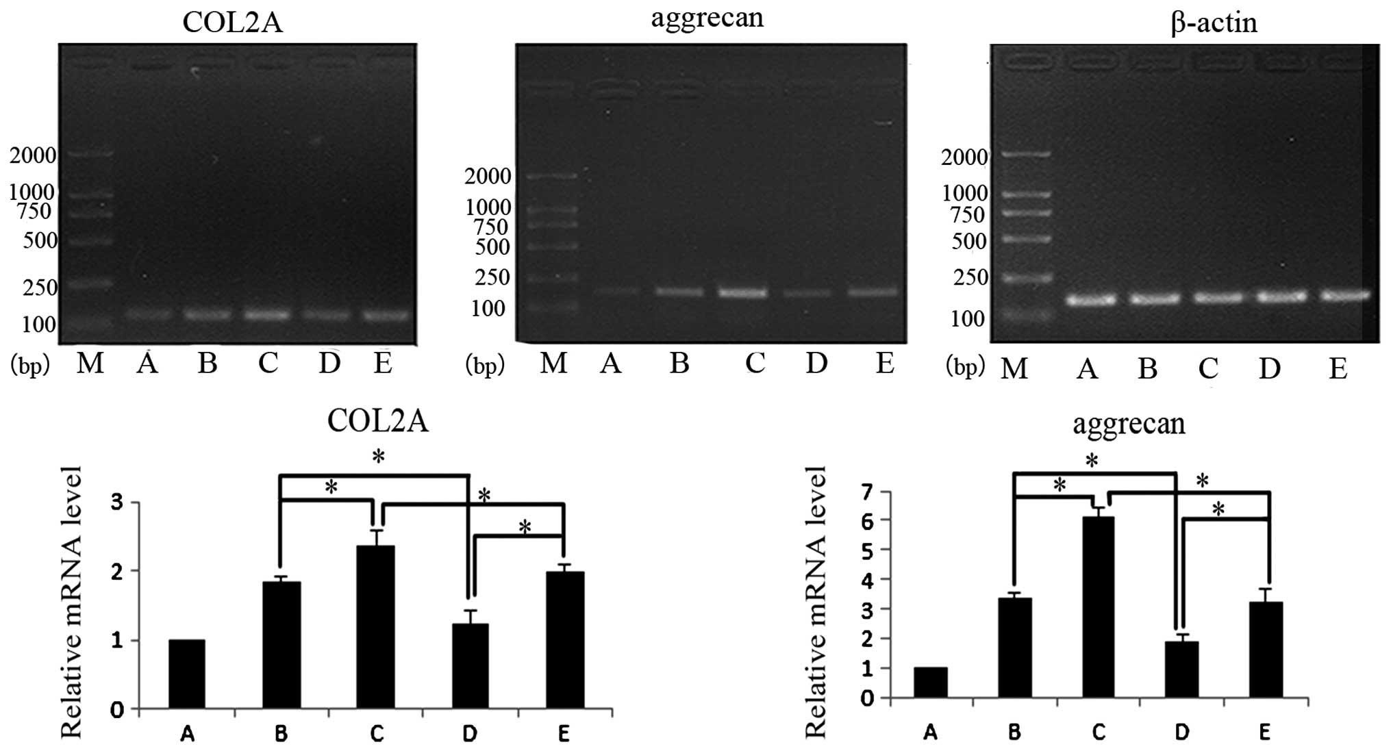
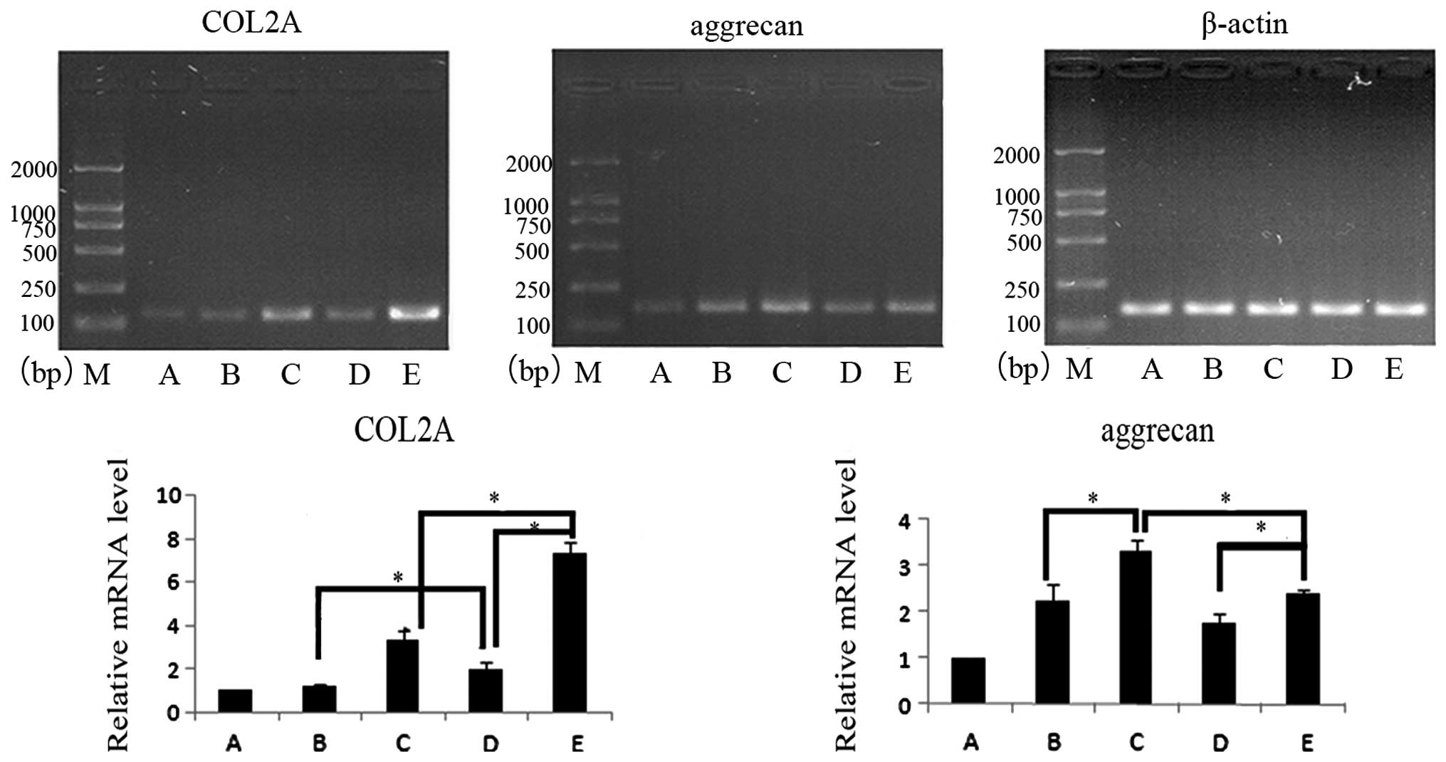
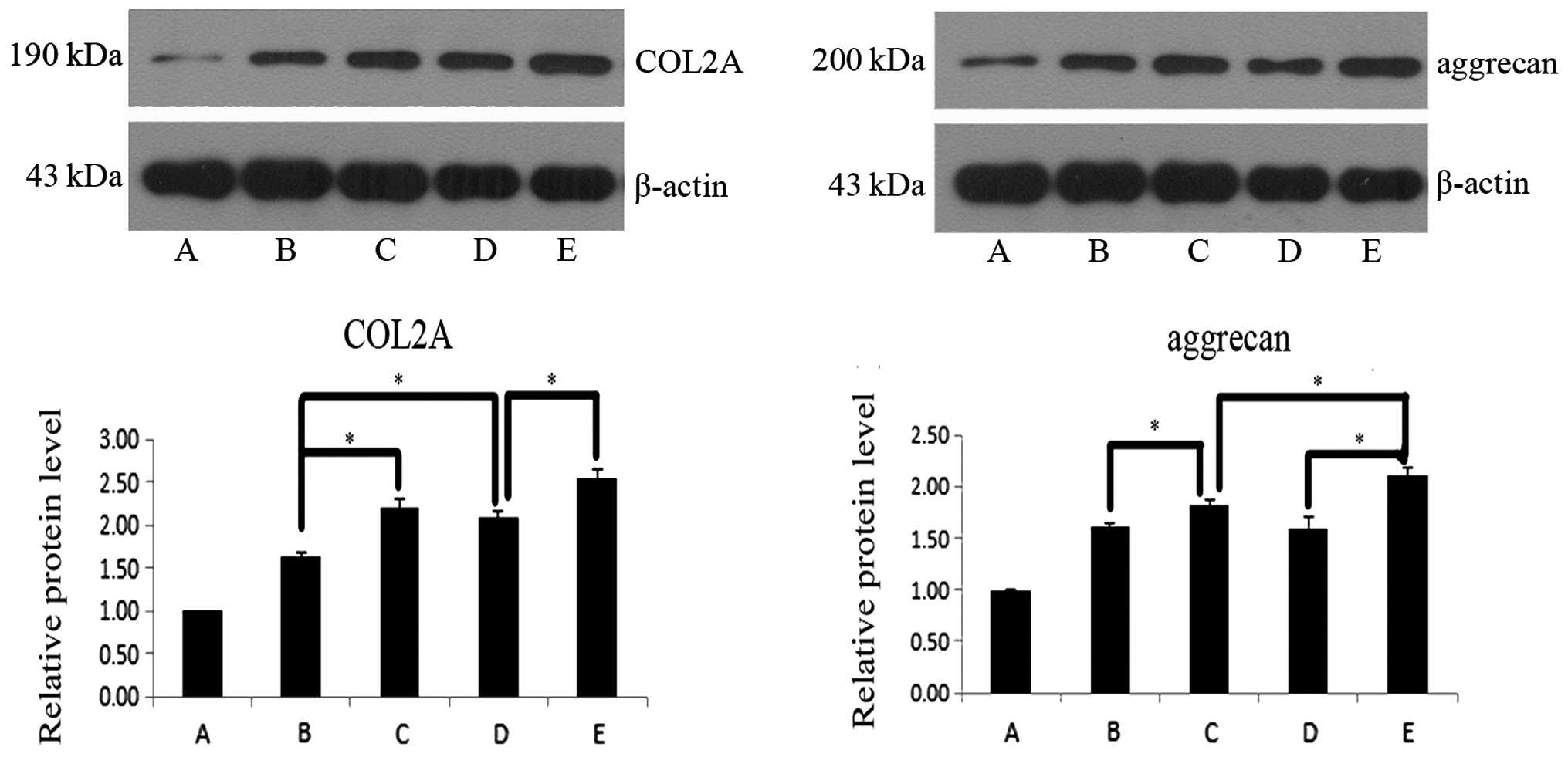
The objective of the current study was to investigate the ability of human umbilical cord blood‑derived mesenchymal stem cells (HUCB‑MSCs) to undergo chondrogenic differentiation, by co‑culture with rabbit chondrocytes. The aim was to obtain more seed cells for tissue engineering research and lay the foundation for the clinical repair of cartilage defects. The studies were performed using isolated rabbit cartilage cells and HUCB‑MSCs in vitro, which were co‑cultured in a 2:1 or 3:1 ratio with or without insulin‑like growth factor‑1 (IGF‑1). Following 7 and 14 days in culture, cell morphology was observed in each group. RNA and protein were extracted to assess the expression levels of aggrecan (ACAN) and collagen type II (COL2A) using quantitative polymerase chain reaction (qPCR) and western blotting, respectively. Groups of cells that were co‑cultured exhibited significantly higher expression levels of ACAN and COL2A mRNA and protein, compared with the reduced effect of IGF‑1 at days 7 and 14 in culture. The addition of IGF‑1 was found to potentiate these effects. Specifically, at day 7, cells co‑cultured at a ratio of 2:1 had a greater induction of ACAN and COL2A compared with cells co‑cultured at a 3:1 ratio. However, following 14 days culture, cells co‑cultured at a 3:1 ratio with additional IGF‑1 exhibited a greater induction of ACAN and COL2A compared with cells co‑cultured at a ratio of 2:1. Human chondrocytes may be successfully induced by co‑culture of HUCB‑MSCs with rabbit chondrocytes, thus providing a theoretical basis to obtain seed cells with the capacity to differentiate into multiple cell types, with low immunogenicity. Notably, these cells may provide a valuable resource for tissue engineering.