Open Access

Atoh1 expression levels define the fate of rat cochlear nonsensory epithelial cells in vitro

  • Authors:
    • Wen‑Wei Luo
    • Juan‑Mei Yang
    • Zhao Han
    • Ya‑Sheng Yuan
    • Hai‑Bin Sheng
    • Xiang Liu
    • Fang‑Lu Chi
  • View Affiliations

  • Published online on: April 30, 2014     https://doi.org/10.3892/mmr.2014.2202
  • Pages: 15-20
  • Copyright: © Luo et al. This is an open access article distributed under the terms of Creative Commons Attribution License [CC BY_NC 3.0].

Metrics: Total Views: 0 (Spandidos Publications: | PMC Statistics: )
Total PDF Downloads: 0 (Spandidos Publications: | PMC Statistics: )


Abstract

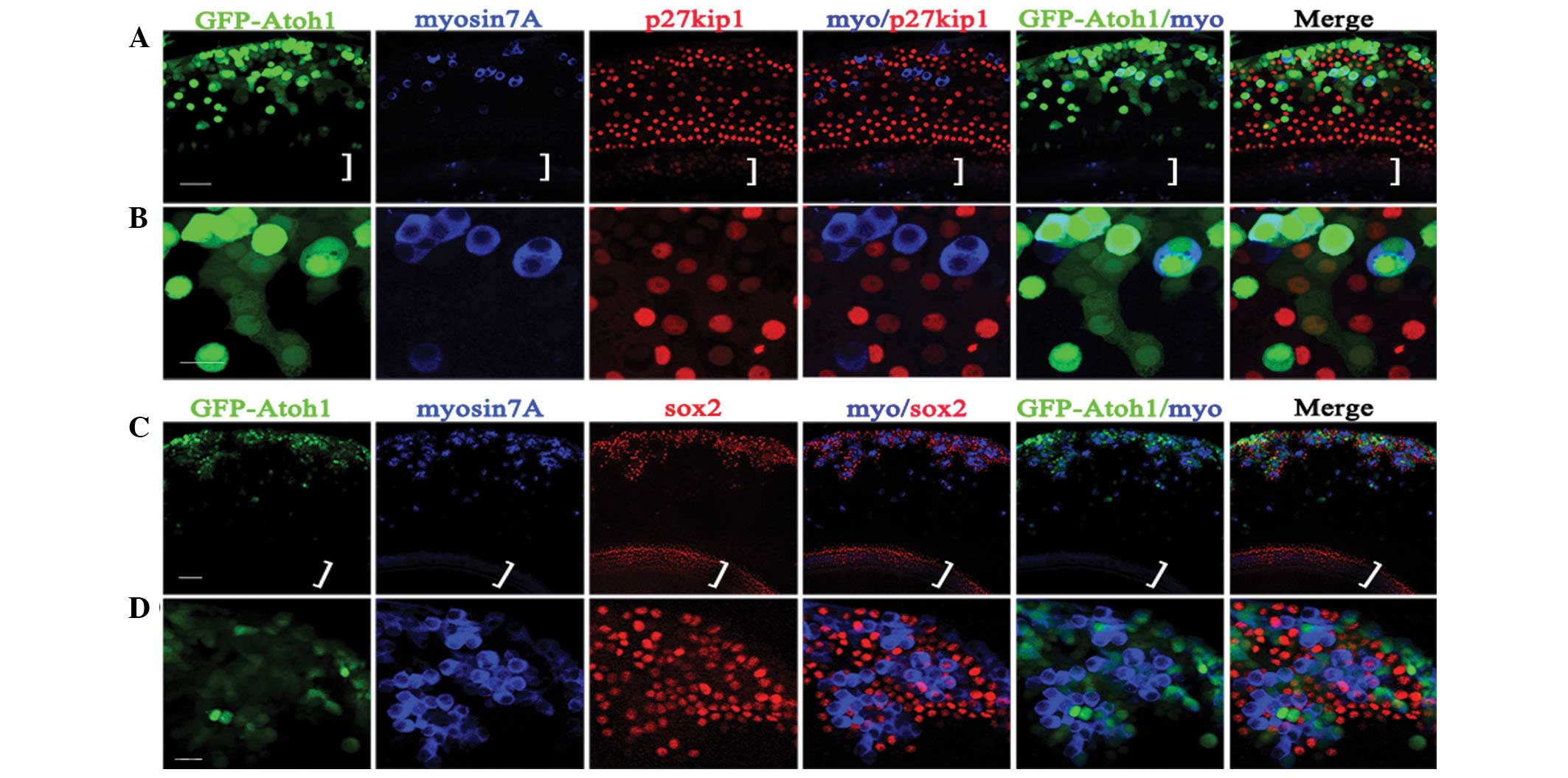
Atonal homolog 1 (Atoh1) is a basic helix‑loop‑helix transcription factor that is essential for inner ear hair cell differentiation. Previous studies have reported that Atoh1 gene transfer induces the production of ectopic hair cell‑like cells (EHCLCs). In the present study, the effect of different Atoh1 expression levels and the duration of EHCLC formation on the lesser epithelial ridge (LER) of cochleae was examined using a human adenovirus serotype 5 (Ad5) vector encoding atoh1 and the reporter gene EGFP. Different Ad5‑EGFP‑atoh1/Ad5‑EGFP virus titers were added to cultured cochlear explants and EHCLCs were detected in the LER at various time points. The results demonstrated that GFP alone did not induce EHCLCs. By contrast, Atoh1 expression induced EHCLCs as early as 2.5‑5 days following EGFP‑atoh1 infection in the LER and depending upon the viral titer, the number of EHCLCs increased with time. Higher Ad5‑EGFP‑atoh1 titers induced enhanced Atoh1 expression, resulting in an increase in EHCLCs. Lower Ad5‑EGFP‑atoh1 titers required more time for EHCLC formation and very low titers of Ad5‑EGFP‑atoh1 induced only weak Atoh1 expression and did not trigger EHCLC formation. In conclusion, the present study utilized an appropriate Ad5‑EGFP‑atoh1 titer range to induce Atoh1 expression and the subsequent production of EHCLCs. The results revealed that the Atoh1 expression level defined the fate of LER cells as either EHCLCs or nonsensory epithelial cells. This evidence may provide an important guideline for future studies into gene therapy strategies for the treatment of deafness.
View Figures
View References

Related Articles

Journal Cover

July-2014
Volume 10 Issue 1

Print ISSN: 1791-2997
Online ISSN:1791-3004

Sign up for eToc alerts

Recommend to Library

Copy and paste a formatted citation
x
Spandidos Publications style
Luo WW, Yang JM, Han Z, Yuan YS, Sheng HB, Liu X and Chi FL: Atoh1 expression levels define the fate of rat cochlear nonsensory epithelial cells in vitro. Mol Med Rep 10: 15-20, 2014
APA
Luo, W., Yang, J., Han, Z., Yuan, Y., Sheng, H., Liu, X., & Chi, F. (2014). Atoh1 expression levels define the fate of rat cochlear nonsensory epithelial cells in vitro. Molecular Medicine Reports, 10, 15-20. https://doi.org/10.3892/mmr.2014.2202
MLA
Luo, W., Yang, J., Han, Z., Yuan, Y., Sheng, H., Liu, X., Chi, F."Atoh1 expression levels define the fate of rat cochlear nonsensory epithelial cells in vitro". Molecular Medicine Reports 10.1 (2014): 15-20.
Chicago
Luo, W., Yang, J., Han, Z., Yuan, Y., Sheng, H., Liu, X., Chi, F."Atoh1 expression levels define the fate of rat cochlear nonsensory epithelial cells in vitro". Molecular Medicine Reports 10, no. 1 (2014): 15-20. https://doi.org/10.3892/mmr.2014.2202