Effects of 5‑fluorouracil and class III phosphoinositide 3‑kinase small interfering RNA combination therapy on SGC7901 human gastric cancer cells
- Authors:
- Bao‑Song Zhu
- Jia‑Lei Sun
- Wei Gong
- Xing‑Ding Zhang
- Yong‑You Wu
- Chun‑Gen Xing
View Affiliations
Affiliations: Department of General Surgery, The Second Affiliated Hospital, Soochow University, Suzhou, Jiangsu 215004, P.R. China, Department of General Surgery, The First Changshu Affiliated Hospital, Soochow University, Changshu, Jiangsu 215500, P.R. China, Cyrus Tang Hematology Center, Soochow University, Suzhou, Jiangsu 215123, P.R. China
- Published online on: November 11, 2014 https://doi.org/10.3892/mmr.2014.2926
-
Pages:
1891-1898
Metrics: Total
Views: 0 (Spandidos Publications: | PMC Statistics: )
Metrics: Total PDF Downloads: 0 (Spandidos Publications: | PMC Statistics: )
This article is mentioned in:
Abstract
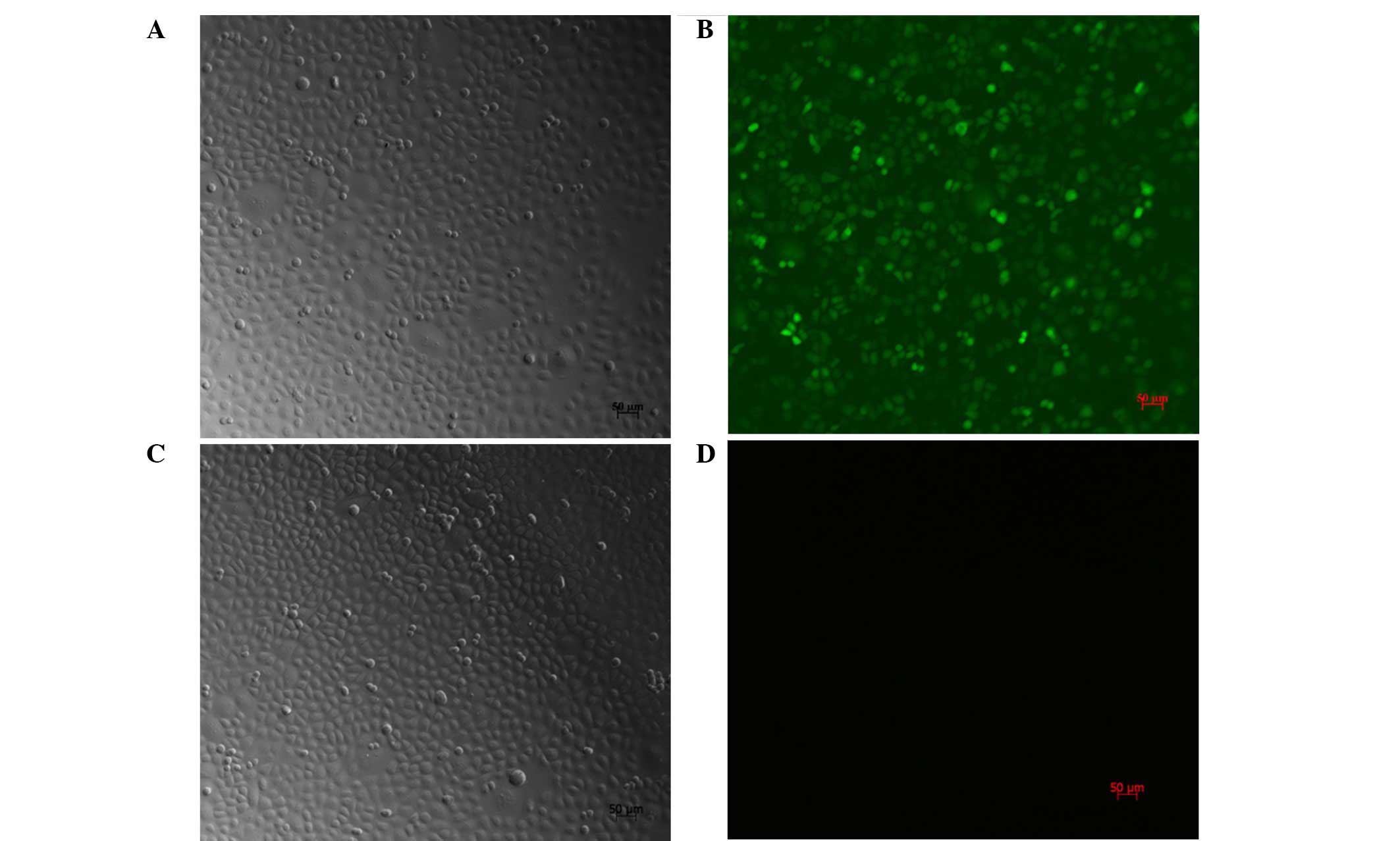
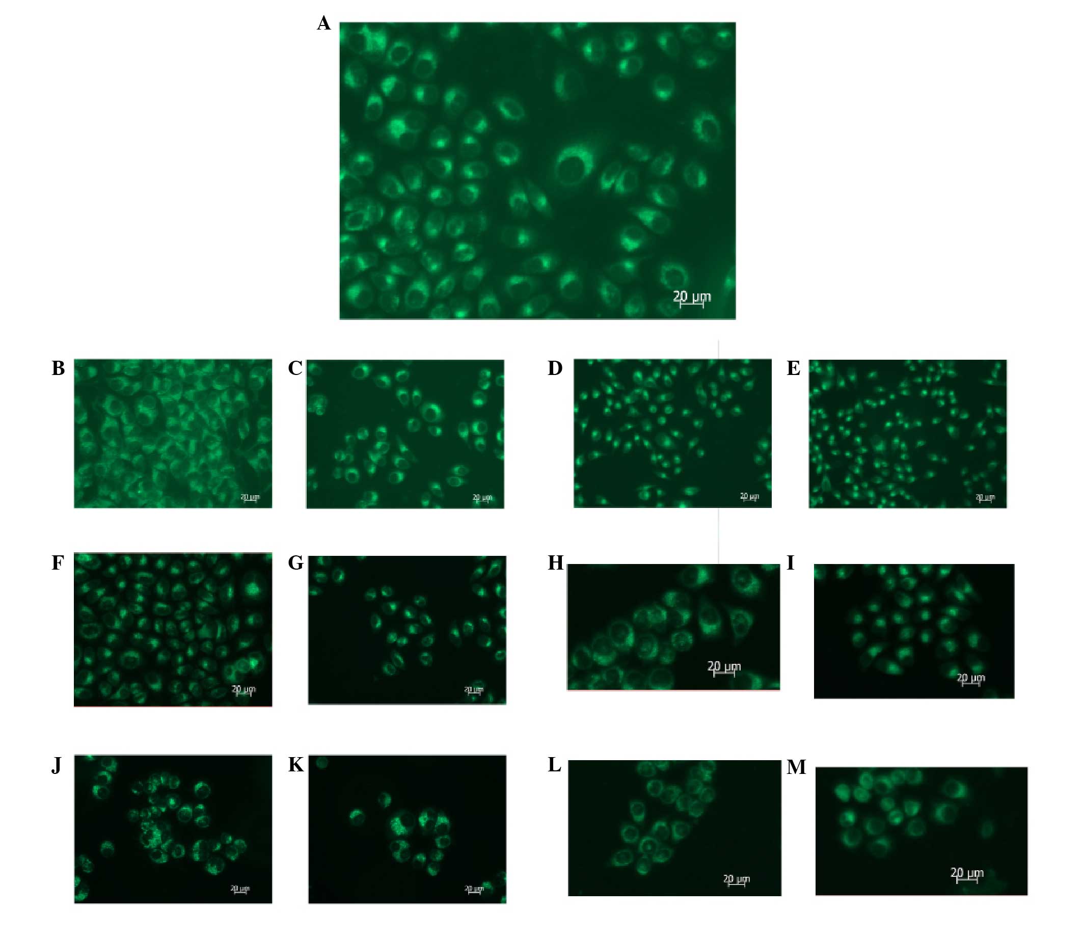
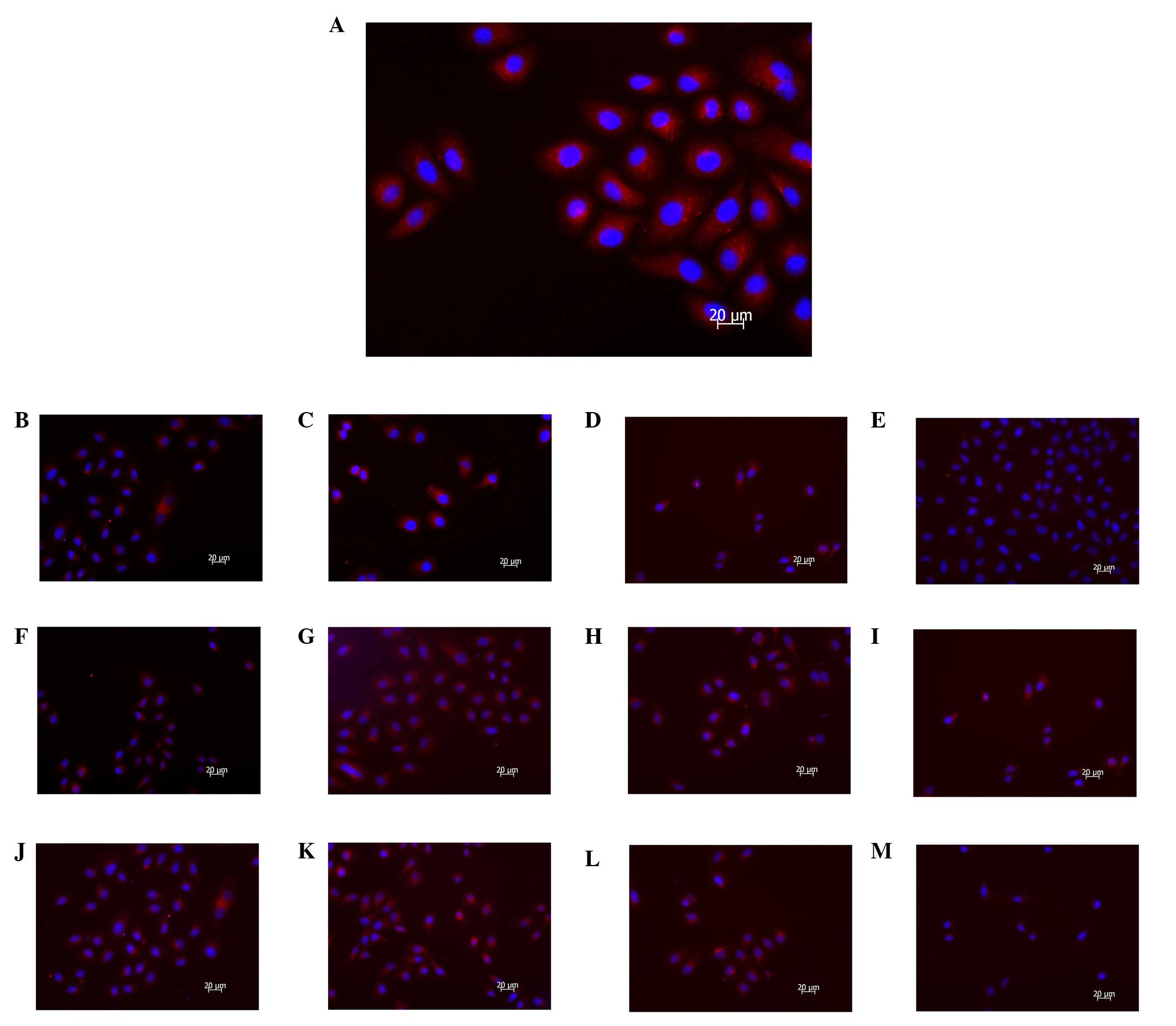
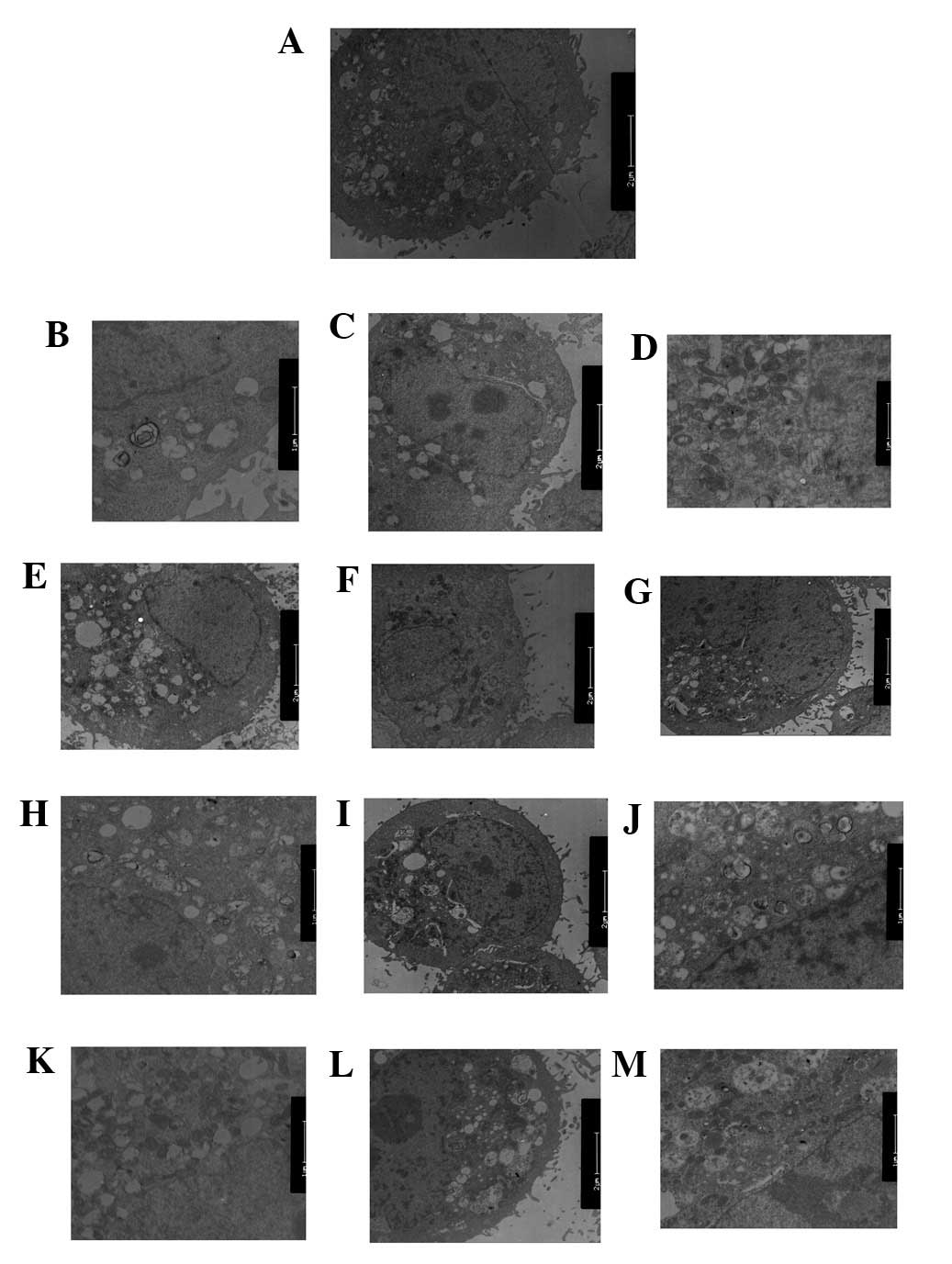
The aim of the present study was to investigate the effects of small interfering RNA‑mediated inhibition of Class III phosphoinositide 3‑kinase (PI3K) signal transduction on the proliferation, apoptosis and autophagy of SGC7901 gastric cancer cells. The present study also aimed to examine the contribution of autophagic inhibition to the antitumor effects of 5‑fluorouracil (5‑FU). A PI3K(III)‑RNA interference (i)‑green fluorescent protein (GFP) recombinant replication adenovirus (AD) and the negative control (NC)‑RNAi‑GFP control AD were constructed and infected into SGC7901 cells. A methyl thiazolyl tetrazolium assay was used to determine the growth rate of the SGC7901 cells. Immunofluorescent staining was used to detect microtubule‑associated protein 1 light chain 3 expression. The mitochondrial membrane potential was measured using the JC‑1 fluorescent probe. Autophagic expression was monitored with MDC staining and transmission electron microscopy. The results revealed that following combination treatment of the SGC7901 gastric cancer cells with 5‑FU + PI3K(III)‑RNAi‑AD, the optical density absorbance values at 24, 48 and 72 h were 0.17±1.64, 0.13±4.64 and 0.11±3.56%, respectively, with cell viability inhibition ratios of 45.89±6.67, 72.57±9.48 and 87.51±4.65%, respectively. As compared with the other treatment groups, the inhibition rate in the combined treatment group was significantly higher (P<0.05). The percentages of the cells with green fluorescence in the combined treatment group were 74.4±3.86 (24 h), 82.3±1.84 (48 h) and 92.5±1.1% (72 h), which were larger than those of the other groups. The percentage of cells with green fluorescence became larger, which indicated that the mitochondrion membrane potential had been reduced to a greater extent. MDC staining revealed that the number of autophagic vacuoles in the cells (measured at 24, 48 and 72 h) decreased gradually with time, with more autophagic vacuoles observed in the cells in the control group at 24 h than those in the other treatment groups. Fewest autophagic vacuoles were identified in the combined treatment group. Using a fluorescence microscope, the immune fluorescence expression of microtubule‑associated proteins 1A/1B light chain 3A, which is the specific protein of autophagy, in the combined treatment group was observed to be significantly downregulated, as compared with the other groups. As determined by transmission electron microscopic observation of the SGC7901 gastric cancer cells, the degree of autophagy in the combined treatment group was significantly reduced, as compared with that of the other treatment groups. In conclusion, following combined treatment with 5‑FU and an inhibitor of class III PI3K signal transduction, the proliferation of SGC7901 cells was significantly suppressed, the mitochondrion membrane potentials were significantly reduced and the expression levels of autophagic markers were significantly downregulated.
View Figures |
Figure 1
|
|
Figure 2
|
|
Figure 3
|
|
Figure 4
|
|
Figure 5
|
|
Figure 6
|
View References
1
|
Crew KD and Neugut AI: Epidemiology of
gastric cancer. World J Gastroenterol. 12:354–362. 2006.PubMed/NCBI
|
2
|
Wu H, Yan Y and Backer JM: Regulation of
class IA PI3Ks. Biochem Soc Trans. 35:242–244. 2007. View Article : Google Scholar : PubMed/NCBI
|
3
|
Yan Y and Backer JM: Regulation of class
III (Vps34) PI3Ks. Biochem Soc Trans. 35:239–241. 2007. View Article : Google Scholar : PubMed/NCBI
|
4
|
Cantley LC: The phosphoinositide 3-kinase
pathway. Science. 296:1655–1657. 2002. View Article : Google Scholar : PubMed/NCBI
|
5
|
Yu L, McPhee CK, Zheng L, Mardones GA,
Rong Y, Peng J, Mi N, Zhao Y, Liu Z, Wan F, et al: Termination of
autophagy and reformation of lysosomes regulated by mTOR. Nature.
465:942–946. 2010. View Article : Google Scholar : PubMed/NCBI
|
6
|
Abedin MJ, Wang D, McDonnell MA, Lehmann U
and Kelekar A: Autophagy delays apoptotic death in breast cancer
cells following DNA damage. Cell Death Differ. 14:500–510. 2007.
View Article : Google Scholar
|
7
|
Carew JS, Nawrocki ST, Kahue CN, Zhang H,
Yang C, Chung L, Houghton JA, Huang P, Giles FJ and Cleveland JL:
Targeting autophagy augments the anticancer activity of the histone
deacetylase inhibitor SAHA to overcome Bcr-Abl-mediated drug
resistance. Blood. 110:313–322. 2007. View Article : Google Scholar : PubMed/NCBI
|
8
|
Mizushima N, Levine B, Cuervo AM and
Klionsky DJ: Autophagy fights disease through cellular
self-digestion. Nature. 451:1069–1075. 2008. View Article : Google Scholar : PubMed/NCBI
|
9
|
Rubinsztein DC: The roles of intracellular
protein-degradation pathways in neurodegeneration. Nature.
443:780–786. 2006. View Article : Google Scholar : PubMed/NCBI
|
10
|
Eisenberg-Lerner A, Bialik S, Simon HU and
Kimchi A: Life and death partners: apoptosis, autophagy and the
cross-talk between them. Cell Death Differ. 16:966–975. 2009.
View Article : Google Scholar : PubMed/NCBI
|
11
|
Biederbick A, Kern HF and Elsässer HP:
Monodansylcadaverine (MDC) is a specific in vivo marker for
autophagic vacuoles. Eur J Cell Biol. 66:3–14. 1995.PubMed/NCBI
|
12
|
Hu Y, Xia XY, Pan LJ, et al: Evaluation of
sperm mitochondrial membrane potential in varicocele patients using
JC-1 fluorescent staining. Zhonghua Nan Ke Xue. 15:792–795.
2009.(In Chinese). PubMed/NCBI
|
13
|
Kapp OH, Mainwaring MG, Vinogradov SN and
Crewe AV: Scanning transmission electron microscopic examination of
the hexagonal bilayer structures formed by the reassociation of
three of the four subunits of the extracellular hemoglobin of
Lumbricus terrestris. Proc Natl Acad Sci USA. 84:7532–7536. 1987.
View Article : Google Scholar : PubMed/NCBI
|
14
|
Biswas G, Guha M and Avadhani NG:
Mitochondria-to-nucleus stress signaling in mammalian cells: Nature
of nuclear gene targets, transcription regulation, and induced
resistance to apoptosis. Gene. 354:132–139. 2005. View Article : Google Scholar : PubMed/NCBI
|
15
|
Kondo Y, Kanzawa T, Sawaya R and Kondo S:
The role of autophagy in cancer development and response to
therapy. Nat Rev Cancer. 5:726–734. 2005. View Article : Google Scholar : PubMed/NCBI
|
16
|
Degtyarev M, De Mazière A, Orr C, et al:
Akt inhibition promotes autophagy and sensitizes PTEN-null tumors
to lysosomotropic agents. J Cell Biol. 183:101–116. 2008.
View Article : Google Scholar : PubMed/NCBI
|
17
|
Lomonaco SL, Finniss S, Xiang C, et al:
The induction of autophagy by gamma-radiation contributes to the
radioresistance of glioma stem cells. Int J Cancer. 125:717–722.
2009. View Article : Google Scholar : PubMed/NCBI
|
18
|
Shingu T, Fujiwara K, Bögler O, et al:
Stage-specific effect of inhibition of autophagy on
chemotherapy-induced cytotoxicity. Autophagy. 5:537–539. 2009.
View Article : Google Scholar : PubMed/NCBI
|