Open Access

Osteogenic cell fractions isolated from mouse tongue muscle

  • Authors:
    • Koji Harada
    • Toyoko Harada
    • Tarannum Ferdous
    • Takanori Takenawa
    • Yoshiya Ueyama
  • View Affiliations

  • Published online on: February 13, 2015     https://doi.org/10.3892/mmr.2015.3350
  • Pages: 31-36
  • Copyright: © Harada et al. This is an open access article distributed under the terms of Creative Commons Attribution License [CC BY_NC 3.0].

Metrics: Total Views: 0 (Spandidos Publications: | PMC Statistics: )
Total PDF Downloads: 0 (Spandidos Publications: | PMC Statistics: )


Abstract

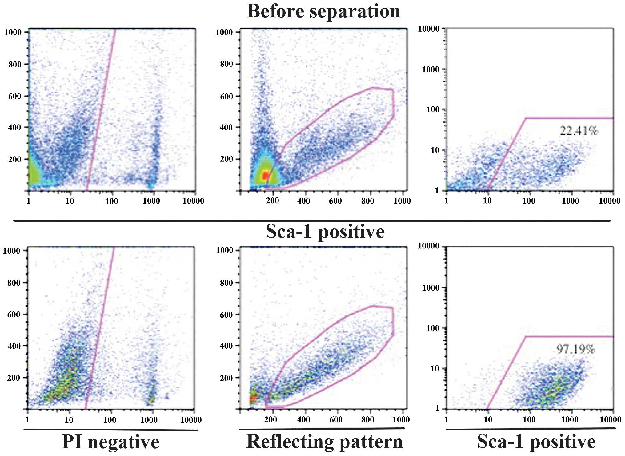
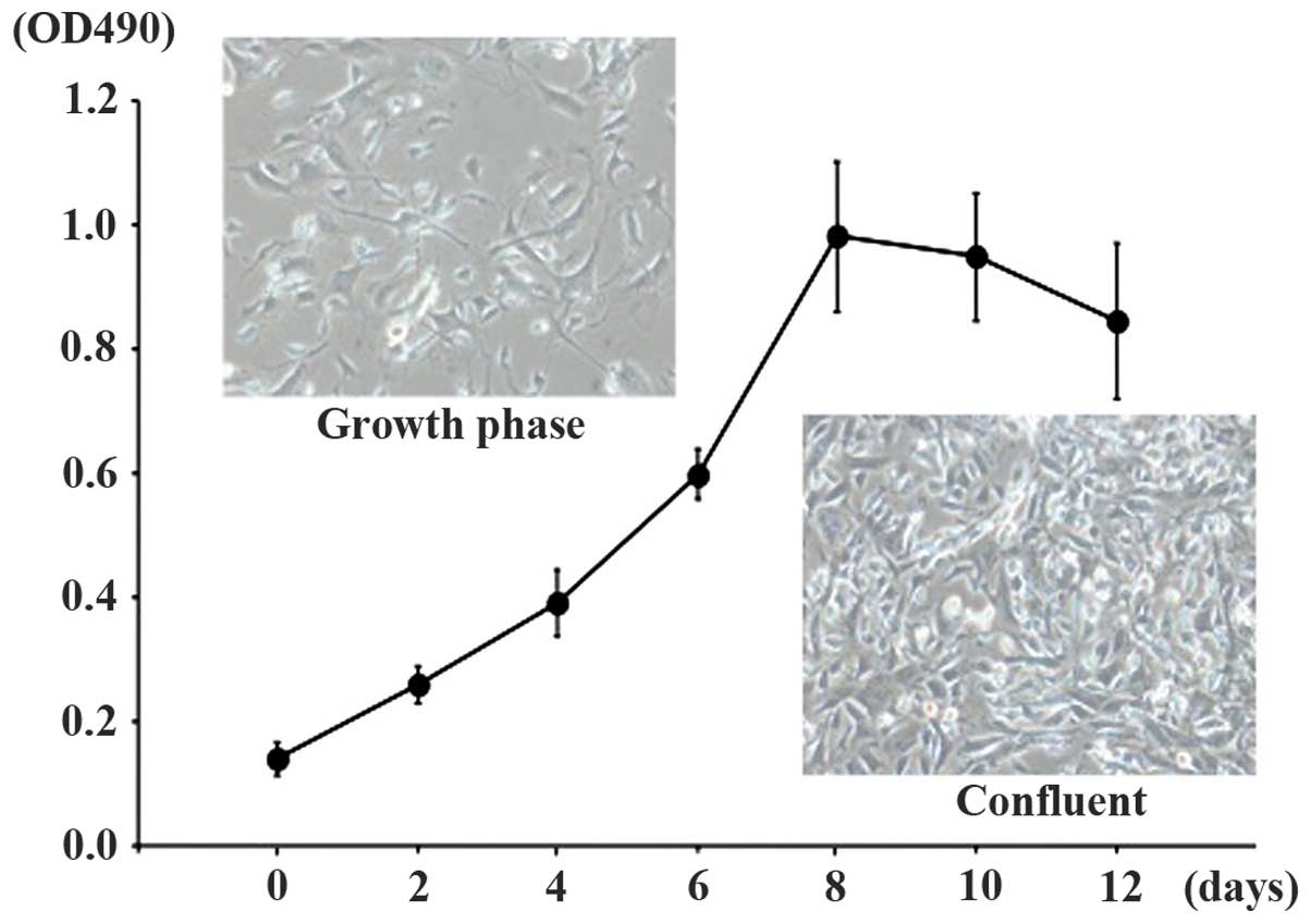
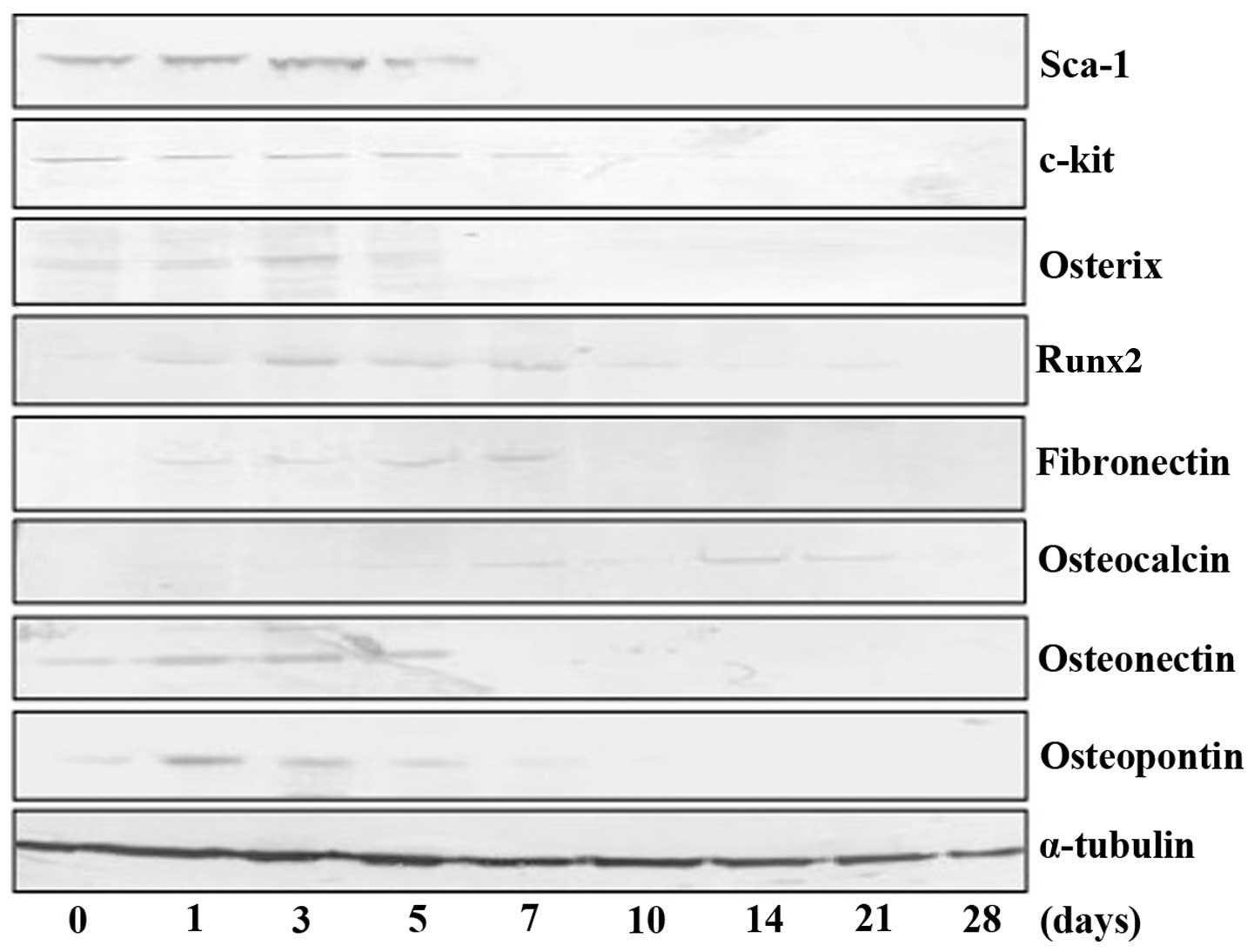
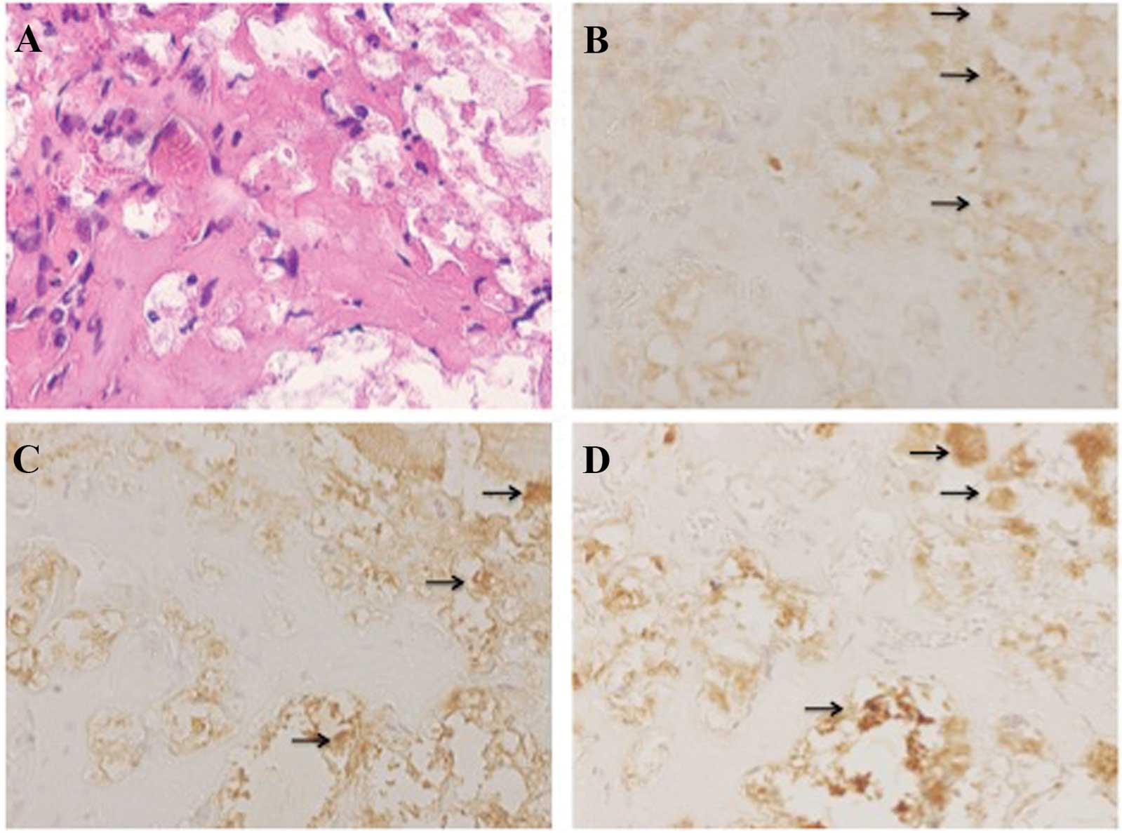
The use of stem cells represents a promising approach for the treatment of bone defects. However, successful treatments rely upon the availability of cells that are easily obtained and that appropriately differentiate into osteoblasts. The tongue potentially represents a source of autologous cells for such purposes. In the present study, the ability of stem cell antigen‑1 (Sca‑1) positive cells derived from tongue muscle to differentiate into osteoblasts was investigated. The tongue muscles were excised from Jcl‑ICR mice and tongue muscle‑derived Sca‑1‑positive cells (TDSCs) were isolated from the tongue muscle using a magnetic cell separation system with microbeads. TDSCs were cultured in plastic dishes or gelatin sponges of β‑tricalcium phosphate (β‑TCP) with bone differentiation‑inducing medium. The expression of osteogenic markers (Runx2, osterix, alkaline phosphatase, fibronectin, osteocalcin, osteonectin and osteopontin) was investigated in cultured TDSCs by western blot analysis. The formation of mineralized matrices was examined using alizarin red S and Von Kossa staining. Bone formation was investigated in cultured TDSCs by hematoxylin‑eosin staining and immunohistochemstry. In the present study, the expression of Sca‑1 in mouse tongue muscle was demonstrated and TDSCs were isolated at high purity. TDSCs differentiated into cells of osteoblast lineage, as demonstrated by the upregulation of osteoblastic marker expression. The formation of mineralized matrices was confirmed by alizarin red S or Von Kossa staining in vitro. Bone formation was observed in the gelatin sponges of β‑TCP, which were subsequently implanted under the skin of the backs of nude mice. These results suggested that TDSCs retain their osteogenic differentiation potential and therefore the tongue muscle may be used as a source of stem cells for bone regeneration.
View Figures
View References

Related Articles

Journal Cover

July-2015
Volume 12 Issue 1

Print ISSN: 1791-2997
Online ISSN:1791-3004

Sign up for eToc alerts

Recommend to Library

Copy and paste a formatted citation
x
Spandidos Publications style
Harada K, Harada T, Ferdous T, Takenawa T and Ueyama Y: Osteogenic cell fractions isolated from mouse tongue muscle. Mol Med Rep 12: 31-36, 2015.
APA
Harada, K., Harada, T., Ferdous, T., Takenawa, T., & Ueyama, Y. (2015). Osteogenic cell fractions isolated from mouse tongue muscle. Molecular Medicine Reports, 12, 31-36. https://doi.org/10.3892/mmr.2015.3350
MLA
Harada, K., Harada, T., Ferdous, T., Takenawa, T., Ueyama, Y."Osteogenic cell fractions isolated from mouse tongue muscle". Molecular Medicine Reports 12.1 (2015): 31-36.
Chicago
Harada, K., Harada, T., Ferdous, T., Takenawa, T., Ueyama, Y."Osteogenic cell fractions isolated from mouse tongue muscle". Molecular Medicine Reports 12, no. 1 (2015): 31-36. https://doi.org/10.3892/mmr.2015.3350