Introduction
Spinal cord injury (SCI) may result in severe
dysfunction in motor neurons (1,2). The
protection of spinal motor neurons following SCI is an important
area of research (3–5). However, despite a degree of
theoretical progress, there is a lack of effective drugs that are
able to improve the motor function of patients following SCI
(6–9). The protective effect of estrogen on
the central nervous system via the estrogen receptor (ER) has been
reported in a number of studies. For example, the use of the ERα
ligand, also termed E2, in the treatment of experimental autoimmune
encephalomyelitis may reduce the severity of this condition
(10). E2 may also reduce
ATP-mediated calcium influx into the primary sensory neurons of
mice (11). Furthermore, E2 may
reduce apoptosis in rat astrocytoma cells via the ER (12). Epidemiological studies have shown
that the probability of females developing SCI is lower than that
of males, and that the degree of neurological recovery in females
is better than in males. Animal experiments have confirmed that
estrogen improves motor function in the limbs of injured animals
(13,14). However, there are numerous ethical
issues in the clinical administration of estrogen, due to its
multiple side effects. Thus, further investigation into the
neuroprotective effects of estrogen is required, in order to
identify novel targets for clinical intervention. The specific
mechanisms underlying estrogen neuroprotection following SCI remain
unclear. ERs are located on the surface layer of the dorsal horn of
the spinal cord, which contains sensory motor neurons, while they
are not found in the ventral horn of the spinal cord, which
contains motor neurons (15,16).
Nonetheless, an improvement in motor function with estrogen
treatment following SCI has been observed in animal models as well
as in clinical studies (1). It has
been shown that estrogen is an antagonist of excitatory
AMPA-mediated toxicity in spinal motor neurons via the indirect
action of ER-containing glial cells (17).
G-protein-coupled receptor 30 (GPR30) is a
membrane-associated estrogen receptor that was originally
identified in the 1990s. Its mode of action and effects are
different from the conventional nuclear receptors, ERα and ERβ, and
it has no homology to these receptors. Thus, GPR30 is a novel
estrogen receptor with an independent effect. Previous studies by
this group have shown that estrogen improves the motor function of
rats with SCI and reduces apoptosis in the spinal cord following
SCI, via the membrane receptor, GPR30, rather than the conventional
ERs (18). GPR30 receptors are
located in the ventral horn of the spinal cord (18), while the classic nuclear ERs are
located in the spinal dorsal horn (19). This suggests that the effect of
estrogen, mediated by GPR30, on spinal motor neurons may be an
important target for neuroprotection following SCI.
In the present study, spinal motor neurons were used
to establish cell damage and animal injury models. E2, G1, G15 and
LY294002 were used as intervention treatments in order to observe
the protective effects of estrogen through GPR30 on spinal motor
neurons, and to explore the mechanisms underlying its effects.
Materials and methods
Culture of spinal motor neurons
In accordance with previous literature (20) as well as our own experience, spinal
motor neurons (Sciencell, Carlsbad, CA, USA) were transported to
the laboratory frozen in liquid nitrogen. After thawing at room
temperature, the neurons were homogeneously inoculated, at a
density of 600–700 cells/mm2, in a cell culture
apparatus coated with poly-Lysine (Sigma-Aldrich, St. Louis, MO,
USA) in medium composed of 482.75 ml Neurobasal medium, 10 ml B-27
(Gibco Life Technologies, Carlsbad, CA, USA), 5 ml fetal bovine
serum and 1.25 ml GlutaMAX stock (Gibco Life Technologies; pH 7.0).
The medium was changed 8 h after inoculation. A second change was
performed at 48 h. At 72 h following inoculation, the cultured
neurons were observed.
Immunofluorescence
Samples were washed with phosphate-buffered saline
(PBS) for 10 min and then fixed with 4% paraformaldehyde for 20
min. The fixed samples were then washed twice with PBS and 0.3%
triton X-100 was added for 10 min, in order to permeabilize the
cell membrane. The samples were then washed twice for 10 min with
PBS, blocked with 800 µl blocking buffer for 60 min, and the
primary antibodies (mouse anti-rat against SMI32 1:1,000
(monoclonal; Covance, Princeton, NJ, USA) or rabbit anti-rat
against GPR30 1:400 (sc-48525-R; polyclonal; Santa Cruz, Dallas,
TX, USA) for 2 h at 37°C was added. Following incubation with the
primary antibodies, the samples were washed 5 times, for 5 min each
time with PBS. The secondary antibodies (rhodamine-conjugated goat
anti-rabbit and FITC-conjugated goat anti-mouse) were then added
and the samples were incubated at 37°C for 1 h. Finally the samples
were washed twice, for 5 min each time with PBS, following which,
DAPI was used to stain the nucleus for 10 min. Following DAPI
staining the samples were washed 4 times, for 5 min each time with
PBS and mounted with anti-quenching resin. The cells were observed
by fluorescence microscopy (Olympus MF53; Olympus, Tokyo,
Japan).
Cell treatment
Estrogen E2 (17β-estradiol, Sigma-Aldrich); GPR30
agonist, G1 (Sigma-Aldrich); GPR30 inhibitor, G15 (Tocris
Bioscience, Ellisvill, MI, USA); and the phosphatidylinositol
3-kinase/protein kinase B (PI3K/Akt) pathway inhibitor, LY294002
(Cayman), were dissolved in dimethyl sulfoxide (DMSO;
Sigma-Aldrich) and added to the medium at the following
concentrations: E2 (1, 10 or 100 nM, or 1 µM), G1 (10 nM),
G15 (10 nM) and LY294002 (10 nM). Equal amounts of DMSO were added
as negative controls.
Establishment of the oxygen-glucose
deprivation model (OGD model)
The cells were place in an incubator containing 95%
nitrogen and 5% CO2. The original culture medium was
replaced by glucose-free Dulbecco’s modified Eagle’s medium (DMEM)
solution (Gibco Life Technologies). Following 3 h of OGD, the
glucose-free DMEM was changed for the original culture medium and
the cells were placed back into an incubator containing 5%
CO2 and 95% air at 37°C. Following an additional culture
period, the corresponding detection indices, including MTT assay,
flow cytometry and western blotting, were performed.
MTT assay procedure
Neuronal growth was detected using a MTT assay. MTT
(Sigma-Aldrich, 50 mg) was dissolved in 10 ml PBS. Following
sterile filtration, this solution was stored at −20°C for
subsequent use. Once grouped, the cells were cultured in 96-well
culture plates. Following a cell culture period of 12, 24, 36 or 48
h, 20 µl of MTT was added into each well and the cells were
cultured for an additional 4 h. After 4 h, unabsorbed MTT was
removed and 150 µl DMSO was added to each well to dissolve
any purple crystals. Following 10 min shaking, the samples were
placed into a microplate reader (Tecan, Mainz, Germany) in order to
measure the optical density at 570 nm.
Flow cytometry
Cells were collected and centrifuged at 1000 × g for
10 min at 4°C. The supernatants were discarded and 1 ml of ice-cold
PBS was added and gently shaken to suspend the cells. The cells
were then centrifuged again at 1000 × g for 10 min at 4°C, and the
supernatants were discarded. The cells were resuspended in 200
µl of Banding buffer (Roche, Basel, Switzerland). To this
buffer, 10 µl Annexin V-FITC and 10 µl PI were added
and gently mixed for 15 min at room temperature in darkness. Flow
Cytometry was used in order to detect cell apoptosis and to
calculate the percentage of cells in early late and total
apoptosis, and of necrotic cells in each group.
Western blot analysis
Following homogenization, the samples were
centrifuged at 28341.3 × g for 1 min at 4°C (10). The supernatants were boiled for 5
min. The samples were separated using an SDS-PAGE gel containing
7.5% polyacrylamide. The protein bands were transferred to PVDF
membranes (GE Healthcare). The membranes were blocked with 5%
non-fat milk in Tris-buffered saline with Tween-20 for 1 h at room
temperature and incubated with anti-GPR30 (1:400; Santa) or
anti-Akt (1:1,000; KangChen, Shanghai, China) antibodies at 4°C
overnight. This was followed by incubation with the appropriate
secondary antibodies. Immunoreactivity was detected using enhanced
chemiluminescence (ECL; GE Healthcare, Buckinghamshire, United
Kingdom) after washing with TBST. Finally, the ECL-exposed films
were digitized. Densitometric quantification was performed using
ImageJ software (National Institutes of Health, Bethesda, MD,
USA).
Animal models and interventions
Healthy male Sprague-Dawley rats (weight, 200–220 g;
The Third Military Medical University, Chongqing, China) were
selected and anesthetized by intraperitoneal injection of 5%
chloral hydrate (400 mg/kg; The Third Military Medical University).
Rats were placed in the prone position on the operating table
following back shaving and routine disinfection. Following
T8-centric longitudinal cutting of the skin and subcutaneous
tissues, the paraspinal muscles were dissected to expose the
spinous process and vertebral plate, using ophthalmic scissors to
cut the spinous process of the T8, and a hemostat to break the
vertebral plate of the T8 along the intervertebral space in order
to fully expose the T8 spinal cord. Sterile cotton was used to
achieve hemostasis, and a 10 g rod was allowed to fall freely from
a height of 1.0 cm in order to induce SCI. The diameter of the
lower end of the rod was 2.5 mm. This was able to produce an injury
with 10 gcf of energy. Following the injury, the paraspinal muscles
and the skin layers were sutured, the wound was disinfected and the
rats were placed under a lamp to warm prior to awakening. The rats
were then housed and fed in single clean cages at room temperature
(20±2°C), with a light/dark cycle of 12 h, and a background noise
level of 40±10 db. The cages were frequently cleaned. Nutrition was
enforced, and when necessary artificial urination and defecation
were employed.
Animals were dosed via the tail vein according to
the previous literature (21–24).
All drugs were dissolved in DMSO and administered once 15 min and
24 h following SCI as follows: E2 (100 µg/kg), G1 (50
µg/kg), G15 (100 µg/kg), and LY294002 (250
µg/kg).
All treatment procedures were approved by the
Institute of Animal Ethics of the Chongqing Southwest Hospital
(Chongqing, China).
Basso, Beattie, Bresnahan (BBB)
scoring
Rats were grouped into the following groups:
Control, DMSO, E2, G1, E2+G15 and E2+LY294002, with five rats in
each group. The average score was calculated from the scores of the
individual rats. Animals were placed on a 2 m-diameter flat, smooth
area of ground and allowed to move freely. The BBB open space
movement score was conducted by two individuals familiar with BBB
scoring, who were not participating in the present study (25). The analysis was double-blind. The
rats were independently observed and recorded for 4 min in order to
determine the number of motions of their hindlimb joints, their
movement range and load level, coordination of the forelimbs and
hindlimbs, and the activities of front paws, hind paws and tails.
Their BBB scores were averaged. There was no filling of the
bladder, perineal inflammation, or hindlimb trauma.
Statistical analysis
Statistical analyses were performed using SPSS 19.0
(SPSS, Inc., Chicago, IL, USA). The data are expressed as the mean
± standard error. Statistical comparisons were performed using
unpaired Student’s t-test or one-way analysis of variance.
P<0.05 was considered to indicated a statistically significant
difference.
Results
Estrogen has a protective effect on
spinal motor neurons, via GPR30, following OGD injury
After the spinal motor neurons were cultured
(Fig. 1 A–C), cell injury was
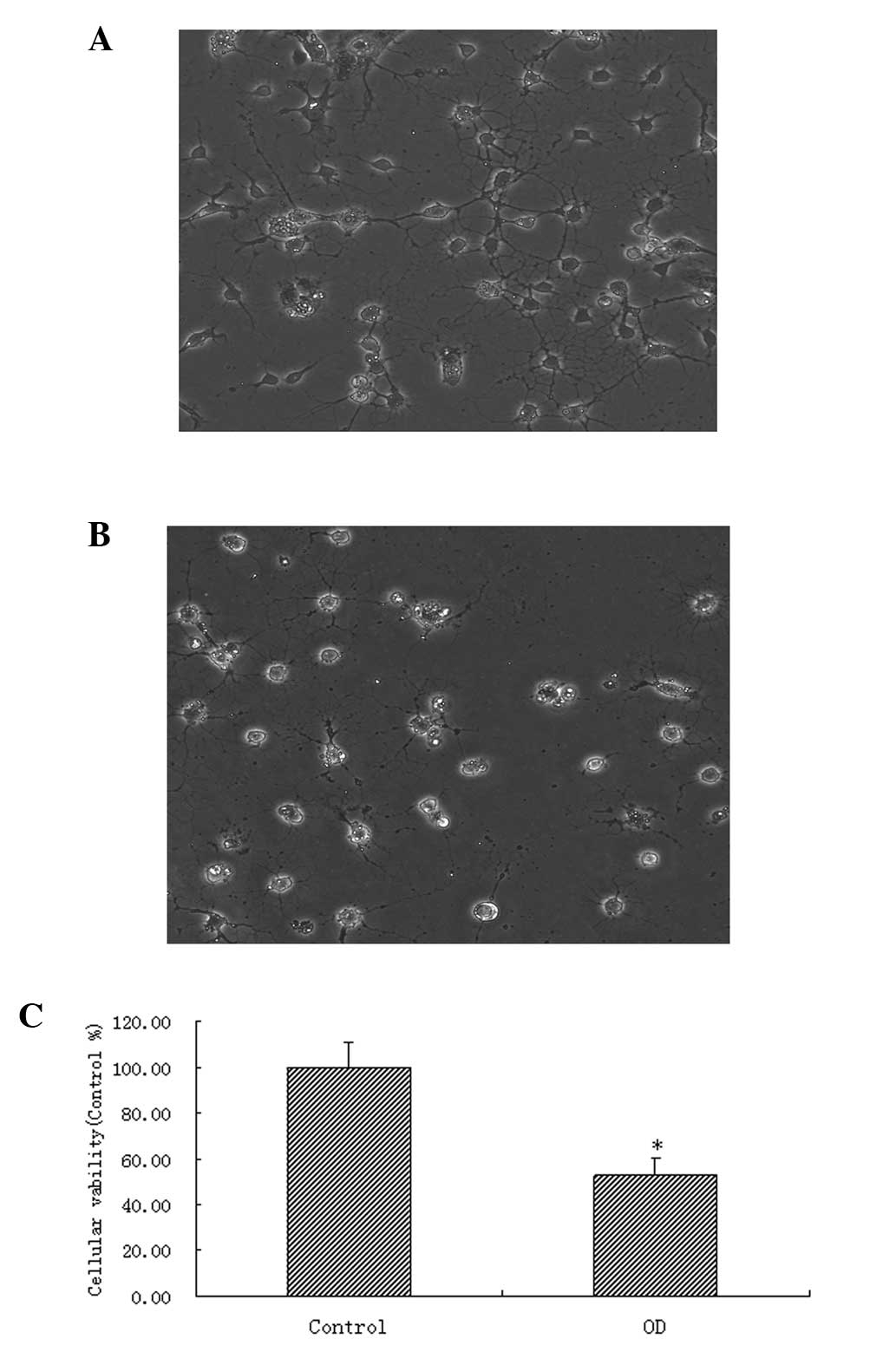
induced (Fig. 2 A–C) by OGD. The
cell activity after 24 h was detected by MTT assay. In accordance
with the previous literature (26), various concentrations of estrogen
in DMSO (1, 10 or 100 nM, or 1 µM) were added to the medium
as interventions for 24 h in order to observe the protective effect
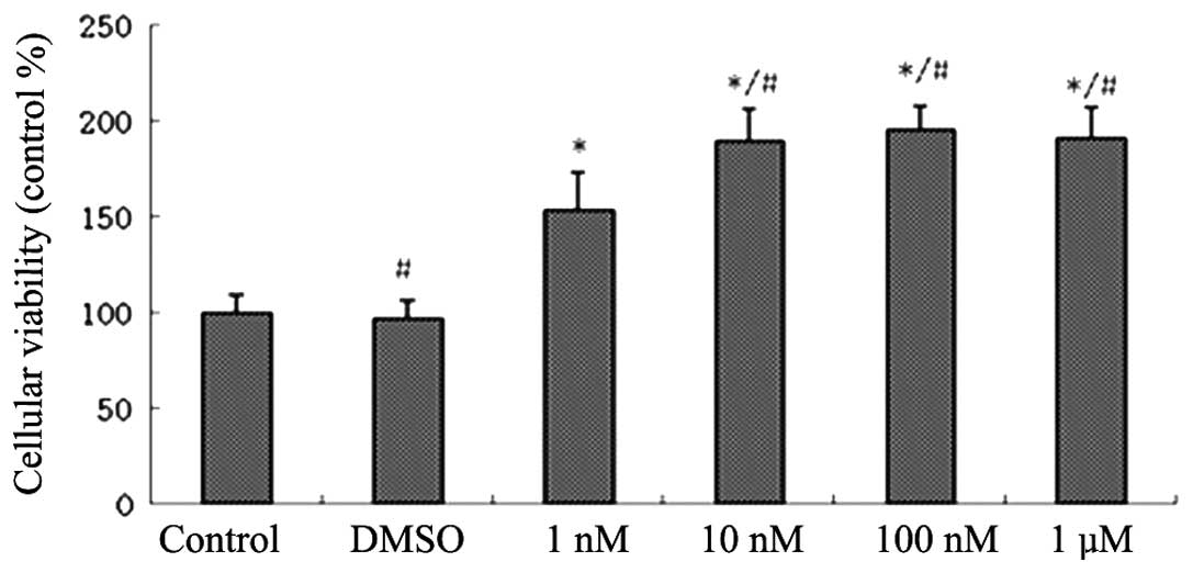
of E2 on spinal motor neurons. The cell viabilities of the E2
groups were higher than those of the DMSO groups. The cell
viabilities of the 10 nM, 100 nM and 1 µM groups were higher
than the viability of the 1 nM group. No significant differences
were detected among the other three groups (Fig. 3). These results suggest that
estrogen may exert a protective effect on rat spinal cord motor
neurons following OGD.
In order to understand whether E2 exerts a
protective effect via GPR30, the GPR30 agonist, G1, and the GPR30
inhibitor, G15, were used 24 hours after injury. The cells were
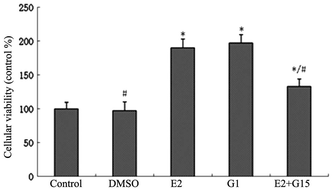
grouped as follows: Control, DMSO, E2, G1 and E2+G15. The MTT assay
showed that the cell viability of the G1 and E2 groups was
significantly higher than that of the DMSO group, while no
significant difference was detected between the G1 group and the E2
group. The cell activity of the E2+G15 group was higher than that
of the DMSO group, while it was significantly lower than that of
the E2 group (Fig. 4). This
indicated that the GPR30 agonist, G1, exerted the same cell
protective effect as E2, while the GPR30 inhibitor, G15, partially
inhibited the neuroprotective effect of E2. In order to determine
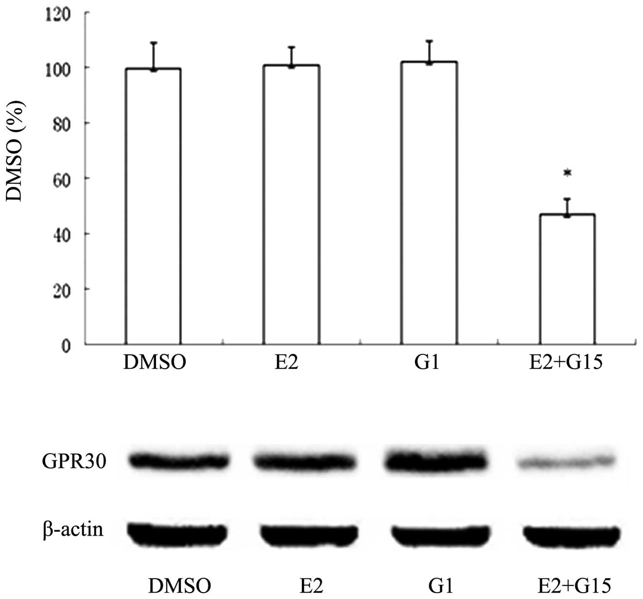
whether G15 indeed inhibited the GPR30 receptor, total protein was
collected from the cells following the MTT assay, and samples were
analyzed using western blotting. The expression of GPR30 was higher
in the DMSO, E2 and G1 groups than in the E2+G15 group (Fig. 5). These results suggested that E2
exerts its neuroprotective effect on spinal motor neurons following
OGD, via GPR30.
E2 protects spinal motor neurons via
inhibition of apoptosis
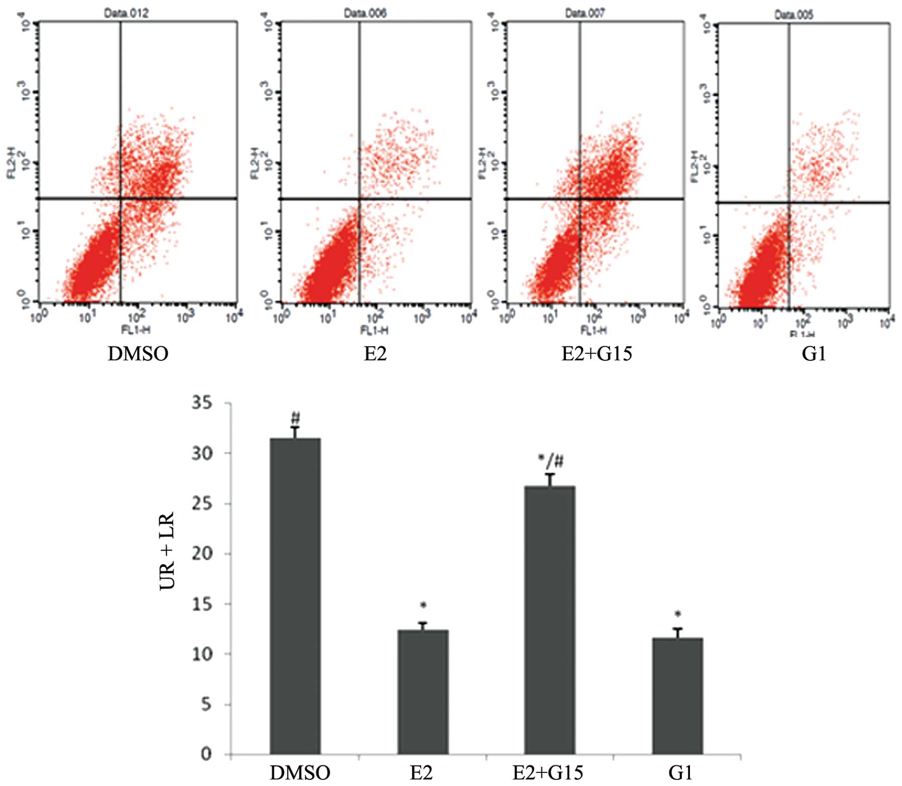
Following treatment with DMSO, E2, G1 or E2+G15 in
cells subjected to OGD for 24 h, flow cytometry was employed. The
results showed that the proportions of apoptotic cells in the E2
and G1 groups were significantly lower than that in the DMSO group.
The proportion of apoptotic cells in the E2+G15 group was
significantly higher than that in the E2 group (Fig. 6), indicating that E2 exerted an
antiapoptotic effect in spinal motor neurons following OGD, via its
effect on GPR30.
PI3K/Akt is the intermediate pathway for
the GPR30-mediated antiapoptotic effect of estrogen
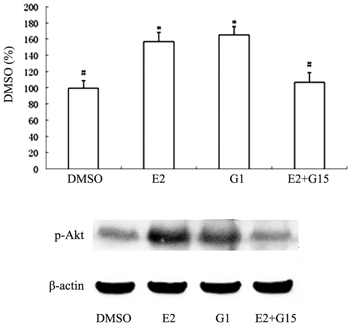
Western blotting was used to detect the expression
of phosphorylated Akt (P-Akt) and its downstream products, using
the same treatment groups. The results demonstrated that P-Akt
expression in the E2 and G1 groups was higher than that in the DMSO
group, while the P-Akt expression in the E2+G15 group was
significantly lower than in the OGD+E2 group (Fig. 7), indicating that estrogen
regulates the activity of the PI3K/Akt pathway through GPR30. In
order to determine whether the PI3K/Akt pathway mediates the
connection between E2 and spinal motor neuron apoptosis, the
PI3K/Akt pathway inhibitor, LY294002, was used to treat the cells.
Flow cytometry showed that the E2+LY294002 group exhibited a higher
proportion of apoptotic cells than the E2 group (Fig. 8), indicating that blocking the
PI3K/Akt pathway weakens the antiapoptotic effect of E2. This
suggests that PI3K/Akt is an intermediate pathway in the
GPR30-mediated antiapoptotic effects of estrogen.
In vivo experiments
The appropriate drugs were given to the rats
following SCI via tail vein injection with either DMSO, E2, G1,
Y294002 or G15. BBB scoring was used to evaluate the motor ability
of the hindlimbs of the rats. Estrogen and GI treatments improved
the hindlimb motor ability of rats following SCI. The G1 group
exhibited a significantly higher BBB score than the E2 group at 21
days. The scores at the majority of time points in the E2+LY294002
group were higher than those in the DMSO group, while the scores at
all time points after 3 days were lower than those in the E2 group
(Fig. 9), which suggested that
LY294002 partially antagonized the neuroprotective effect of E2.
The scores of the E2+G15 group exhibited a similar trend to those
of the E2 group.
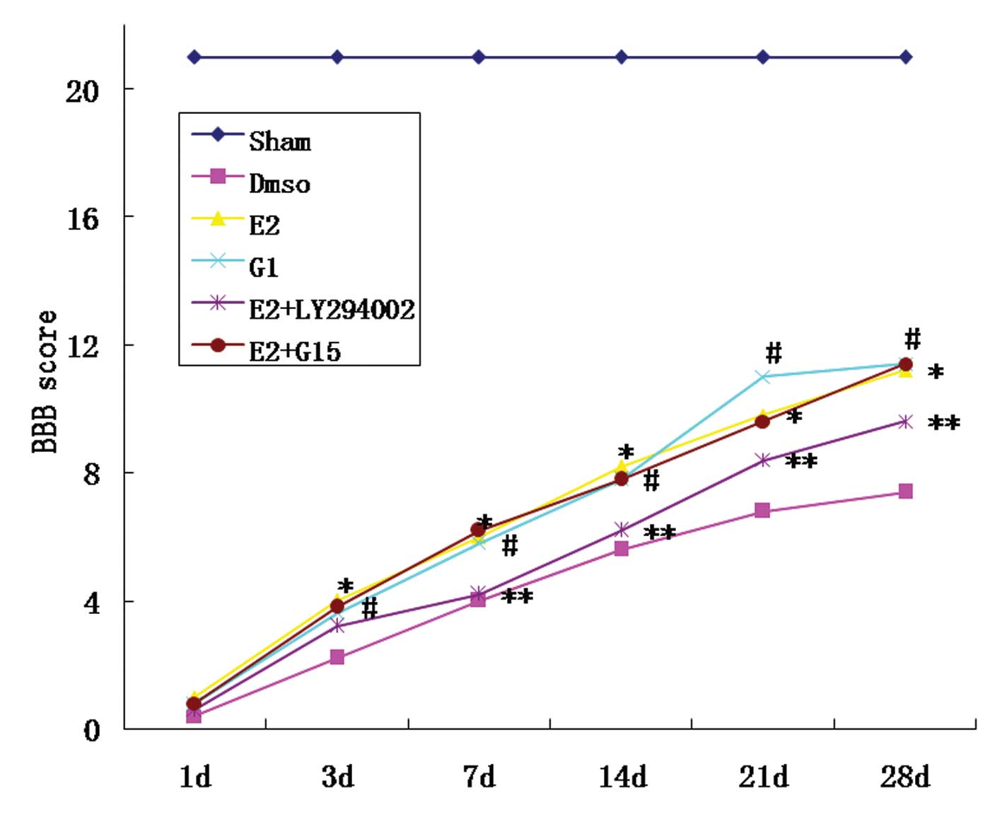 | Figure 9BBB score results. The scores of the
E2 group were higher than those for the DMSO group, at 3 days
following SCI (*P<0.05; E2 group, vs. DMSO group).
The scores of the G1 group were higher than those of the DMSO
group, at 3 days following SCI (#P<0.05; G1 group,
vs. DMSO group), with a higher score on day 21 than that of the E2
group (#P<0.05; G1 group, vs. DMSO group). The scores
of the E2+LY294002 group at 3, 14, 21 and 28 days following SCI
were higher than the scores for the E2 group
(**P<0.05; E2+y294002 group, vs. DMSO group). The
scores of the E2+G15 group were similar to those of the E2 group.
BBB, Basso, Beattie, Bresnahan score; DMSO, dimethyl sulfoxide;
SCI, spinal cord injury. |
Discussion
A previous study by this group confirmed that
estrogen exerts a neuroprotective effect following SCI, via GPR30
(18). It was hypothesized that
apoptosis within the spinal cord may increase following SCI, and
that estrogen administration may decrease this apoptosis. In the
present study a direct protective effect of estrogen on spinal
motor neurons was observed and the mechanisms underlying this
effect were subsequently further investigated.
After cells were thawed and cultured to stability
for 72 h, they were confirmed as spinal motor neurons by the
neurofilament marker, SMI32 (Fig.
1). In addition to mechanical trauma, ischemia and hypoxia are
the primary pathological processes that occur following SCI, the
persistence of which has been confirmed in observations during
clinical autopsy and in microvasculature perfusion following SCI,
in animals in which the neuron-containing gray matter of the spinal
cord dominates (27). These
secondary processes contribute the evolution of the pathological
changes of rats with spinal cord injury involving mainly the grey
matter (28,29). A linear correlation between the
severity of SCI and loss of blood flow in the spinal cord has been
demonstrated (30). In the present
study, a classical model of OGD was established in order to
simulate hypoxic-ischemic injury following SCI. This model was easy
to establish, controllable and reproducible. In the current study,
marked neuronal swelling was observed following OGD. In addition,
the neuronal projections were beaded, with a number exhibiting
breakage. These morphological changes were consistent with the
changes in cell morphology that are commonly observed following SCI
(26). The MTT results also
confirmed that cell activity following OGD injury was lower than
that in the control group (Fig.
2).
As there have been no previous studies showing that
estrogen acts directly on cultured spinal motor neurons, a series
of gradient concentrations of estrogen were designed in the present
study, with reference to the concentrations used in previous
literature for hippocampal neurons (31–33).
The concentration of estrogen was varied in order to examine its
protective effect on spinal motor neurons following OGD injury. The
MTT results demonstrated that only a concentration of estrogen
>1 nM exerted a protective effect on cells. Concentrations of
estrogen ≥10 nM significantly increased the percentage of viable
cells, compared with the 1 nM group, with no significant difference
among the three groups treated with the higher doses (Fig. 3). This finding was consistent with
the estrogen concentrations that have been shown in previous
studies to be effective in neurons in other locations and with
other types of injury (31–33).
G15 is a recently identified competitive antagonist of GPR30, which
interacts specifically with GPR30 and has no affinity for the
classic ERs. G15 has become an effective and convenient tool for
use in GPR30 studies (34). By
using the GPR30 agonist, G1, and the GPR30 antagonist, G15, it was
observed that G1 exerted the same cell protective effect as
estrogen, while G15 partially offset thIs protective effect
(Fig. 4), indicating that estrogen
exerts its neuroprotective effect on spinal motor neurons through
its action GPR30. These results explain in part why estrogen
improves the motor function of animal limbs following SCI (13,14).
In a previous animal study by this group, it was
observed that apoptosis in the spinal cord increases in the early
stages following SCI (18). It has
been reported in a number of previous studies that estrogen may
exert an antiapoptotic effect following SCI by enhancing
antiapoptotic gene expression, while reducing caspase-3 activity
and inhibiting calcium activation (21,35–37).
However, the pathways involved in mediating these effects remain to
be elucidated. To the best of our knowledge, the present results
demonstrate for the first time that estrogen reduces spinal motor
neuron apoptosis following OGD by a direct effect on spinal motor
neurons, via GPR30 (Fig. 6). This
represents a potential approach for exploring the protective effect
of estrogen on spinal motor neurons. There are a number of pathways
upstream of apoptosis, such as the death receptor-mediated pathway,
the mitochondrial pathway and the endoplasmic reticulum pathway
(38,39). The PI3K/Akt pathway is involved in
the inhibition of apoptosis and the promotion of proliferation in
cells, via its effect on the activated state of multiple downstream
effectors. It has been observed in a number of studies that GPR30
may regulate the PI3K/Akt pathway. For example, it is involved in
estrogen-driven, GPR30-mediated endometrial carcinoma cell
proliferation via the PI3K/Akt pathway (40). Furthermore, GPR30 receptors on the
endoplasmic reticulum of tumor cells cause rapid calcium
mobilization through activation of the PI3K/Akt pathway (41). Finally, using the specific GPR30
agonist, STX, to treat H-38 cells and choriocarcinoma cells
significantly activates the PI3K/Akt pathway, thereby increasing
intracellular phosphatidyl alcohol, while ERα and ERβ receptor
agonists do not produce such effects (42). The present results demonstrated
that G1 and estrogen increased the expression of PI3K/Akt pathway
metabolites and the phosphorylation of Akt (P-Akt), while G15
partially offset these effects (Fig.
7). In addition, the PI3K/Akt pathway antagonist, LY294002, was
used in order to block the protective effect of estrogen on spinal
motor neurons (Fig. 8). These
results suggest that estrogen exerts an antiapoptotic effect on
spinal motor neurons through the regulation of the PI3K/AKt
pathway, via GPR30.
In order to investigate whether the above results
occur in vivo, E2, G1, G15 and LY294002 were administered to
rats following SCI, via tail vein injections. E2 and G1 improved
hindlimb motor function in rats from an early stage following SCI.
LY294002 antagonized the neuroprotective effect of estrogen at a
later stage following SCI. No significant difference between E2
gorup and E2+G15 group were observed in the animal studies. This
may be related to the following aspects: ER interference cannot be
excluded in vivo; G15 may pass through the blood-brain
barrier; or there may be an impact of the complex internal
environment of the body. The antagonism by LY294002 on the
protective effect of estrogen, also indicates that the PI3K/Akt
pathway is the downstream molecular pathway for GPR30-mediated
estrogen neuroprotection.
The present study is only a preliminary exploration
of the mechanisms underlying the estrogen protective effect on
spinal motor neurons. Due to time limitations, only certain aspects
of the mechanisms underlying the effects on apoptosis and the
PI3K/Akt pathway were investigated. The interactions of each part
of the mechanism require further elucidation.
Estrogen exerts a protective effect on spinal motor
neurons following OGD injury. Estrogen exhibits a protective role
against apoptosis through its action on the membrane receptor,
GPR30. PI3K/Akt is one of the antiapoptotic pathways of estrogen by
way of GPR30. Protection of spinal cord motor function following
SCI is an important focus in clinical practice, and spinal motor
neurons are the primary cells involved in this process. The present
study illustrates a preliminary mechanism for the protection effect
of estrogen on motor neurons, and identifies targets for clinical
intervention that may be further explored in future studies.
Acknowledgments
The present study was supported by National Natural
Science Foundation of China (grant no. 81301058).
References
|
1
|
Thuret S, Moon LD and Gage FH: Therapeutic
interventions after spinal cord injury. Nat Rev Neurosci.
7:628–643. 2006. View
Article : Google Scholar : PubMed/NCBI
|
|
2
|
Fehlings MG and Hawryluk GW: Scarring
after spinal cord injury. J Neurosurg Spine. 13:165–167. 2010.
View Article : Google Scholar : PubMed/NCBI
|
|
3
|
Crowe MJ, Bresnahan JC, Shuman SL, et al:
Apoptosis and delayed degeneration after spinal cord injury in rats
and monkeys. Nat Med. 3:73–76. 1997. View Article : Google Scholar : PubMed/NCBI
|
|
4
|
Liu XZ, Xu XM, Hu R, et al: Neuronal and
glial apoptosis after traumatic spinal cord injury. J Neurosci.
17:5395–5406. 1997.PubMed/NCBI
|
|
5
|
Grossman SD, Rosenberg LJ and Wrathall JR:
Temporal-spatial pattern of acute neuronal and glial loss after
spinal cord contusion. Exp Neurol. 168:273–282. 2001. View Article : Google Scholar : PubMed/NCBI
|
|
6
|
Hu R, Zhou J, Luo C, et al: Glial scar and
neuroregeneration: Histological, functional, and magnetic resonance
imaging analysis in chronic spinal cord injury. J Neurosurg Spine.
13:169–180. 2010. View Article : Google Scholar : PubMed/NCBI
|
|
7
|
Macias CA, Rosengart MR, Puyana JC, et al:
The effects of trauma center care, admission volume, and surgical
volume on paralysis after traumatic spinal cord injury. Ann Surg.
249:10–17. 2009. View Article : Google Scholar
|
|
8
|
Samantaray S, Sribnick EA, Das A, et al:
Neuroprotective efficacy of estrogen in experimental spinal cord
injury in rats. Ann NY Acad Sci. 1199:90–94. 2010. View Article : Google Scholar : PubMed/NCBI
|
|
9
|
Fu ES and Tummala RP: Neuroprotection in
brain and spinal cord trauma. Curr Opin Anaesthesiol. 18:181–187.
2005. View Article : Google Scholar
|
|
10
|
Elloso MM, Phiel K, Henderson RA, et al:
Suppression of experimental autoimmune encephalomyelitis using
estrogen receptor-selective ligands. J Endocrinol. 185:243–252.
2005. View Article : Google Scholar : PubMed/NCBI
|
|
11
|
Cho T and Chaban VV: Interaction between
P2X3 and ERα/ERβ in ATP-mediated calcium signaling in mice sensory
neurons. J Neuroendocrinology. 24:789–797. 2012. View Article : Google Scholar
|
|
12
|
Sribnick EA, Ray SK and Banik NL: Estrogen
prevents glutamate-induced apoptosis in C6 glioma cells by a
receptor-mediated mechanism. Neuroscience. 137:197–209. 2006.
View Article : Google Scholar
|
|
13
|
Furlan JC, Krassioukov AV and Fehlings MG:
The effects of gender on clinical and neurological outcomes after
acute cervical spinal cord injury. J Neurotrauma. 22:368–381. 2005.
View Article : Google Scholar : PubMed/NCBI
|
|
14
|
Webb AA, Chan CB, Brown A and Saleh TM:
Estrogen reduces the severity of autonomic dysfunction in spinal
cord-injured male mice. Behav Brain Res. 171:338–349. 2006.
View Article : Google Scholar : PubMed/NCBI
|
|
15
|
Vanderhorst VG, Gustafsson JA and Ulfhake
B: Estrogen receptor-alpha and -beta immunoreactive neurons in the
brainstem and spinal cord of male and female mice: Relationships to
monoaminergic, cholinergic, and spinal projection systems. J Comp
Neurol. 488:152–179. 2005. View Article : Google Scholar : PubMed/NCBI
|
|
16
|
Vanderhorst VG, Terasawa E and Ralston HJ
III: Estrogen receptor-alpha immunoreactive neurons in the
brainstem and spinal cord of the female rhesus monkey:
Species-specific characteristics. Neuroscience. 158:798–810. 2009.
View Article : Google Scholar
|
|
17
|
Platania P, Seminara G, Aronica E, et al:
17B-estradiol rescues spinal motoneurons from AMPA-induced
toxicity: A role for glial cells. Neurobiol Dis. 20:461–470. 2005.
View Article : Google Scholar : PubMed/NCBI
|
|
18
|
Hu R, Sun HD, Zhang Q, et al: G-protein
coupled estrogen receptor 1 mediated estrogenic neuroprotection
against spinal cord injury. Crit Care Med. 40:3230–3237. 2012.
View Article : Google Scholar : PubMed/NCBI
|
|
19
|
McEwen BS: Invited review: Estrogens
effects on the brain: Multiple sites and molecular mechanisms. J
Appl Physiol. 91:2785–2801. 2001.PubMed/NCBI
|
|
20
|
Yang X, Tomita T, Wines-Samuelson M,
Beglopoulos V, Tansey MG, Kopan R and Shen J: Notch1 signaling
influences v2 interneuron and motor neuron development in the
spinal cord. Dev Neurosci. 28:102–117. 2006. View Article : Google Scholar : PubMed/NCBI
|
|
21
|
Yune TY, Kim SJ, Lee SM, et al: Systemic
administration of 17beta-estradiol reduces apoptotic cell death and
improves functional recovery following traumatic spinal cord injury
in rats. J Neurotrauma. 21:293–306. 2004. View Article : Google Scholar : PubMed/NCBI
|
|
22
|
Lu CL, Hsieh JC, Dun NJ, et al: Estrogen
rapidly modulates 5-hydroxytrytophan-induced visceral
hypersensitivity via GPR30 in rats. Gastroenterology.
137:1040–1050. 2009. View Article : Google Scholar : PubMed/NCBI
|
|
23
|
Dennis MK, Burai R, Ramesh C, et al: In
vivo effects of a GPR30 antagonist. Nature ChemBiol. 5:421–427.
2009.
|
|
24
|
Du DS, Ma XB, Zhang JF, et al: The
protective effect of capsaicin receptor-mediated genistein
postconditioning on gastric ischemia-reperfusion injury in rats.
Dig Dis Sci. 55:3070–3077. 2010. View Article : Google Scholar : PubMed/NCBI
|
|
25
|
Basso DM, Beattie MS and Bresnahan JC: A
sensitive and reliable locomotor rating scale for open field
testing in rats. J Neurotrauma. 12:1–21. 1995. View Article : Google Scholar : PubMed/NCBI
|
|
26
|
Yu HP, Hsieh YC, Suzuki T, et al: The
PI3K/Akt pathway mediates the nongenomic cardioprotective effects
of estrogen following trauma-hemorrhage. Ann Surg. 245:971–977.
2007. View Article : Google Scholar : PubMed/NCBI
|
|
27
|
Tator CH: Upadate on the pathophysiology
and pathology of acute spinal cord injury. Brain Pathol. 5:407–413.
1995. View Article : Google Scholar : PubMed/NCBI
|
|
28
|
Ambrozaitis E, Spakauskas B and Vaitkaitis
D: Pathophysiology of acute spinal cord injury. Medicina (Kaunas).
255–261. 2006.In Lithuanian.
|
|
29
|
Fiford RJ, Bilston LE, Waite P and Lu J: A
vertebral dislocation model cord injury in rats. J Neurotrauma.
21:451–458. 2004. View Article : Google Scholar : PubMed/NCBI
|
|
30
|
Akdemir H, Paşaoğlu A, Oztürk F, et al:
Histopathology of experiment spinal cord trauma. Comparison of
treatment with TRH, naloxone, and dexamethasone. Res Exp Med
(Berl). 92:177–183. 1992. View Article : Google Scholar
|
|
31
|
Nguyen TV, Jayaraman A, Quaglino A and
Pike CJ: Androgens selectively protect against apoptosis in
hippocampal neurones. J Neuroendocrinol. 22:1013–1022. 2010.
View Article : Google Scholar : PubMed/NCBI
|
|
32
|
Meda C, Vegeto E, Pollio G, et al:
Oestrogen prevention of neural cell death correlates with decreased
expression of mRNA for the pro-apoptotic protein nip-2. J
Neuroendocrinol. 12:1051–1059. 2000. View Article : Google Scholar : PubMed/NCBI
|
|
33
|
Revankar CM, Cimino DF, Sklar LA, et al: A
transmembrane intracellular estrogen receptor mediates rapid cell
signaling. Science. 307:1625–1630. 2005. View Article : Google Scholar : PubMed/NCBI
|
|
34
|
Dennis MK, Burai R, Ramesh C, et al: In
vivo effects of a GPR30 antagonist. Nat Chem Biol. 5:421–427. 2009.
View Article : Google Scholar : PubMed/NCBI
|
|
35
|
Yune TY, Park HG, Lee JY and Oh TH:
Estrogen-induced Bcl-2 expression after spinal cord injury is
mediated through phosphoinositide- 3-kinase/Akt-dependent CREB
activation. J Neurotrauma. 25:1121–1131. 2008. View Article : Google Scholar : PubMed/NCBI
|
|
36
|
Sribnick EA, Matzelle DD, Ray SK and Banik
NL: Estrogen treatment of spinal cord injury attenuates calpain
activation and apoptosis. J Neurosci Res. 84:1064–1075. 2006.
View Article : Google Scholar : PubMed/NCBI
|
|
37
|
Das A, Smith JA, Gibson C, et al: Estrogen
receptor agonists and estrogen attenuate TNF-α-induced apoptosis in
VSC4.1 motoneurons. J Endocrinol. 208:171–182. 2011. View Article : Google Scholar
|
|
38
|
Danial NN and Korsmeyer SJ: Cell death:
Critical control points. Cel. 116:205–219. 2004. View Article : Google Scholar
|
|
39
|
Ferrington DA, Tran TN, Lew KL, et al:
Different death stimuli evoke apoptosis via multiple pathways in
retinal pigment epithelial cells. Exp Eye Res. 83:638–650. 2006.
View Article : Google Scholar : PubMed/NCBI
|
|
40
|
Zhang J, Yang Y, Zhang Z, et al: Gankyrin
plays an essential role in estrogen-driven and GPR30-mediated
endometrial carcinoma cell proliferation via the PTEN/PI3K/AKT
signaling pathway. Cancer Lett. 339:279–287. 2013. View Article : Google Scholar
|
|
41
|
Revankar CM, Mitchell HD, Field AS, Burai
R, Corona C, Ramesh C, Sklar LA, Arterburn JB and Prossnitz ER:
Synthetic estrogen derivatives demonstrate the functionality of
intracellular GPR30. ACS Chem Biol. 2:536–544. 2007. View Article : Google Scholar : PubMed/NCBI
|
|
42
|
Lin BC, Suzawa M, Blind RD, et al:
Stimulating the GPR30 estrogen receptor with a novel tamoxifen
analogue activates SF-1 and promotes endometrial cell
proliferation. Cancer Res. 69:5415–5423. 2009. View Article : Google Scholar : PubMed/NCBI
|


