Introduction
Glaucoma remains the leading cause of irreversible
blindness globally, and the apoptosis and/or death of retinal
ganglion cells (RGCs) induced by abnormally high intraocular
pressure (IOP) is the predominant pathophysiological basis of
glaucoma (1). Lowering the
elevated IOP in the eyes is the only effective treatment for
glaucoma at present, however, a substantial proportion of patients
with glaucoma experience visual field deterioration even following
a significant IOP reduction, indicating progression of the disease
(2). Neuroprotection aiming to
rescue RGCs from degeneration is, therefore, considered to be
important in glaucoma treatment in addition to IOP reduction.
Leukemia inhibitory factor (LIF), a member of the
interleukin (IL)-6 family, has been revealed to be a potential
neuroprotective cytokine (3).
Members of the IL-6 family do not share sequence homology, however,
they are able to activate the same receptor, glycoprotein 130
(gp130) (4). Binding of LIF to its
low-affinity LIF receptor (LIFR), results in the tyrosine
phosphorylation of LIF, followed by the formation of a heterodimer.
The heterodimer binds to its high-affinity receptor gp130 and
exerts a variety of physiological effects via the activation of
certain signaling pathways, including the Janus kinase/signal
transducers and activators of transcription (JAK/STAT) pathway and
mitogen-activated protein kinase (MAPK) pathways, including the
extracellular signal-regulated kinase (ERK) and/or
phosphatidylinositol-3 kinase (PI3K) pathways (5,6).
LIF has been reported to prevent the death of
axotomized sensory neurons in the dorsal root ganglia and promote
peripheral regeneration in rats (7,8). A
previous study also demonstrated that endogenous LIF has an axon
protective capacity in acute experimental autoimmune
encephalomyelitis in mice (9).
In the eye, LIF protects photoreceptor cells against
degeneration and extends the lifespan of photoreceptors following
light-induced retinal damage (10,11).
LIF is also reported to promote the in vitro generation,
survival and maturation of oligodendrocytes of the rat optic nerve
(12). Furthermore, in a rat
glaucoma model, upregulation in the gene expression of LIF is
detected in early optic nerve head injury (13), suggesting its potential role in the
pathophysiology of glaucoma. However, the potential neuroprotective
effect of LIF on RGCs in glaucoma remains to be fully elucidated.
In the present study, the expression levels of LIF, LIFR and the
downstream signaling pathway of LIF (STAT3, Akt and ERK1/2) were
investigated in the rat retina following acute IOP elevation.
Materials and methods
Establishment of a rat acute ocular
hypertension model
All experimental procedures were performed in
accordance with the ARVO Statement for the Use of Animals in
Ophthalmic and Vision Research and the experimental protocol was
approved by the experimental animal ethics committee of Xiamen
University (Xiamen, China). A total of 60 Sprague-Dawley rats
(200–250 g) were purchased from the Shanghai Laboratory Animal
Center (Shanghai, China). The rats were maintained on a 12-h
light-dark cycle and were dark-adapted for at least 2 h prior to
experiments. The animals had ad libitum access to food
(standard lab chow) and water. The rats were injected
intraperitoneally with chloral hydrate (10 mg/100 g; Sinopharm
Chemical Reagent Co., Ltd., Shanghai, China) to ensure the animals
remained immobile. Pupil dilatation was achieved using 0.5%
tropicamide (Alcon, Fort Worth, TX, USA). Following topical
administration of 0.5% proparacaine (Alcon), the anterior chamber
was cannulated with a 7-scalp acupuncture, which was connected to a
container carrying 500 ml sterile normal saline. The IOP was
increased to 110 mmHg by elevating the saline reservoir to 150 cm
above the eye for 60 min. The body temperature of the rat was
maintained at 37°C with a blanket. The opposite eye of each animal
served as the normal control. The animals were sacrificed
intraperitoneally with chloral hydrate (20 mg/100 g) 12 h, 24 h, 2,
3 or 7 days (n=10/group) following termination of the increase in
IOP.
In situ staining of apoptotic cells
The rats (n=3/group) were anesthetized with an
intraperitoneal injection of chloral hydrate (10 mg/100 g) and
perfused intracardially with cold 4% paraformaldehyde (Sinopharm
Chemical Reagent Co., Ltd.) in 0.1 mol/l phosphate-buffered saline
(PBS; pH 7.4). The eyes were immediately enucleated and the globes
were postfixed in 4% paraformaldehyde in 0.1 mol/l PBS for 2 h at
4°C. The cornea and the lens of the eye were removed, and the
remaining eye cups were placed in the same fixative overnight.
Prior to embedding in paraffin (Beijing Solarbio Science &
Technology Co., Ltd., Beijing, China), the eye cups were immersed
in 70%, 95% and 100% ethyl alcohol in series, and then embedded
with paraffin. Paraffin sections of 5-µm thickness were then
cut with a semi-motorized rotary microtome (RM2245; Leica,
Germany). To assess the end-stage apoptosis of the tissue, in
situ terminal deoxynucleotidyl transferase dUTP nick end
labeling (TUNEL) was performed on the retinal tissues using an
assay kit (DeadEnd Fluorometric TUNEL system G3250; Promega
Corporation, Madison, WI, USA), according to the manufacturer's
instructions. The cellular nuclei were stained with DAPI (Vector
Laboratories, Inc., Burlingame, CA, USA), and the apoptotic cells
were examined under a laser confocal microscope (Fluoview 1000;
Olympus, Tokyo, Japan). The cellular nuclei and apoptotic cells
were counted in three sections from each sample. As a positive
control, sections were incubated in DNase I (0.5 µg/ml)
prior to addition of the equilibration buffer. As a negative
control, distilled water was used instead of the TdT reaction
mixture.
Quantification of RGCs in rat retinal
flat mounts
Retrograde staining of the RGCs of the two eyes was
achieved by injecting a fluorescent dye into the superior
colliculus bilaterally. The rats (n=3/group) were placed in a
stereotactic apparatus (RWD Life Science Co. Ltd., Shenzhen,
China), following intraperitoneal injection of 10 mg/100 g chloral
hydrate to ensure the animals remained immobile and the skin of the
skull was incised. The brain surface was exposed by perforating the
parietal bone with a dental drill to facilitate dye injection.
Fluorogold (FG; Fluorochrome LLC, Denver, CO, USA) was injected
(4%; 3.0 µl each) at a point 5.00 mm caudal to the bregma
and 1.00 mm lateral to the midline on the two sides, to a depth of
5.00 mm from the surface of the skull.
Subsequently, 10 days after the injection of FG into
the superior colliculus, the eyes were enucleated following
sacrifice of the animals with an overdose of intraperitoneal 10%
chloral hydrate. The eyes were fixed in 4% paraformaldehyde in PBS
for 1 h in the dark at 4°C. The anterior segments were removed and
the eye cups were fixed in 4% paraformaldehyde/PBS for 30 min in
the same conditions. A total of four radial cuts were made in the
periphery of the eye cup, and the retina was carefully separated
from the retinal pigment epithelium. A small cut was also made in
the peripheral corner of the superior retinal portion in order to
correctly identify retinal orientation.
The retina were then flat mounted on a glass slide
and preserved in the dark at 4°C. The retinal flat mounts were
viewed using a fluorescence microscope (Leica DM2500; Leica
Microsystems GmbH, Wetzlar, Germany; magnification, ×100). The
FG-labeled RGCs were manually counted in a blinded-manner, by
another investigator, in each quadrant at a distance of 1 mm radial
from the optic nerve. The total quantity of RGCs per mm2
in all four quadrants were calculated. Cells with an irregular
shape, intense dye staining, or a smaller or larger size than
typical RGCs were considered to be non-RGC cells, including
microglia.
RNA extraction and reverse
transcription-quantitative polymerase chain reaction (RT-qPCR)
Total RNA was extracted from the retina using TRIzol
reagent (Invitrogen Life Technologies, Carlsbad, CA, USA). Reverse
transcription was performed using Oligo 18T primers and RT reagents
(Takara Bio Inc., Shiga, Japan), according to the manufacturer's
instructions. RT-qPCR was performed with mRNA-specific primers. The
following primer sequences were used: LIF, forward
5′-tcaactggctcaactcaacg-3′ and reverse
5′-aaaggtgggaaatccgtcat-3′; and LIFR, forward
5′-gctgacttctcgacctccac-3′ and reverse
5′-ccagttccagtggtgacctt-3′. The qPCR reactions were
performed on the StepOne Plus real-time PCR system (Applied
Biosystems, Foster City, CA, USA) with SYBR Premix Ex Taq (Takara
Bio Inc.) at 95°C for 10 min, followed by 40 cycles of 95°C for 10
sec, 57°C for 30 sec and 75°C for 10 sec. Melt curve analysis was
performed immediately between 65°C and 95°C. All reactions were
performed in triplicate and average threshold cycle (Ct) values
>35 were considered to be negative.
Western blot analysis
The total protein from the retina at different
time-points was extracted using cold radioimmunoprecipitation assay
buffer (Beijing Solarbio Science & Technology Co., Ltd.). The
protein concentration was determined with a bicinchoninic acid
protein assay (Pierce Biotechnology, Inc., Rockford, IL, USA).
Equal quantities (3 mg/ml; 10 µl) of proteins extracted from
the lysates were subjected to electrophoresis on 8% or 9% SDS-PAGE
and then electrophoretically transferred onto polyvinylidene
difluoride membranes (EMD Millipore, Billerica, MA, USA). Following
blocking for 1 h in 2% bovine serum albumin, the membranes were
incubated with primary antibodies against LIF (1:200; rabbit
polyclonal anti-rat; Santa Cruz Biotechnology, Inc., Santa Cruz,
CA, USA), LIFR (1:200; rabbit polyclonal anti-rat; Santa Cruz
Biotechnology, Inc.), STAT3 (1:200, rabbit polyclonal anti-rat;
Santa Cruz Biotechnology, Inc.), P-STAT3 (1:200; rabbit polyclonal
anti-rat; Santa Cruz Biotechnology, Inc.), AKT (1:2,000; rabbit
polyclonal anti-rat; Cell Signaling Technology, Danvers, MA, USA),
p-AKT (1:1,000; rabbit polyclonal anti-rat; Cell Signaling
Technology), ERK (1:1,000; rabbit polyclonal anti-rat; Cell
Signaling Technology), p-ERK (1;1,000; rabbit polyclonal anti-rat;
Cell Signaling Technology) and β-actin (1:10,000; Sigma-Aldrich,
St. Louis, MO, USA) overnight at 4°C. Following three washes with
Tris-buffered saline containing 0.05% Tween-20 for 10 min each, the
membranes were incubated with horseradish peroxidase-conjugated
goat anti-rabbit IgG (1:10,000; Bio-Rad Laboratories, Inc.,
Hercules, CA, USA) for 1 h at room temperature. The specific bands
were visualized using enhanced chemiluminescence reagents (Lulong
Inc., Xiamen, China) and recorded using a transilluminator
(ChemiDoc XRS, Bio-Rad Laboratories, Inc.). The data were analyzed
using Image J software (National Institutes of Health, Bethesda,
MD, USA). All experiments were performed at least three times with
similar results.
Statistical analysis
All data are expressed as the mean ± standard error
of the mean and were analyzed using one-way analysis of variance
followed by Tukey's multiple comparison test with SPSS version 13.0
software (SPSS, Inc., Chicago, IL, USA). P<0.05 was considered
to indicate a statistically significant difference.
Results
Retinal damage is induced by acute ocular
hypertension
To assess the changes in retinal histopathology
following acute ocular hypertension, hematoxylin and eosin staining
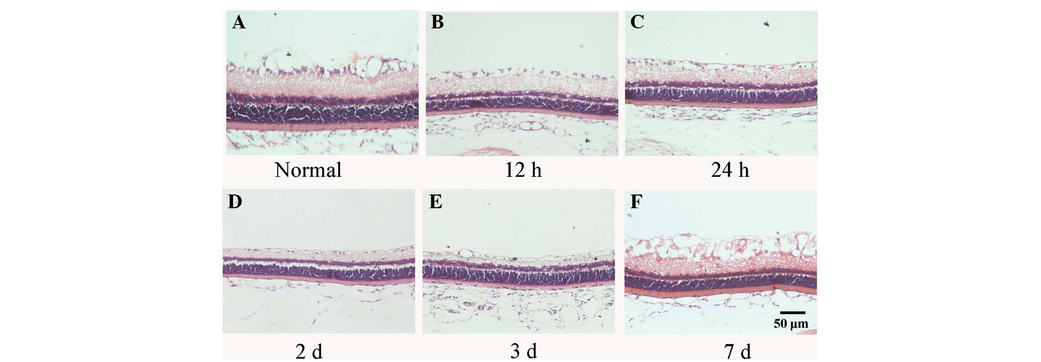
was performed on the tissues. At 12 h, 24 h, 2, 3 and 7 days
post-reperfusion, the thickness of the inner nuclear layer (INL)
and the inner plexiform layer (IPL) gradually decreased, compared
with the normal retina. The cell arrangement of the RGCs and the
INL was irregular, with a significant reduction in the number of
RGCs (Fig. 1). At 7 days
post-reperfusion, cytoplasmic vacuolation occurred in the RGCs and
cells were absent in the IPL (Fig.
1).
Apoptosis of the retinal cells is induced
by acute ocular hypertension
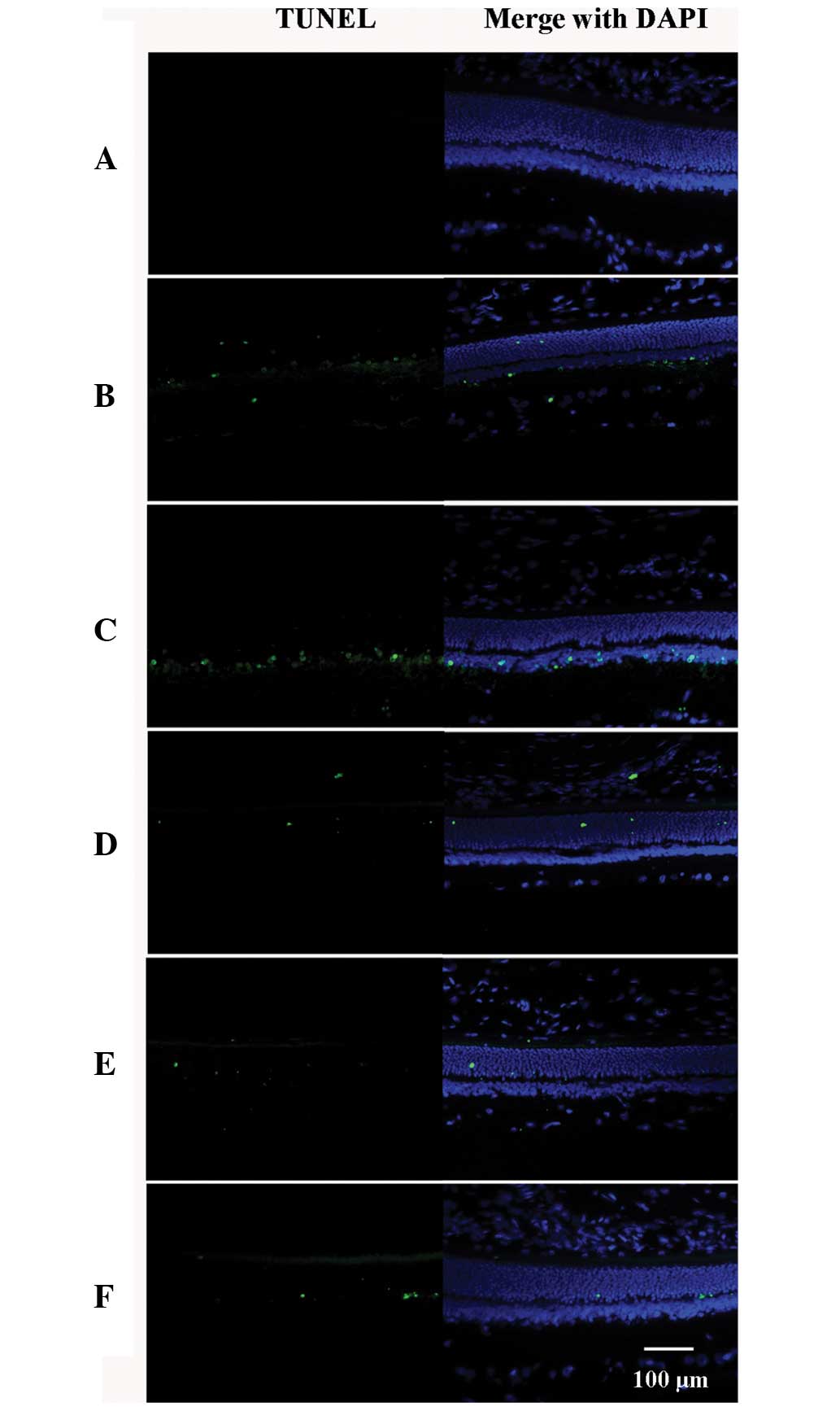
TUNEL staining of normal retinas and retinas at 12
h, 24 h, 2, 3 or 7 days post-reperfusion after 60 min acute ocular
hypertension is shown in Fig. 2.
TUNEL-positive cells were absent in the normal retina (Fig. 2A), but were observed in the outer
nuclear layer (ONL) and the INL in the group subjected to acute
ocular hypertension at 12 h post-reperfusion (Fig. 2B–F), and the number of apoptotic
cells peaked 24 h after reperfusion.
RGC loss occurs following acute ocular
hypertension
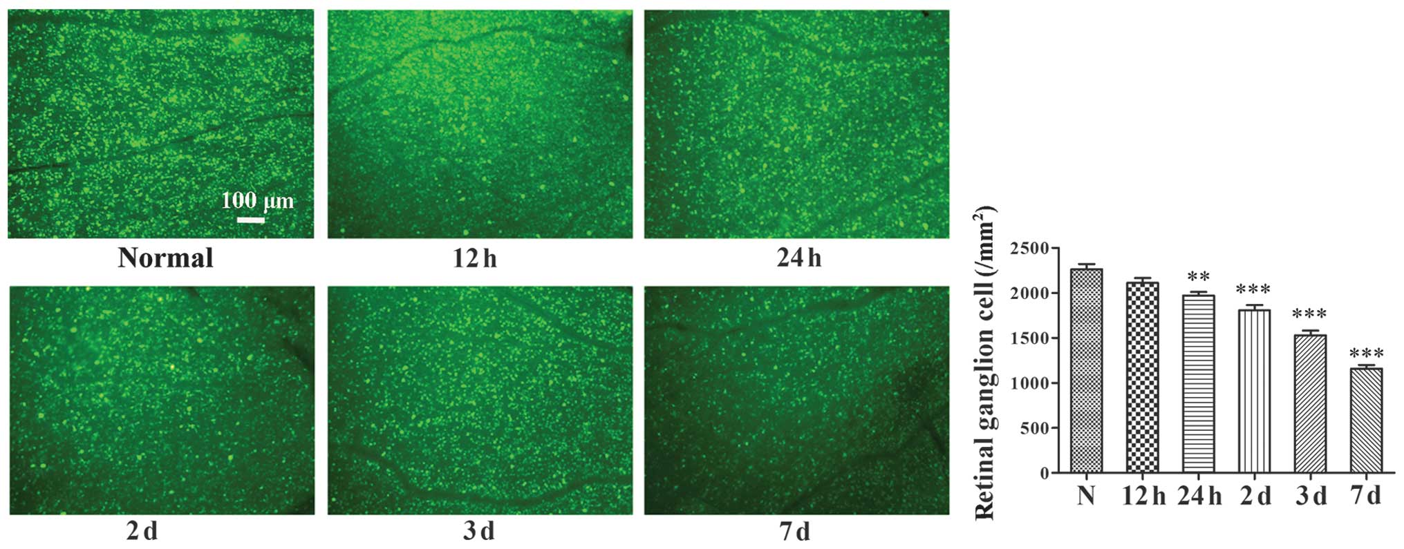
The results of the FG retrograde staining of RGCs
are shown in Fig. 3. Following
acute ocular hypertension, significant reductions in the numbers of
RGCs were found at 24 h, 2, 3 and 7 days post-retinal
reperfusion.
Expression levels of LIF and LIFR in the
rat retina following acute ocular hypertension
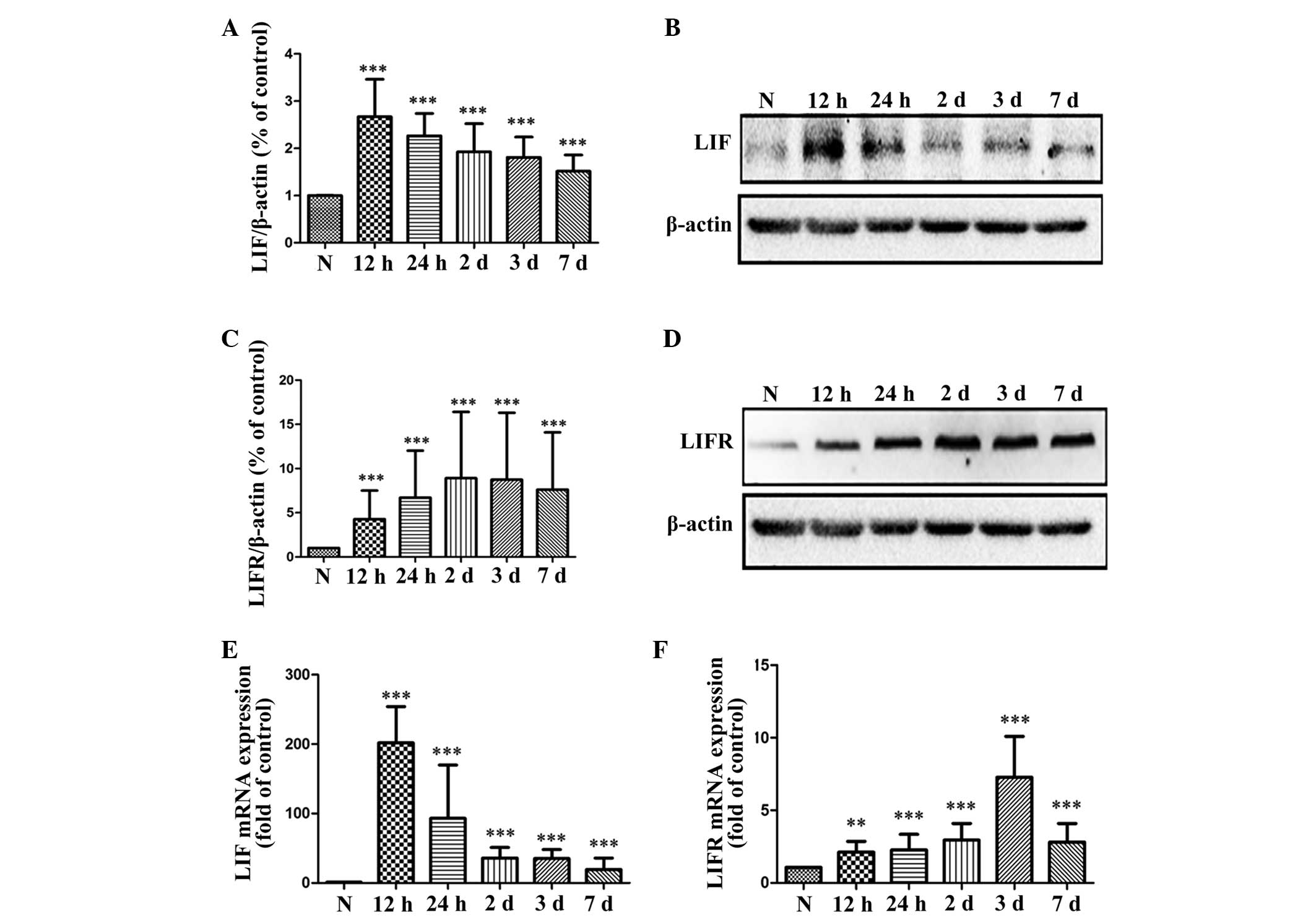
Western blot analysis was performed to assess the
protein levels of LIF and LIFR, and RT-qPCR was performed to
determine the mRNA expression levels of LIF and LIFR following
acute ocular hypertension in the rat retina (Fig. 4). The western blot analysis
demonstrated that the protein expression of LIF was significantly
increased at 12 h, 24 h, 2, 3 and 7 days, and peaked 12 h after the
IOP increase was terminated (Fig. 4A
and B). The protein level of LIFR was also significantly
higher, compared with the normal control at 12 h post-retinal
reperfusion, and peaking 3 days after reperfusion (Fig. 4C and D). The mRNA expression levels
of LIF and LIFR revealed the same patterns as the protein
expression of LIF and LIFR in the rat retina following acute ocular
hypertension (Fig. 4E and F).
Assessment of the activation of STAT3,
Akt and ERK in the rat retina
Activation of the downstream pathways of LIF were
assessed by investigating the expression levels of the
phosphorylated forms of STAT3, Akt and ERK (Fig. 5). The results revealed that, at 12
h post-retinal reperfusion, the levels of P-STAT3 were
significantly higher than that in the normal retina, and the
highest levels of P-STAT3 were observed 24 h after retinal
reperfusion (Fig. 5A and B).
Upregulation in the phosphorylation of Akt was also observed, which
peaked 12 h after retinal reperfusion (Fig. 5A and C). In contrast to P-STAT3 and
P-Akt, the level of P-ERK1/2 was reduced from 12 h after retinal
reperfusion in the rat retina (Fig. 5A
and D).
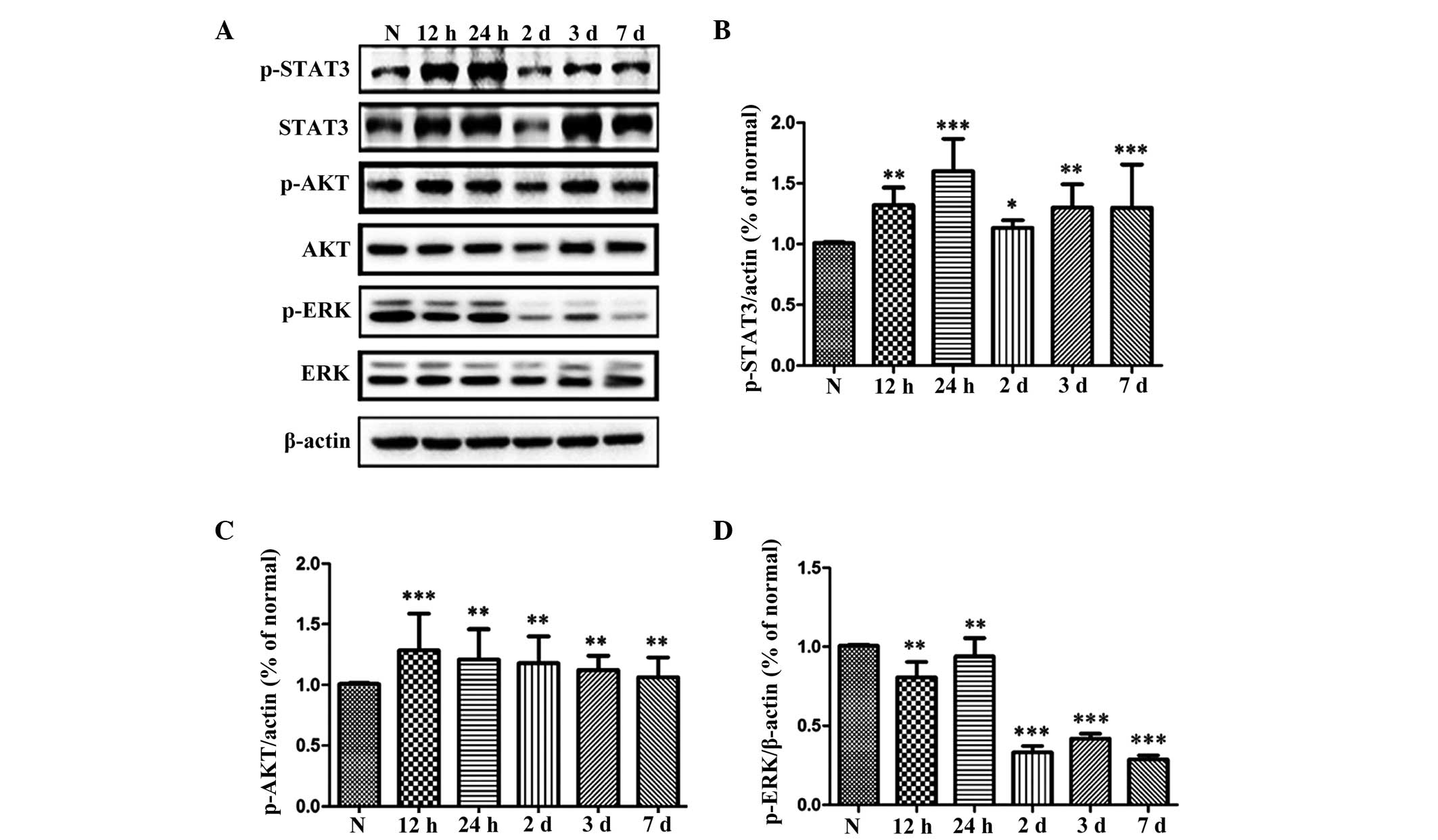 | Figure 5Expression levels of STAT3, Akt, ERK
and their corresponding phosphorylated forms in the rat retina
following acute ocular hypertension. The protein expression of
STAT3, Akt, ERK and their corresponding phosphorylated forms was
tested by (A) western blotting, and then (B–D) quantitative
analysis, respectively. Data are expressed as the mean ± standard
error of the mean. *P<0.05, **P<0.01
and ***P<0.001, vs. N group. N, normal control;
STAT3, signal transducers and activators of transcription 3; ERK,
extracellular signal-regulated kinase; p-, phosphorylated. |
Discussion
Glaucoma is a group of neurodegenerative diseases
characterized by structural damage to the optic nerve and visual
field loss, which are induced by pathologically raised IOP,
resulting in the death of RGCs. In previous decades, efforts have
been made to establish a stable and replicable animal model of
glaucoma to investigate the pathology and treatment of the damaged
nerve. By cannulating the anterior chamber with a needle connected
to a reservoir containing sterile saline, the IOP of an animal can
be raised to an abnormally high level and can be higher than the
ocular perfusion pressure, preventing ocular blood flow and causing
retinal ischemia, resulting in the death of RGCs. The apoptosis of
RGCs induced by acute ocular hypertension is considered to
simulate, at least partially, the pathophysiology of acute
angle-closure glaucoma (14). In
the present study, the results revealed thinning of the rat retina
following acute ocular hypertension, particularly in the layers of
the IPL, INL and RGCs. TUNEL-positive cell staining in the INL and
ONL suggested the existence of cell apoptosis in the retina.
LIF is a multifunctional, pleiotropic cytokine
(15) with a wide variety of
physiological functions in the body, promoting the proliferation of
primordial germ cells and spermatocyte differentiation (16,17),
inhibiting the differentiation of embryonic stem cells (18,19)
and being involved in bone formation (20,21).
Furthermore, as a neurotrophic factor, LIF is reported to enhance
the regeneration of axotomized sciatic nerves, accelerating the
repair of the injured sensory nerve, promoting olfactory sensory
neuron regeneration and corneal nerve recovery following laser
in situ keratomileusis (22–25).
In the retina, LIF is produced predominantly in the
Müller cells. Previous reports have demonstrated that the
expression of LIF is markedly upregulated in the mouse retina when
light damage is induced with bright cyclic light preconditioning
(26,27). The present study demonstrated that
the number of apoptotic cells increased significantly 12 h after
acute ocular hypertension and peaked at 24, followed by a decrease.
The apoptotic cells were located predominantly in the INL 12 h and
24 h after termination of ocular hypertension, while from day 2,
the apoptotic cells were scattered in the ONL (Fig. 2). A significant reduction in the
number of RGCs was noted at 24 h after acute ocular hypertension.
The protein and mRNA expression levels of LIF were upregulated
following acute ocular hypertension, peaking at 12 h. The
expression pattern of LIF was consistent with the change in the
number of apoptotic cells in the retina (Fig. 4). The expression of LIFR was
increased steadily following acute ocular hypertension-induced
ischemia and peaked after 3 days. An explanation for this
inconsistency in the expression of LIFR and LIF was that the LIFR
is able to bind with several cytokines in addition to LIF,
including ciliary neurotrophic factor (CNTF), cardiotrophin-1 and
oncostatin M, which, in turn alter the expression of LIFR (28–31).
Notably, the expression of CNTF has been reported to increase
following acute ocular hypertension in the rat retina (32).
It has been reported that LIF is involved in the
protection of photoreceptors against light-induced injury,
predominantly through activation of the JAK-STAT3 pathway (10,11).
In the present study, the results of the western blot analysis
demonstrated increased expression of P-STAT3 in the retina
following acute ocular hypertension. In addition, the Akt pathway
has been reported to have potent effects on neuroprotection in the
retina. When phosphorylated, the Akt kinase promotes cell survival
via the inactivation of apoptosis-associated components in the
cell. Inactivation of the Akt signaling pathway is considered to be
responsible for photoreceptor apoptosis in the mouse model of
retinal degeneration (33). LIF
has been reported to activate the PI3K signaling pathways,
including Akt in cardiac myocytes (3,6,34,35).
In the present study, the increase in the expression of LIF was
associated with elevated P-Akt levels in the retina following
retinal ischemia induced by acute ocular hypertension. These
results suggested that LIF may mediate the processes of injury and
repair in the retina through activation of the JAK-STAT3 and Akt
signaling pathways.
As an MAPK pathway, the ERK pathway has been
reported to be involved in cell proliferation and differentiation
(36) when activated by mitogenic
stimuli, including growth factors, cytokines, and phorbol esters.
It has been reported that ERK activation is necessary for synaptic
plasticity and memory in the central nervous system (37). In the rat retina, Roth et al
(38) revealed that the expression
of P-ERK peaked between 1 and 6 h following ischemia, followed by a
decrease. Only weak expression levels of P-ERK were detected
between days 3 and 7. The number of apoptotic cells in the RGC and
photoreceptor layers are markedly decreased following the
inhibition of ERK activation (38). In the present study, significant
downregulation of the expression of P-ERK was observed in the rat
retina following an acute increase in IOP. These observations were
consistent with the findings of Roth et al (38), described above. Namura et al
also demonstrated that intravenous injection of the MAPK/ERK kinase
inhibitor, U0126, protects brain tissues from damage following
forebrain and focal cerebral ischemia (39). However, the mechanisms responsible
for cell death following ERK activation require further
investigation.
In conclusion, the dynamic changes in the expression
levels of LIF and LIFR observed in the present study suggested that
LIF may be important in the process of degeneration/protection
following acute ocular hypertension-induced retinal ischemia via
the JAK-STAT3 and Akt signaling pathways.
Acknowledgments
The authors would like to thank Dr Zhen Zhang and Dr
Yanfeng Chen for their assistance in animal experiments. This study
was supported by the National Natural Science Foundation of China
(grant no. 81170841) and Xiamen Science and Technology Project
(grant nos. 3502Z20116011 and 3502Z20134040).
References
|
1
|
Ji JZ, Elyaman W, Yip HK, Lee VW, Yick LW
and Hugon J: The possible involvement of STAT3 pathway. Eur J
nerurosci. 19:265–272. 2004. View Article : Google Scholar
|
|
2
|
Ciotu IM, Stoian I, Gaman L, Popescu MV
and Atanasiu V: Biochemical changes and treatment in glaucoma. J
Med Life. 8:28–31. 2015.PubMed/NCBI
|
|
3
|
Heinrich PC, Behrmann I, Haan S, Hermanns
HM, Müller-Newen G and Schaper F: Principles of interleukin
(IL)-6-type cytokine signalling and its regulation. Biochem J.
374:1–20. 2003. View Article : Google Scholar : PubMed/NCBI
|
|
4
|
Yamauchi-Takihara K: Gp130-mediated
pathway and let ventricular remodeling. J Card Fail. 8(6 Suppl):
S374–S378. 2002. View Article : Google Scholar
|
|
5
|
Auernhammer CJ and Melmed S:
Leukemia-inhibitory factor-neuroimmnue modulator of endocrine
function. Endocr Rev. 21:313–345. 2000.PubMed/NCBI
|
|
6
|
Oh H, Fujio Y, kunisada K, Hirota H,
Matsui H, Kishimoto T and Yamauchi-Takihara K: Activation of
phosphatidylinositol 3-kinase through glycoprotein 130 induces
protein kinase B and p70 s6 kinase phosphorylation in cardiac
myocytes. J Biol Chem. 273:9703–9710. 1998. View Article : Google Scholar : PubMed/NCBI
|
|
7
|
Cheema SS, Richards L, Murphy M and
Bartlett PF: Leukemia inhibitory factor prevents the death of
axotomised sensory neurons in the dorsal root ganglia of the
neonatal rat. J Neurosci Res. 37:213–218. 1994. View Article : Google Scholar : PubMed/NCBI
|
|
8
|
Mckay Hart A, Wiberg M and Terenghi G:
Exogenous leukemia inhibitory factor enhances nerve regeneration
after late secondary repair using a bioartificial nerve conduit. Br
J plast surg. 56:444–450. 2003. View Article : Google Scholar : PubMed/NCBI
|
|
9
|
Gresle MM, Alexandrou E, Wu Q, Egan G,
Jokubaitis V, Ayers M, Jonas A, Doherty W, Friedhuber A, Shaw G, et
al: Leukemia inhibitory factor protects axons in experimental
autoimmune encephalomyelitis via an oligodendrocyte-independent
mechanism. PLoS One. 7:e473792012. View Article : Google Scholar : PubMed/NCBI
|
|
10
|
Bürgi S, Samardziia M and Grimm C:
Endogenous leukemia inhibitory factor protects photoreceptor cells
against light-induced degeneration. Mol Vis. 15:1631–1637.
2009.PubMed/NCBI
|
|
11
|
Joly S, Lange C, Thiersch M, Samardzija M
and Grimm C: Leukemia inhibitory factor extends the lifespan of
injured photo-receptors in vivo. J Neurosci. 28:13765–13774. 2008.
View Article : Google Scholar : PubMed/NCBI
|
|
12
|
Mayer M, Bhakoo K and Noble M: Ciliary
nerrotrophic factor and leukemia inhibitory factor promote the
generation, maturation and survival of oligodendrocytes in vitro.
Development. 120:143–153. 1994.PubMed/NCBI
|
|
13
|
Johnson EC, Doser TA, Cepurna WO, Dyck JA,
Jia L, Guo Y, Lambert WS and Morrison JC: Cell proliferation and
interleukin-6-type cytokine signaling are implicated by gene
expression responses in early optic nerve head injury in rat
glaucoma. Invest Ophthalmol Vis Sci. 52:504–518. 2011. View Article : Google Scholar :
|
|
14
|
Johnson TV and Tomarev SI: Rodent models
of glaucoma. Brain Res Bull. 81:349–358. 2010. View Article : Google Scholar :
|
|
15
|
Metcalf D: The unsolved enigmas of
leukemia inhibitory factor. Stem Cells. 21:5–14. 2003. View Article : Google Scholar : PubMed/NCBI
|
|
16
|
Piguet-Pellorce C, Dorval-Coiffec I, Pham
MD and Jégou B: Leukemia inhibitory factor expression and
regulation within the testis. Endocrinonlogy. 141:1136–1141. 2000.
View Article : Google Scholar
|
|
17
|
Sariola H: The neurotrophic factors in
non-neuronal tissues. Cell Mol Life Sci. 58:1061–1066. 2001.
View Article : Google Scholar : PubMed/NCBI
|
|
18
|
Williams RL, Hilton DJ, Pease S, Willson
TA, Stewart CL, Gearing DP, Wagner EF, Metcalf D, Nicola NA and
Gough NM: Myeloid leukemia inhibitory factor maintains the
developmental potential of embryonic stem cells. Nature.
336:684–687. 1988. View
Article : Google Scholar : PubMed/NCBI
|
|
19
|
Smith AG, Health JK, Donaldson DD, Wong
GG, Moreau J, Stahl M and Rogers D: Inhibition of pluripotential
embryonic stem cell differentiation by purified polypeptides.
Nature. 336:688–690. 1988. View
Article : Google Scholar : PubMed/NCBI
|
|
20
|
Reid LR, Lowe C, Cornish J, Skinner SJ,
Hilton DJ, Willson TA, Gearing DP and Martin TJ: Leukemia
inhibitory factor: A novel bone-active cytokine. Endocrinology.
126:1416–1420. 1990. View Article : Google Scholar : PubMed/NCBI
|
|
21
|
Dazai S, Akita S, Hirano A, Rashid MA,
Naito S, Akino K and Fujii T: Leukemia inhibitory factor enhances
bone formation in calvarial bone defect. J Craniofac Srug.
11:513–520. 2000. View Article : Google Scholar
|
|
22
|
Dowsing BJ, Hayes A, Bennett TM, Morrison
WA and Messina A: Effects of LIF dose and laminin plus fibronectin
on axotomized sciatic nerves. Muscle Nerve. 23:1356–1364. 2000.
View Article : Google Scholar : PubMed/NCBI
|
|
23
|
Cafferty WB, Gardiner NJ, Gavazzi I,
Powell J, McMahon SB, Heath JK, Munson J, Cohen J and Thompson SW:
Leukemia inhibitory factor determines the growth status of injured
adult sensory neurons. J Neurosci. 21:7161–7170. 2001.PubMed/NCBI
|
|
24
|
Bauer S, Rasika S, Han J, Mauduit C,
Raccurt M, Morel G, Jourdan F, Benahmed M, Moyse E and Patterson
PH: Leukemia inhibitory factor is a key signal for injury-induced
neurogenesis in the adult mouse olfactory epithelium. J Neurosci.
23:1792–1803. 2003.PubMed/NCBI
|
|
25
|
Pan S, Li L, Xu Z and Zhao J: Effect of
leukemia inhibitory factor on corneal nerve regeneration of rabbit
eyes after laser in situ keratomileusis. Neurosci Lett. 499:99–103.
2011. View Article : Google Scholar : PubMed/NCBI
|
|
26
|
Chollangi S, Wang J, Martin A, Quinn J and
Ash JD: Preconditioning-induced protection from oxidative injury is
mediated by leukemia inhibitory factor receptor (LIFR) and its
ligands in the retina. Neurobiol Dis. 34:535–544. 2009. View Article : Google Scholar : PubMed/NCBI
|
|
27
|
Ueki Y, Le YZ, Chollangi S, Muller W and
Ash JD: Preconditioning-induced protection of photoreceptors
requires activation of the signal-transducing receptor gp130 in
photoreceptors. Proc Natl Acad Sci USA. 106:21389–21394. 2009.
View Article : Google Scholar : PubMed/NCBI
|
|
28
|
Davis S, Aldrich TH, Stahl N, Pan L, Taga
T, Kishimoto T, Ip NY and Yancopoulos GD: LIRR beta and gp130 as
heterodimerizing signal transducers of the tripartite CNTF
receptor. Science. 260:1805–1808. 1993. View Article : Google Scholar : PubMed/NCBI
|
|
29
|
Pennica D, Shaw KJ, Swanson TA, Moore MW,
Shelton DL, Zioncheck KA, Rosenthal A, Taga T, Paoni NF and Wood
WI: Cardiotrophin-1. Biological activities and binding to the
leukemia inhibitory factor receptor/gp130 signaling complex. J Biol
Chem. 270:10915–10922. 1995.PubMed/NCBI
|
|
30
|
Gearing DP and Bruce AG: Oncostatin M
binds the high-affinity leukemia inhibitory factor receptor. New
Biol. 4:61–65. 1992.PubMed/NCBI
|
|
31
|
Plun-Favreau H, Perret D, Diveu C, Froger
J, Chevalier S, Lelièvre E, Gascan H and Chabbert M: Leukemia
inhibitory factor (LIF), cardiotrophin-1 and oncostatin M share
structural binding determinants in the immunoglobulin-like domain
of LIF receptor. J Biol Chem. 278:27169–27179. 2003. View Article : Google Scholar : PubMed/NCBI
|
|
32
|
Ju WK, Lee MY, Hofmann HD, Kirsch M, Oh
SJ, Chung JW and Chun MH: Increased expression of ciliary
neurotrophic factor receptor alpha mRNA in the ischemic rat retina.
Neurosci Lett. 283:133–136. 2000. View Article : Google Scholar : PubMed/NCBI
|
|
33
|
Jomary C, Cullen J and Jones SE:
Inactivation of the Akt survival pathway during photoreceptor
apoptosis in the retinal degeneration mouse. Invest Ophthalmol Vis
Sci. 47:1620–1629. 2006. View Article : Google Scholar : PubMed/NCBI
|
|
34
|
Hideshima T, Nakamura N, Chauhan D and
Anderson KC: Biologic sequelae of interleukin-6 induced PI3-K/Akt
signaling in multiple myeloma. Oncogene. 20:5991–6000. 2001.
View Article : Google Scholar : PubMed/NCBI
|
|
35
|
Jee SH, Chiu HC, Tsai TF, Tsai WL, Liao
YH, Chu CY and Kuo ML: The phosphotidyl inositol 3-kinase/Akt
signal pathway is involved in interleukin-6-mediated Mcl-1
upregulation and anti-apoptosis activity in basal cell carcinoma
cells. J Invest Dermatol. 119:1121–1127. 2002. View Article : Google Scholar : PubMed/NCBI
|
|
36
|
Chen J, Fujii K, Zhang L, Roberts T and Fu
H: Raf-1 promotes cell survival by antagonizing apoptosis
signal-regulating kinase 1 through a MEK-ERK independent mechanism.
Proc Natl Acad Sci USA. 98:7783–7788. 2001. View Article : Google Scholar : PubMed/NCBI
|
|
37
|
Sweatt JD: The neuronal MAP kinase
cascade: A biochemical signal integration system subserving
synaptic plasticity and memory. J Neurochem. 76:1–10. 2001.
View Article : Google Scholar : PubMed/NCBI
|
|
38
|
Roth S, Shaikh AR, Hennelly MM, Li Q,
Bindokas V and Graham CE: Mitogen-activated protein kinases and
retinal ischemia. Invest Ophthalmol Vis Sci. 44:5383–5395. 2003.
View Article : Google Scholar : PubMed/NCBI
|
|
39
|
Namura S, Iihara K, Takami S, Nagata I,
Kikuchi H, Matsushita K, Moskowitz MA, Bonventre JV and
Alessandrini A: Intravenous administration of MEK inhibitor U0126
affords brain protection against forebrain ischemia and focal
cerebral ischemia. Proc Natl Acad Sci USA. 98:11569–11574. 2001.
View Article : Google Scholar : PubMed/NCBI
|


