Introduction
The airway epithelium is pivotal to host defense
against foreign microbial pathogens. There is increasing evidence
that epithelial alterations are associated with multiple airway
diseases, including asthma, chronic obstructive pulmonary diseases
(COPD), obliterative bronchiolitis and cystic fibrosis (1–5).
Following injury to the epithelium, airway progenitor cells are a
crucial part of the repair process (6). These progenitor cells interact with
their microenvironment to determine their proliferation,
differentiation and capacity for self-renewal. The growth of these
progenitor cells is reported to be supported by the production of
growth factors by stromal cells (7). Additionally, the research of Lee
et al (8) suggests that
endothelial cells direct the specification and differentiation of
airway progenitor cells. Furthermore, parabronchial smooth muscle
cells activate airway epithelial progenitor cells to undergo a
post-injury epithelial to mesenchymal transition (9). Vimentin-positive lung fibroblasts can
create a niche for airway progenitor cells (10). Airway progenitor cells cross talk
with their microenvironmental elements through direct contact or
autocrine and/or paracrine signals. These include Wnt-β-catenin
signaling, BMP signaling, Notch signaling and TGF-β signaling
(7,11–13).
The TGF-β signal is overactivated in response to
pulmonary fibrosis (14,15). Moreover, TGF-β has been reported to
be upregulated in COPD and allergic asthma (16). Inhibition of TGF-β signaling with
SB431542 promotes the proliferation of airway epithelial progenitor
cells (7). TGF-β signal in
fibroblasts can act FGF10 to regulate epithelial stem cell growth
(7). It was shown that TGF-β
inhibits HGF expression in mesenchymal cells through a TGF-β
inhibitory element (17). HGF acts
as a ligand with its receptor tyrosine kinase, c-Met to fulfill its
function (18). HGF modulates the
function of Smad4 through activating the Ras/MAPK pathway (19). However, the role of Smad4 in the
regulation of airway progenitor cells has not been addressed.
In the present study, the authors adopted an in
vitro epithelial-fibroblast co-culture assay developed
previously (20). It was observed
that TGF-β inhibits the proliferation of airway progenitor cells
through the TGF-β receptors on fibroblasts. Inhibition of the
TGF-β/TGFR2 pathway alters the secretory properties of fibroblasts,
including production of HGF. Deletion of Smad4 resulted in an
increase in the colony forming ability of airway progenitor
cells.
Materials and methods
Ethics statement
Experimental mice were maintained under
pathogen-free conditions in Tianjin Haihe Hospital's animal
facility, and the permit number is SYXK (Jin) 2016-0002. Adult mice
between the ages of 2–4 months old were sacrificed for experiments
according to protocols approved by the Haihe Hospital Animal Care
and Use Committee (Tianjin, China). All surgery was performed under
1% sodium pentobarbital 50 mg/kg intraperitoneal injection
anesthesia, and all efforts were made to minimize suffering.
Mice
β-actin-GFP, Sftpc-Cre, TGFβR2f/f,
and Smad4f/f mice were donated by Stripp B.R. (Cedars-Sinai
Medical Center, Los Angeles, CA, USA). The authors crossed
Sftpc-Cre mice with TGFβR2f/f mice to generate
Sftpc-Cre; TGFβR2f/f mice. Additionally, Sftpc-Cre
mice and Smad4f/f mice were crossed to generate
Sftpc-Cre; Smad4f/f mice.
Fractionation of airway epithelial
progenitor cells
Lung cell suspensions were prepared using an
elastase digestion and stained for fluorescence-activated cell
sorting (FACS), as previously described (20). Briefly, cells were resuspended in
Hanks' balanced saline solution buffer supplemented with 2% fetal
bovine serum, 0.1 mM EDTA, 10 mM HEPES, 100 IU/ml penicillin and
100 µg/ml streptomycin (HBSS+). Cells were then stained
with the primary antibodies on ice for 45 min. The following
antibodies were employed: EpCAM-PE-Cy7 (25-5791-80, 1:100),
CD31-Biotin (13-0311-81, 1:40), CD34-Biotin (13-0341-81, 1:10),
CD45-Biotin (13-0451-81, 1:100), Sca-1-APC (17-5981-81, 1:100), and
CD24-PE (12-0242-81, 1:20) (all from eBioscience, Inc., San Diego,
CA, USA). Cells were subsequently stained with the secondary
antibody on ice for 40 min using streptavidin-APC-Cy7 (47-4317-82,
1:100; eBioscience, Inc.). Dead cells were identified using
7-aminoactinomycin D staining (BD Biosciences, Franlkin Lakes, NJ,
USA).
Cell cultures and treatment
MLg cells (CCL-206; American Type Culture
Collection, Manassas, VA, USA) were cultured in Dulbecco's modified
Eagle's medium (DMEM; Gibco; Thermo Fisher Scientific, Inc.,
Waltham, MA, USA). For the experiments, 6-well culture plates
(Thermo Fisher Scientific, Inc.) were seeded with MLg cells at
5,000 cells/well and maintained at 37°C in a humidified atmosphere
with 5% CO2. MLg cells were then exposed to 10 µM
SB431542 (Sigma-Aldrich; Merck KGaA, Darmstadt, Germany) for 48 h.
Cultures were visualized under an OLYMPUS IX73 (Olympus
Corporation, Tokyo, Japan) inverted microscope before being
harvested for reverse transcription-quantitative polymerase chain
reaction (RT-qPCR) analysis.
Matrigel cultures of airway progenitor
cells
Mouse airway progenitor cells were co-cultured with
MLg cells in Matrigel, as described previously (20). In brief, sorted airway progenitor
cells were mixed with MLg cells in growth factor-reduced Matrigel
(BD Biosciences) and basic medium (BM) at a 1:1 ratio. The basic
medium consisted of DMEM/F12 medium (Cellgro, Manassas, VA, USA),
10% FBS (Invitrogen; Thermo Fisher Scientific, Inc.),
Insulin-Transferrin-Selenium supplement (Sigma-Aldrich; Merck
KGaA), 100 IU/ml penicillin and 100 µg/ml streptomycin. The cell
mixture was placed in 24-well Transwell filter inserts (BD
Biosciences) in a 24-well flat-bottom culture plate containing
culture medium (BM + SB431542). Cultures were incubated at 37°C in
a humidified atmosphere with 5% CO2, and medium was
replaced every other day. Colony-forming efficiency (CFE) was
determined by counting the number of colonies with a diameter of
≥100 µm in each culture and representing this number as a
percentage of seeded progenitor cells.
Total RNA isolation and RT-qPCR
Total RNA was extracted from MLg cells with TRIzol
reagent (Invitrogen; Thermo Fisher Scientific, Inc.) following the
manufacturer's instructions. RNA concentration was assessed and
cDNA was synthesized using SuperScript III reagents (Invitrogen;
Thermo Fisher Scientific, Inc.) with Oligo-dT and random hexamer
priming (Takara Bio, Inc., Otsu, Japan). RT-qPCR was performed
using SYBR Green SuperMix (Applied Biosystems; Thermo Fisher
Scientific, Inc.) with the Light Cycler 96 Real-Time PCR system
(Roche Diagnostics, Indianapolis, IN, USA) under the following
reaction conditions: Initial heating cycle of 95°C for 2 min; and
40 cycles of denaturation at 95°C for 25 sec, primer annealing at
60°C for 25 sec and extension at 72°C for 20 sec. Melting curves
were used to clarify the identity of amplicons, and the
housekeeping gene, β-actin, served as an internal control. The
relative mRNA expression levels of targeted genes were calculated
using the comparative threshold cycle (CT) method (21) normalized to β-actin mRNA in the
same sample. Primers were designed as follows: β-actin forward,
5′-GGCCAACCGTGAAAAGATGA-3′ and reverse,
5′-CAGCCTGGATGGCTACGTACA-3′; Hgf forward,
5′-CCTGGTGTTTCACAAGCAATC-3′ and reverse,
5′-CATGGGACCTCTGTAGCTTTC-3′. β-actin was used as a housekeeping
gene.
Microarray analysis
EpCAM+ lung epithelial cells or
EpCAM+Sca-1+ airway progenitor cells were
sorted and pooled for total RNA extraction using RNeasy Mini kit
(Qiagen Sciences, Inc., Gaithersburg, MD, USA). RNA (0.1 µg) was
used for Affymetrix microarray analysis using mouse genome 430 2.0
arrays (Affymetrix, Inc., Santa Clara, CA, USA). Data were
annotated with Affymetrix Expression Console software (Affymetrix,
Inc.). Pathway analysis was performed by online Gather KEGG
analysis (Kyoto Encyclopedia of Genes and Genomes; http://gather.genome.duke.edu/). A Bayes factor
was also included in the consideration of KEGG pathway
association.
Statistical analysis
The data were analyzed using SPSS software (version
13.0; SPSS, Inc., Chicago, IL, USA). Data are expressed as the mean
± standard error of the mean. The significance of the results was
assessed using Student's t test between paired groups. P<0.05
was considered to indicate a statistically significant
difference.
Results
TGF-β regulates mouse airway
progenitor cells through its receptors on MLg cells
To investigate the role of fibroblasts in the
regulation of mouse airway progenitor cells by TGF-β, the authors
fractionated mouse airway progenitor cells from β-actin-GFP
mice using a FACS-based strategy. Lung cells from
β-actin-GFP mice were isolated by elastase digestion. The
cells were stained with fluorescent antibodies and a viability dye.
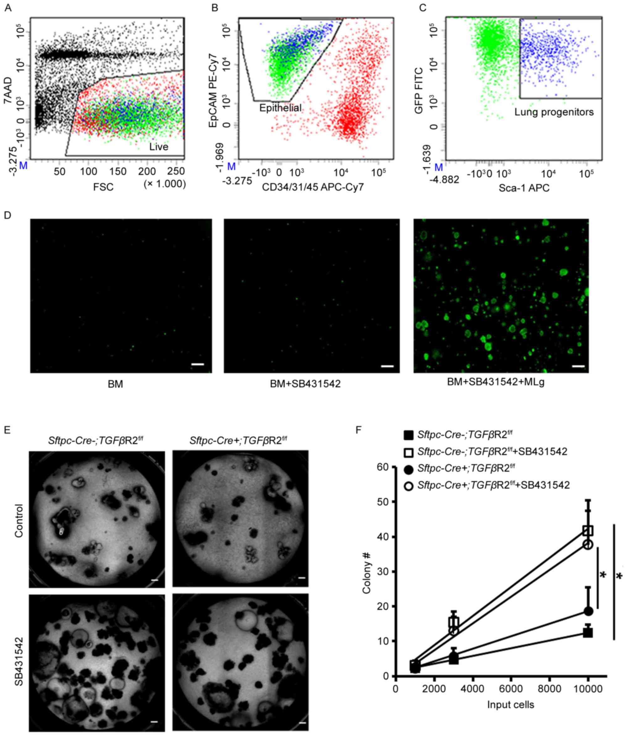
Dead cells were detected by 7-AAD staining (Fig. 1A). Endothelial, stromal and
hematopoietic cells were excluded by surface staining for CD31,
CD34 and CD45 (Fig. 1B). Airway
epithelial progenitor cells, also positive for GFP, were further
enriched by surface EpCAM and Sca-1 staining (Fig. 1B and C). A significant number of
colonies were formed in presence of both SB431542 and MLg cells
compared to stromal-free and SB431542 alone cultures (Fig. 1D), which was consistent with the
authors' previous findings (20).
The authors further examined the TGF-β role in the regulation of
airway progenitor cells using Sftpc-Cre+;
TGFβR2f/f mice, in which TGFβR2 is absent in their airway
progenitor cells. In vitro cultures of airway progenitor
cells in presence of MLg cells indicated that the colony-forming
ability was comparable between Sftpc-Cre−;
TGFβR2f/f and Sftpc-Cre+; TGFβR2f/f
(Fig. 1E and F). SB431542 enhanced
the colony-forming ability of airway progenitor cells in both
Sftpc-Cre−; TGFβR2f/f and
Sftpc-Cre+; TGFβR2f/f (Fig. 1E and F). These data suggested that
TGF-β exerts its regulatory role in the colony-forming ability of
airway progenitor cells by acting on MLg cells, rather than on
airway progenitor cells.
Inhibition of TGF-β signaling promotes
the secretion of HGF in fibroblasts
Fibroblasts have been shown to promote airway
epithelial progenitor cells by producing growth factors. HGF
expression was measured in the absence or presence of SB431542.
Within 48 h, the number of MLg cells increased by approximately
tenfold in the control group (Fig.
2A). There was no difference in the number of MLg cells between
control and SB431542 treatment (Fig.
2B). Additionally, the morphology of MLg cells did not differ
between the control and SB431542 treatment (Fig. 2A). Using RT-qPCR, the authors
observed that the mRNA expression of Hgf in the SB431542
treatment was higher than that in the control (Fig. 2C). These data suggested that
activation of the TGF-β signal pathway inhibits the production of
growth factor HGF in fibroblasts.
Stromal-derived HGF exerts its function through its
specific receptor c-Met. Following this, the c-Met expression was
determined in airway progenitor cells, alveolar type 2 (AT2) cells,
ciliated cells, and MLg cells using quantitative RT-PCR (Fig. 3A). Mouse AT2 cells and ciliated
cells were fractionated as previously (20). c-Met expression was indicated to be
more abundant in lung epithelial cells than in stromal cells
(Fig. 3A). AT2 cells express more
c-Met than airway progenitor cells and ciliated cells in mouse lung
(Fig. 3A). In vitro 3-D
Matrigel culture indicated that HGF promotes the growth of airway
progenitor cells in absence of MLg cells (Fig. 3B), suggesting that HGF acts
directly on airway progenitor cells to promote their growth.
Smad4 regulates the proliferation of
airway progenitor cells
Smad elements are implicated in downstream TGF-β
signal, and have been shown to be modulated by HGF (22). Smad elements were hypothesized to
play a role in the regulation of airway progenitor cells by HGF.
Microarray analysis was used to identify enriched transcripts of
airway progenitor cells as compared to EpCAM+ lung
epithelial cells. The top 20 genes enriched in airway progenitor
cells were listed in Table I.
Notably, TGF-β transcript is enriched by 3.5 fold over total lung
epithelial cells (Table I). In
addition, the authors sorted the list of transcripts by their
relative expression level, and 1213 transcripts were observed that
read 1800 or greater which indicate their potential expression in
real samples (Table II). As
expected, Scgb1a1, Cyp2f2 and Plunc were presented in the list of
highly expressed gene list of airway progenitor cells (Table II). Smad4 was in the list even it
did not show up in Table II.
Gathered analysis of top genes in airway progenitor cells indicated
that Smad4 signaling pathway may participate in the regulation of
airway progenitor cells proliferation (Table III). In addition, Xenopus
fork head domain factor 3 and direct repeat 4 transcription factors
may also serve a role in regulating airway progenitor cells. These
data suggested that Smad4 may be a regulator of airway progenitor
cells.
 | Table I.Top genes expressed in airway
progenitor cells compared to total lung epithelial cells. |
Table I.
Top genes expressed in airway
progenitor cells compared to total lung epithelial cells.
| Gene ID | Gene name | Gene symbol |
Sca-1+/EpCAM |
|---|
| 742 | Proline/serine-rich
coiled-coil 1 | Psrc1 | 12.0 |
| 12623 | Carboxylesterase
1 | Ces1 | 8.6 |
| 232400 | cDNA sequence
BC048546 | BC048546 | 7.4 |
| 17984 | Necdin | Ndn | 7.4 |
| 67005 | Polymerase (RNA)
III (DNA directed) polypeptide K | Polr3k | 4.8 |
| 140474 | Mucin 4 | Muc4 | 4.7 |
| 23886 | Growth
differentiation factor 15 | Gdf15 | 4.6 |
| 22270 | Uroplakin 3A | Upk3a | 4.3 |
| 13078 | Cytochrome P450,
family 1, subfamily b, polypeptide 1 | Cyp1b1 | 3.7 |
| 11670 | Aldehyde
dehydrogenase family 3, subfamily A1 | Aldh3a1 | 3.6 |
| 27280 | Pleckstrin
homology-like domain, family A, member 3 | Phlda3 | 3.6 |
| 70337 | Iodotyrosine
deiodinase | Iyd | 3.6 |
| 21808 | Transforming growth
factor, β | TGF-β | 3.5 |
| 399638 | RIKEN cDNA
A030001D16 gene | A030001D16Rik | 3.5 |
| 215446 | Ectonucleoside
triphosphate diphosphohydrolase 3 | Entpd3 | 3.4 |
| 18104 | NAD(P)H
dehydrogenase, quinone 1 | Nqo1 | 3.4 |
| 217830 | RIKEN cDNA
9030617O03 gene | 9030617O03Rik | 3.4 |
| 77914 | Keratin associated
protein 17-1 | Krtap17-1 | 3.4 |
| 70536 | Glutaminyl-peptide
cyclotransferase (glutaminyl cyclase) | Qpct | 3.4 |
| 19363 | RAD51-like 1 (S.
cerevisiae) | Rad51l1 | 3.3 |
| Table II.Highly expressed genes in airway
progenitor cells. |
Table II.
Highly expressed genes in airway
progenitor cells.
| Entrez gene | Gene name | Gene symbol |
Ave.Sca1+ |
Ave.EpCAM+ |
|---|
| 287 | Secretoglobin,
family 1A, member 1 (uteroglobin) | Scgb1a1 | 33358 | 33006 |
| 13107 | Cytochrome P450,
family 2, subfamily f, polypeptide 2 | Cyp2f2 | 31168 | 26482 |
| 12409 | Carbonyl reductase
2 | Cbr2 | 28700 | 24926 |
| 117158 | Secretoglobin,
family 3A, member 2 | Scgb3a2 | 28154 | 25734 |
| 15439 | Haptoglobin | Hp | 25201 | 19952 |
| 67701 | WAP four-disulfide
core domain 2 | Wfdc2 | 24106 | 19162 |
| 22070 | Tumor protein,
translationally-controlled 1 | Tpt1 | 23147 | 22975 |
| 13627 | Eukaryotic
translation elongation factor 1 alpha 1 | Eef1a1 | 21612 | 21224 |
| 68662 | Secretoglobin,
family 3A, member 1 | Scgb3a1 | 21599 | 19061 |
| 56615 | Microsomal
glutathione S-transferase 1 | Mgst1 | 21468 | 18682 |
| 19241 | Thymosin, β 4, X
chromosome | Tmsb4x | 21214 | 22595 |
| 20387 | Surfactant
associated protein A1 | Sftpa1 | 20590 | 27427 |
| 22190 | Ubiquitin C | Ubc | 20572 | 18226 |
| 18843 | Palate, lung, and
nasal epithelium associated | Plunc | 20304 | 13823 |
| 20341 | Selenium binding
protein 1 | Selenbp1 | 20207 | 15170 |
| 14319 | Ferritin heavy
chain 1 | Fth1 | 19942 | 17755 |
| 100042862 | Predicted gene
4076 | Gm4076 | 19843 | 18573 |
| 14281 | FBJ osteosarcoma
oncogene | Fos | 19779 | 18595 |
| 65019 | Ribosomal protein
L23 | Rpl23 | 19029 | 18042 |
| 14319 | Ferritin heavy
chain 1 | Fth1 | 18279 | 15370 |
| Table III.Potential transcription factors
active in airway progenitor cells by Gather analysis. |
Table III.
Potential transcription factors
active in airway progenitor cells by Gather analysis.
| n | Gene name | Total gene (n) | Bayes factor | P-value | Genes |
|---|
| 1 | SMAD4 | 88 | 3.9 | 0.0004 | AW210596, Abcc3,
Acas2, Acp5, Adhfe1, Afp, Aox1, Arhgap24, Asgr1, Atp2b2, BC004853,
BC010843, BC018371, BC024561, Btbd11, Bucs1, C030018L16Rik, Cbs,
Ccng1, Ces1, Chn2, Col4a6, Cyp1b1, Cyp2b10, Cyp4f15, D430038H04Rik,
Daf1, Dapk2, Dp1l1, Edg7, Efna5, Entpd3, Epdr2, Ephx1, Fmo2, Fmo3,
Foxq1, Gabrp, Gdpd2, Gmnn, Gpr120, Gpx2, Gpx3, Grasp, Gss, Gsta3,
Gstk1, Gstm2, Gsto1, Hod, Hpgd, Hspa5bp1, Ifit1, Itih5, Kdr, Lnx1,
Ltf, Ly6a, Mscp, Nupr1, Oact1, Palmd, Pgam2, Pgd, Pglyrp1, Phlda3
Polr3k, Ppp2r2b, Prdx6, Qpct, Rab6b, Rarb, Rbp4, Samd4, Sema3a
Serpinf1, Slc23a1, Stat4, Stxbp6, Sulf2, Tacstd2, Tgfb2, Tmem40,
Tnfrsf21, Tnrc9, Trim3, Trim30 U46068 |
| 2 | XFD3 | 7 | 2.3 | 0.002 | Abca4, Ace2, Ces1,
Epdr2, Hpgd, Igf1r, Lmyc1 |
| 3 | DR4 | 148 | 1.6 | 0.005 | 1810008K03Rik,
4933428I03Rik, 5430413I02Rik, 9030408N13Rik, A630065K24Rik,
AW210596, Abca4, Abcc3, Acas2, Ace2, Acp5, Acpp, Adhfe1, Afp,
Aldh3a1, Aqp4, Arhgap24, Armcx2, Asgr1, Atp2b2, Atp6v0d2, Azgp1,
BC004853, BC010843, BC018371, BC024561, BC048546, Bax, Btbd11,
Bucs1, C030018L16Rik, Cbs, Cckar, Ccng1, Ces1, Chn2, Cldn10,
Cldn23, Cnga2, Col23a1, Col4a6 Cyp1b1, Cyp2b10, Cyp4f15,
D430038H04Rik, Daf1, Dapk2, Dp1l1, Dtna, Edg7, Efna5, Enah, Entpd3,
Epdr2, Ephx1, Fign, Fmo1, Fmo2, Foxq1, Frem1, Gabrp, Gcnt2, Gdf15,
Gdpd2, Ggh, Gmnn, Gpr120, Gpx2, Gpx3, Grasp, Gss, Gsta2, Gsta3,
Gstk1, Gstm1, Gstm2, Gsto1, Hck, Hmox1, Hod, Hpgd, Hspa5bp1, Igf1r,
Il1f9, Il6ra, Itih2, Itih5, Kcnk2, Kdr, Kitl, Ldh2, Lif, Lmyc1,
Lnx1, Ltf, Ly6a, MGC25972, Mgat3, Mia1, Mscp, Ndn, Nfe2l2, Notch3,
Nqo1, Nrarp, Oact1, Palmd, Pgam2, Pgd, Pglyrp1, Phlda3, Pir,
Polr3k, Pparg, Prdx6, Ptger4, Qpct, Rab6b, Rad51l1, Rarb, Rbp4,
Rpgrip1, Samd4, Scnn1b, Scnn1g, Sdpr, Sec8l1, Sema3a, Serpinf1,
Slc1a1, Slc23a1, Slc38a1, Slc9a9, Sphk1, Stat4, Stxbp6, Sulf2,
Tacstd2, Tgfb2, Tmem40, Tmie, Tnfrsf21, Tnrc9, Trim30, Ttpa,
U46068, Upk3a, Zfhx1a |
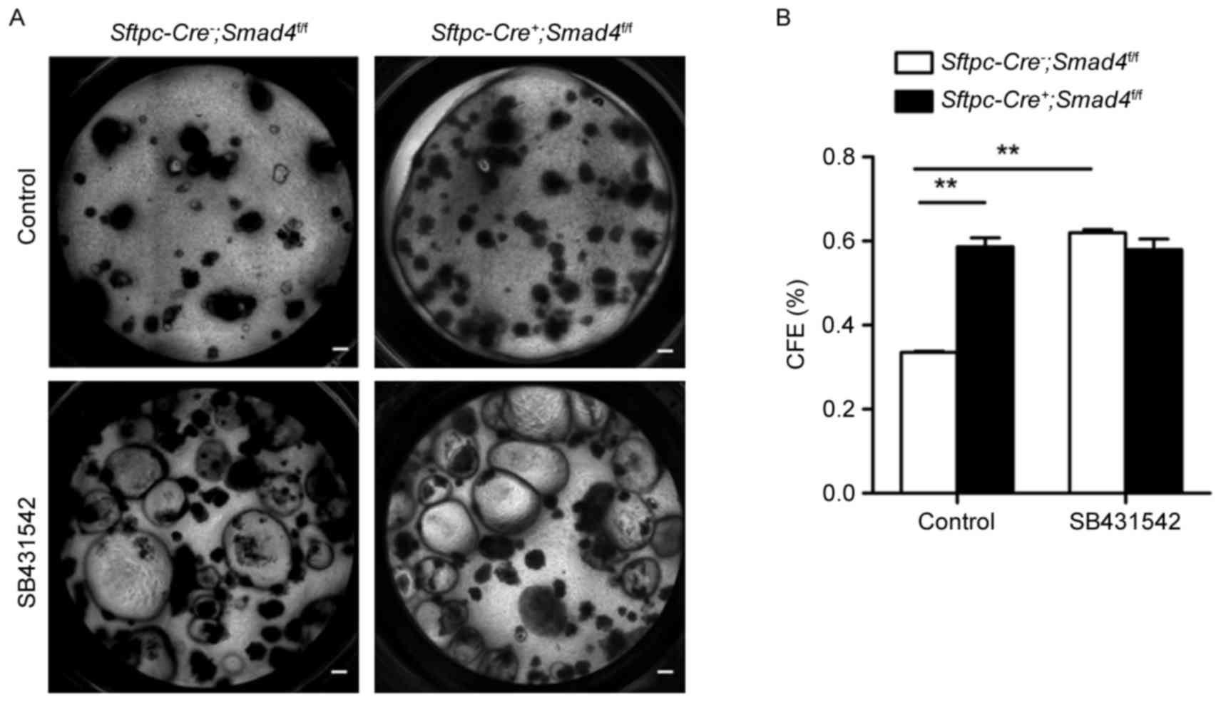
To examine this, the authors used a transgenic mouse
model, Sftpc-Cre−; Smad4f/f, in
which Smad4 is absent in all lung epithelia. Airway progenitor
cells from Sftpc-Cre−; Smad4f/f
or Sftpc-Cre+; Smad4f/f mice
were sorted and cultured in the presence of MLg cells. More
epithelial colonies were observed in the
Sftpc-Cre+; Smad4f/f group
compared to the Sftpc-Cre−;
Smad4f/f control group in the absence of SB431542
(Fig. 4A and B), suggesting that
Smad4 plays a negative role in the proliferation of airway
progenitor cells. This change was absent, however, between these
two groups in the presence of SB431542 (Fig. 4A and B). These data suggest that
TGF-β activation is required for the regulation of airway
progenitor cells by Smad4.
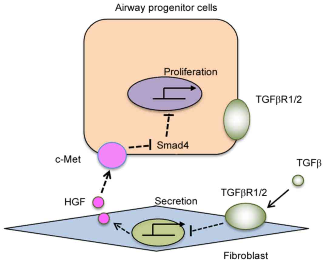
It was proposed, therefore, that the TGFβ/TGFβR1/2
axis plays an inhibitory role in the expression and/or secretion of
growth factors HGF in fibroblasts (Fig. 5). Fibroblast-derived HGF, through
its receptor c-Met on airway progenitor cells, supports the
proliferation of airway epithelial progenitor cells (Fig. 5). Smad4 may serve as a negative
regulator, given its deletion promotes the proliferation of airway
progenitor cells (Fig. 5).
Epithelial TGFβR1/2 seems not to be involved in the regulation of
airway progenitor cell proliferation.
Discussion
In the present study, the authors demonstrated that
airway progenitor cell proliferation is not affected by inhibition
of the TGF-β signal or deletion of the TGF-β receptor 2 on
progenitor cells in particular. Rather, inhibition of TGF-β
signaling specifically in fibroblasts contributes to the
proliferation of airway progenitor cells, likely due to the
inducible role of TGF-β signaling in the secretion of fibroblast
growth factors HGF.
TGF-β signal controls lung progenitor cell fate by
altering the microenvironment of the lung progenitor cells niche
(23). The TGF-β signal has been
implicated in the regeneration of airway epithelium following
injury. For instance, the upregulation of TGF-β stimulates
extracellular matrix secretion and activates the profibrotic
signaling pathway in lung fibrosis (24). Li et al (25) demonstrated that deletion of TGF-βR2
on the epithelia prevented mice from bleomycin-induced lung
fibrosis. Furthermore, TGF-β signaling promotes differentiation of
club cells and alveolar type 2 cells (26–28).
These results strongly suggest that TGF-β signaling reduces the
proliferation of epithelial progenitor cells and inhibits the
regeneration of injured epithelium. In the present study, the
authors found that TGF-β indirectly regulates airway progenitor
cells. More specifically, TGF-β ligand binds to a cell membrane
serine/threonine kinase heteromeric receptor complex, which is
composed of type 1 and type 2 receptors. TGFβ1R is known to be
inhibited by SB431542 (29).
Stimulation of TGFβ1R leads to phosphorylation of Smad2 and Smad3,
which combine with Smad4 and translocate into the nucleus (30). Smad plays a critical role in the
TGF-β1/Smad signal pathway. Inhibition of TGF-β1/Smad3 signaling
induces alveolar repair in the lung of hyperoxic mice (31). It also has been shown that the
deletion of Smad4 promotes lung cancer growth and metastasis
(32). The authors observed that
the proliferation of airway progenitor cells was also promoted in
absence of Smad4. Conversely, Smad4 can direct the repair of
epithelium under microenvironmental signals.
Growth factors are critical in the regulation of
airway epithelial cells by TGF-β. McQualter et al (7) found that inhibition of TGF-β can
restore stromal cell epithelial-supportive capacity, and upregulate
the expression of FGF10 in stromal cells (7). In addition, colonies of airway
progenitor cells were apparent in cultures supplemented with FGF10
and HGF in stromal-free conditions by Mungunsukh and Day (33). Similar to FGF10, HGF expression is
suppressed by TGF-β signaling post-transcription in a human adult
fibroblast culture. In contrast, HGF expression is upregulated by
knocking out TGF-β receptor 2 in mammary fibroblasts (34). Consistent with these data, the
authors observed that inhibition of TGF-β promotes HGF expression.
These findings suggest that blocking TGF-β activates HGF, after
which HGF binds to its receptor c-Met on epithelial and endothelial
cells to fulfill its functions.
Theoretically, HGF and FGF have been demonstrated to
inhibit Smad4 function through activating the Ras/MAPK pathway
(19). In the present study, the
authors observed that as compared with fibroblasts, airway
progenitor cells express c-Met abundantly. c-Met may mediate HGF's
role in regulating airway progenitor cells which may be dependent
on Smad4. Therefore, the TGFβ-TGFβR1/2-HGF-Smad4 axis may play a
role in airway epithelial homeostasis.
Nonetheless, there are still some limitations of the
current study. The relationship between HGF and Smad4 was not
detected directly, which can be addressed by creating
Sftpc-Cre+; Met f/f mice to
understand the interactions between c-Met and Smad4 in the
regulation of airway epithelial regeneration.
In conclusion, the study suggests that TGF-β
represses the proliferation of airway progenitor cells via the
TGF-β receptor on fibroblasts, thereby altering their secretory
properties including the secretion of HGF. Additionally, Smad4 may
play a mechanistic role in the regulation of airway progenitor
cells by HGF.
Acknowledgements
The current work was supported by the National
Natural Science Foundation of China (grant nos. 31471121, 81728001
and 81773394) and the Natural Science Foundation of Tianjin City
(grant nos. 13JCYBJC40000 and 17JCYBJC24700), and and Key Projects
of Health and Family Planning Commission of Tianjin (grant nos.
16KG163 and 16KG164).
References
|
1
|
O'Koren EG, Hogan BL and Gunn MD: Loss of
basal cells precedes bronchiolitis obliterans-like pathological
changes in a murine model of chlorine gas inhalation. Am J Respir
Cell Mol Biol. 49:788–797. 2013. View Article : Google Scholar : PubMed/NCBI
|
|
2
|
Trinh NT, Bardou O, Privé A, Maillé E,
Adam D, Lingée S, Ferraro P, Desrosiers MY, Coraux C and Brochiero
E: Improvement of defective cystic fibrosis airway epithelial wound
repair after CFTR rescue. Eur Respir J. 40:1390–1400. 2012.
View Article : Google Scholar : PubMed/NCBI
|
|
3
|
Lambrecht BN and Hammad H: The airway
epithelium in asthma. Nat Med. 18:684–692. 2012. View Article : Google Scholar : PubMed/NCBI
|
|
4
|
Proud D and Leigh R: Epithelial cells and
airway diseases. Immunol Rev. 242:186–204. 2011. View Article : Google Scholar : PubMed/NCBI
|
|
5
|
Tzortzaki EG and Siafakas NM: A hypothesis
for the initiation of COPD. Eur Respir J. 34:310–315. 2009.
View Article : Google Scholar : PubMed/NCBI
|
|
6
|
Hogan BL, Barkauskas CE, Chapman HA,
Epstein JA, Jain R, Hsia CC, Niklason L, Calle E, Le A, Randell SH,
et al: Repair and regeneration of the respiratory system:
Complexity, plasticity, and mechanisms of lung stem cell function.
Cell Stem Cell. 15:123–138. 2014. View Article : Google Scholar : PubMed/NCBI
|
|
7
|
McQualter JL, McCarty RC, Van der Velden
J, O'Donoghue RJ, Asselin-Labat ML, Bozinovski S and Bertoncello I:
TGF-β signaling in stromal cells acts upstream of FGF-10 to
regulate epithelial stem cell growth in the adult lung. Stem Cell
Res. 11:1222–1233. 2013. View Article : Google Scholar : PubMed/NCBI
|
|
8
|
Lee JH, Bhang DH, Beede A, Huang TL,
Stripp BR, Bloch KD, Wagers AJ, Tseng YH, Ryeom S and Kim CF: Lung
stem cell differentiation in mice directed by endothelial cells via
a BMP4-NFATc1-thrombospondin-1 axis. Cell. 156:440–455. 2014.
View Article : Google Scholar : PubMed/NCBI
|
|
9
|
Volckaert T, Dill E, Campbell A, Tiozzo C,
Majka S, Bellusci S and De Langhe SP: Parabronchial smooth muscle
constitutes an airway epithelial stem cell niche in the mouse lung
after injury. J Clin Invest. 121:4409–4419. 2011. View Article : Google Scholar : PubMed/NCBI
|
|
10
|
Ruiz EJ, Oeztuerk-Winder F and Ventura JJ:
A paracrine network regulates the cross-talk between human lung
stem cells and the stroma. Nat Commun. 5:31752014. View Article : Google Scholar : PubMed/NCBI
|
|
11
|
Tadokoro T, Gao X, Hong CC, Hotten D and
Hogan BL: BMP signaling and cellular dynamics during regeneration
of airway epithelium from basal progenitors. Development.
143:764–773. 2016. View Article : Google Scholar : PubMed/NCBI
|
|
12
|
Lynch TJ, Anderson PJ, Xie W, Crooke AK,
Liu X, Tyler SR, Luo M, Kusner DM, Zhang Y, Neff T, et al: Wnt
signaling regulates airway epithelial stem cells in adult murine
submucosal glands. Stem Cells. 34:2758–2771. 2016. View Article : Google Scholar : PubMed/NCBI
|
|
13
|
Rock JR, Gao X, Xue Y, Randell SH, Kong YY
and Hogan BL: Notch-dependent differentiation of adult airway basal
stem cells. Cell Stem Cell. 8:639–648. 2011. View Article : Google Scholar : PubMed/NCBI
|
|
14
|
Kim KK, Kugler MC, Wolters PJ, Robillard
L, Galvez MG, Brumwell AN, Sheppard D and Chapman HA: Alveolar
epithelial cell mesenchymal transition develops in vivo during
pulmonary fibrosis and is regulated by the extracellular matrix.
Proc Natl Acad Sci USA. 103:pp. 13180–13185. 2006; View Article : Google Scholar : PubMed/NCBI
|
|
15
|
Larson-Casey JL, Deshane JS, Ryan AJ,
Thannickal VJ and Carter AB: Macrophage Akt1 kinase-mediated
mitophagy modulates apoptosis resistance and pulmonary fibrosis.
Immunity. 44:582–596. 2016. View Article : Google Scholar : PubMed/NCBI
|
|
16
|
Yang YC, Zhang N, Van Crombruggen K, Hu
GH, Hong SL and Bachert C: Transforming growth factor-beta1 in
inflammatory airway disease: A key for understanding inflammation
and remodeling. Allergy. 67:1193–1202. 2012. View Article : Google Scholar : PubMed/NCBI
|
|
17
|
Plaschke-Schlütter A, Behrens J, Gherardi
E and Birchmeier W: Characterization of the scatter
factor/hepatocyte growth factor gene promoter. Positive and
negative regulatory elements direct gene expression to mesenchymal
cells. J Biol Chem. 270:830–836. 1995. View Article : Google Scholar : PubMed/NCBI
|
|
18
|
Hoot KE, Oka M, Han G, Bottinger E, Zhang
Q and Wang XJ: HGF upregulation contributes to angiogenesis in mice
with keratinocyte-specific Smad2 deletion. J Clin Invest.
120:3606–3616. 2010. View
Article : Google Scholar : PubMed/NCBI
|
|
19
|
Korc M: Smad4: Gatekeeper gene in head and
neck squamous cell carcinoma. J Clin Invest. 119:3208–3211.
2009.PubMed/NCBI
|
|
20
|
Teisanu RM, Chen H, Matsumoto K, McQualter
JL, Potts E, Foster WM, Bertoncello I and Stripp BR: Functional
analysis of two distinct bronchiolar progenitors during lung injury
and repair. Am J Respir Cell Mol Biol. 44:794–803. 2011. View Article : Google Scholar : PubMed/NCBI
|
|
21
|
Livak KJ and Schmittgen TD: Analysis of
relative gene expression data using real-time quantitative PCR and
the 2(-Delta Delta C(T)) method. Methods. 25:402–408. 2001.
View Article : Google Scholar : PubMed/NCBI
|
|
22
|
Shukla MN, Rose JL, Ray R, Lathrop KL, Ray
A and Ray P: Hepatocyte growth factor inhibits epithelial to
myofibroblast transition in lung cells via Smad7. Am J Respir Cell
Mol Biol. 40:643–653. 2009. View Article : Google Scholar : PubMed/NCBI
|
|
23
|
Quantius J, Schmoldt C, Vazquez-Armendariz
AI, Becker C, El Agha E, Wilhelm J, Morty RE, Vadász I, Mayer K,
Gattenloehner S, et al: Influenza virus infects epithelial
stem/progenitor cells of the distal lung: Impact on Fgfr2b-driven
epithelial repair. PLoS Pathog. 12:e10055442016. View Article : Google Scholar : PubMed/NCBI
|
|
24
|
Gauldie J, Kolb M, Ask K, Martin G,
Bonniaud P and Warburton D: Smad3 signaling involved in pulmonary
fibrosis and emphysema. Proc Am Thorac Soc. 3:pp. 696–702. 2006;
View Article : Google Scholar : PubMed/NCBI
|
|
25
|
Li M, Krishnaveni MS, Li C, Zhou B, Xing
Y, Banfalvi A, Li A, Lombardi V, Akbari O, Borok Z and Minoo P:
Epithelium-specific deletion of TGF-β receptor type II protects
mice from bleomycin-induced pulmonary fibrosis. J Clin Invest.
121:277–287. 2011. View Article : Google Scholar : PubMed/NCBI
|
|
26
|
Jain R, Barkauskas CE, Takeda N, Bowie EJ,
Aghajanian H, Wang Q, Padmanabhan A, Manderfield LJ, Gupta M, Li D,
et al: Plasticity of Hopx(+) type I alveolar cells to regenerate
type II cells in the lung. Nat Commun. 6:67272015. View Article : Google Scholar : PubMed/NCBI
|
|
27
|
Zhao L, Yee M and O'Reilly MA:
Transdifferentiation of alveolar epithelial type II to type I cells
is controlled by opposing TGF-β and BMP signaling. Am J Physiol
Lung Cell Mol Physiol. 305:L409–L418. 2013. View Article : Google Scholar : PubMed/NCBI
|
|
28
|
Xing Y, Li C, Li A, Sridurongrit S, Tiozzo
C, Bellusci S, Borok Z, Kaartinen V and Minoo P: Signaling via Alk5
controls the ontogeny of lung clara cells. Development.
137:825–833. 2010. View Article : Google Scholar : PubMed/NCBI
|
|
29
|
Prime SS, Pring M, Davies M and Paterson
IC: TGF-beta signal transduction in oro-facial health and
non-malignant disease (part I). Crit Rev Oral Biol Med. 15:324–336.
2004. View Article : Google Scholar : PubMed/NCBI
|
|
30
|
Mori S, Matsuzaki K, Yoshida K, Furukawa
F, Tahashi Y, Yamagata H, Sekimoto G, Seki T, Matsui H, Nishizawa
M, et al: TGF-beta and HGF transmit the signals through
JNK-dependent Smad2/3 phosphorylation at the linker regions.
Oncogene. 23:7416–7429. 2004. View Article : Google Scholar : PubMed/NCBI
|
|
31
|
Kayalar O and Oztay F: Retinoic acid
induced repair in the lung of adult hyperoxic mice, reducing
transforming growth factor-β1 (TGF-β1) mediated abnormal
alterations. Acta Histochem. 116:810–819. 2014. View Article : Google Scholar : PubMed/NCBI
|
|
32
|
Liu J, Cho SN, Akkanti B, Jin N, Mao J,
Long W, Chen T, Zhang Y, Tang X, Wistub II, et al: ErbB2 Pathway
Activation upon Smad4 loss promotes lung tumor growth and
metastasis. Cell Rep. pii:S2211–S1247. 2015.
|
|
33
|
Mungunsukh O and Day RM: Transforming
growth factor-β1 selectively inhibits hepatocyte growth factor
expression via a micro-RNA-199-dependent posttranscriptional
mechanism. Mol Biol Cell. 24:2088–2097. 2013. View Article : Google Scholar : PubMed/NCBI
|
|
34
|
Cheng N, Chytil A, Shyr Y, Joly A and
Moses HL: Transforming growth factor-beta signaling-deficient
fibroblasts enhance hepatocyte growth factor signaling in mammary
carcinoma cells to promote scattering and invasion. Mol Cancer Res.
6:1521–1533. 2008. View Article : Google Scholar : PubMed/NCBI
|