Vacquinol‑1 induces apoptosis in hepatocellular carcinoma cell
- Authors:
- Published online on: May 3, 2018 https://doi.org/10.3892/mmr.2018.8957
- Pages: 557-563
Metrics: Total
Views: 0 (Spandidos Publications: | PMC Statistics: )
Total PDF Downloads: 0 (Spandidos Publications: | PMC Statistics: )
Abstract
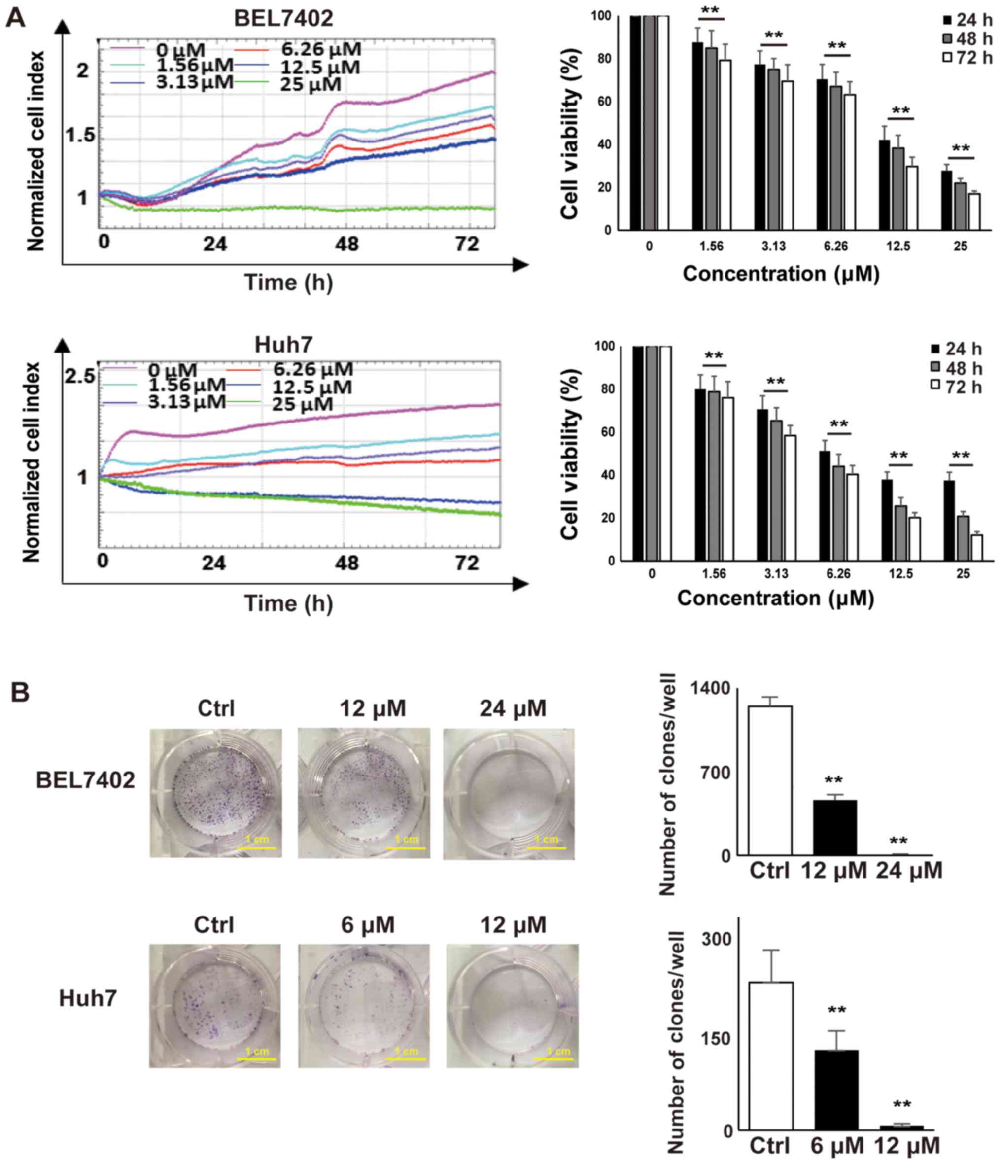
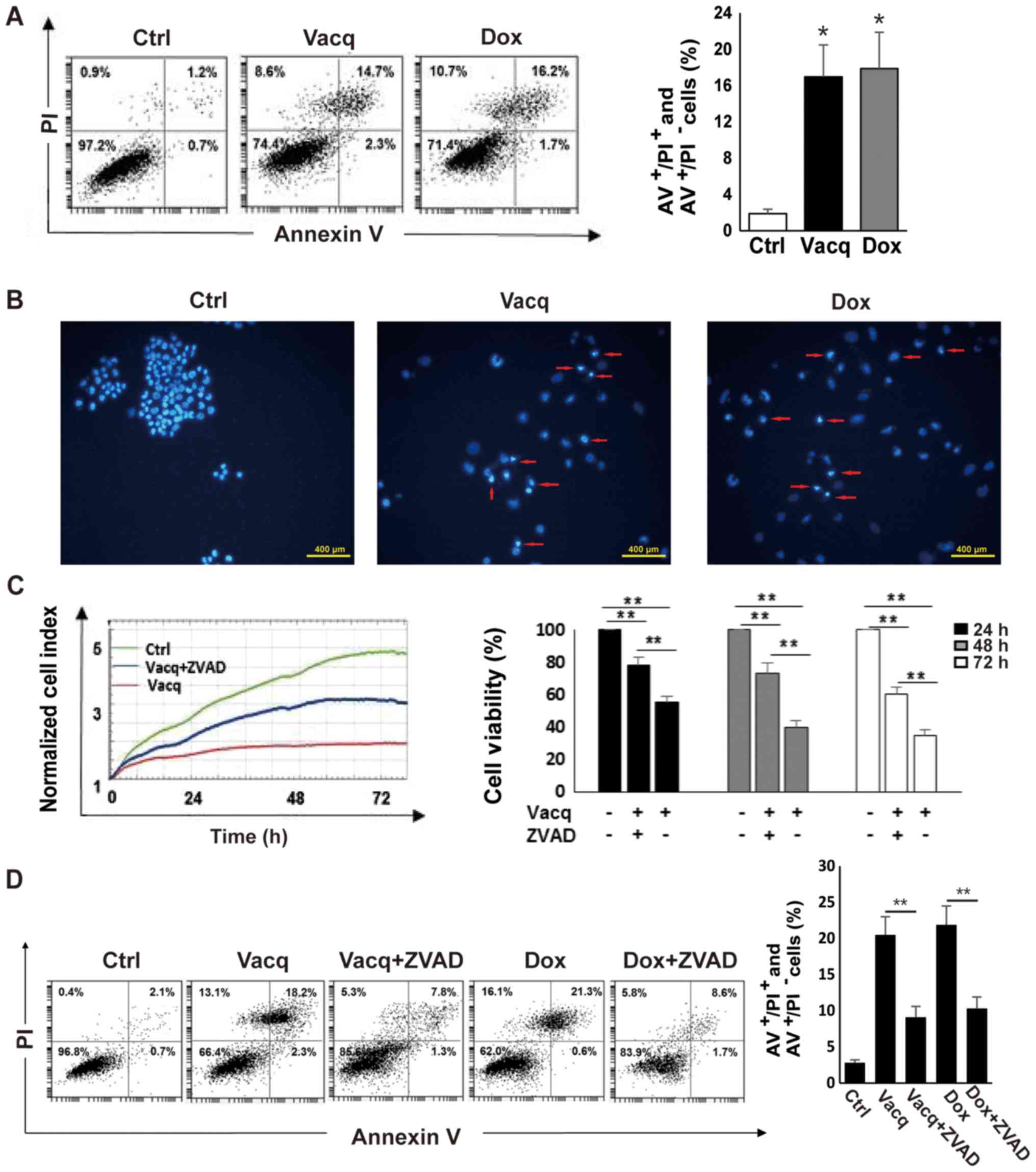
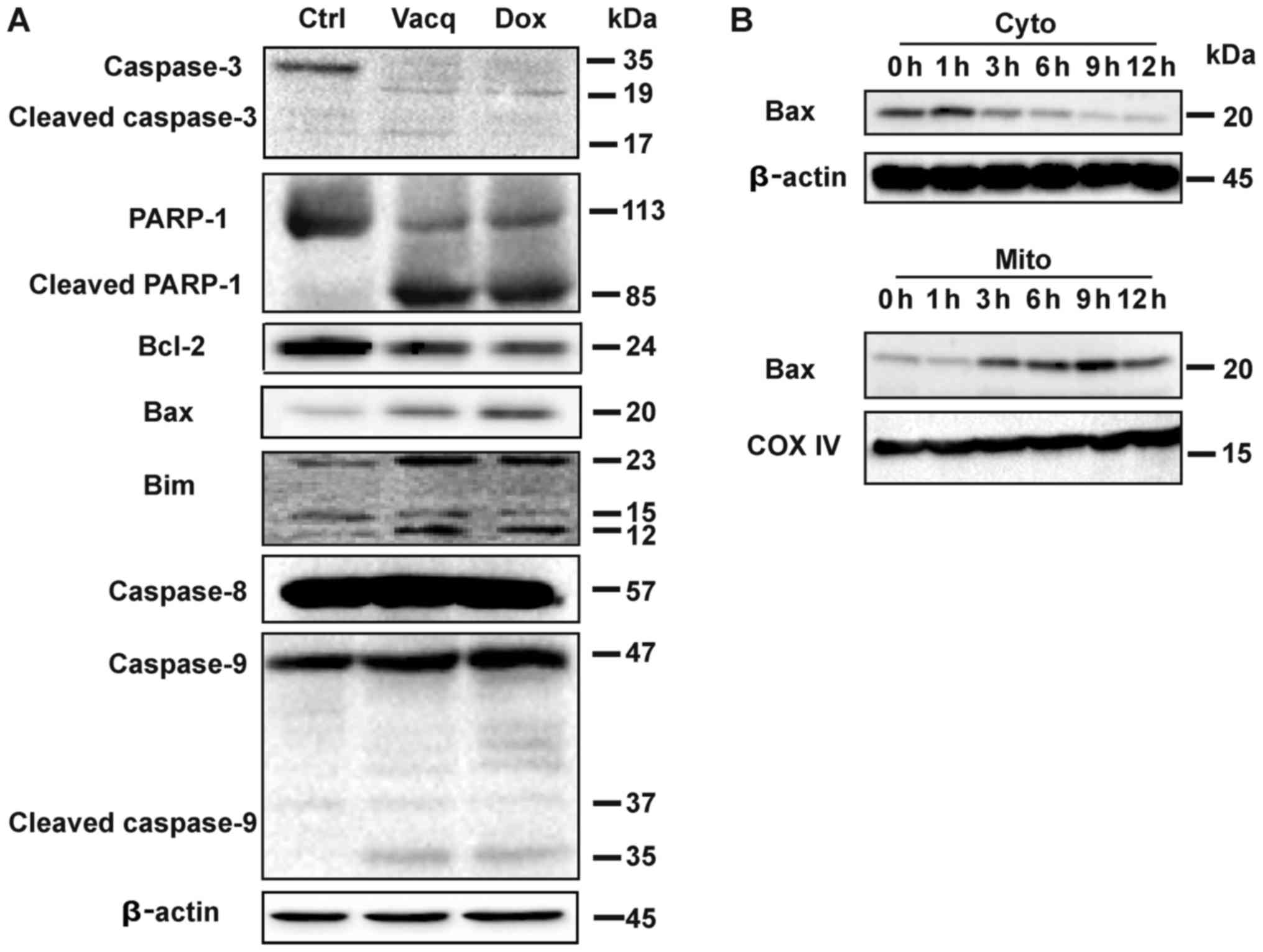
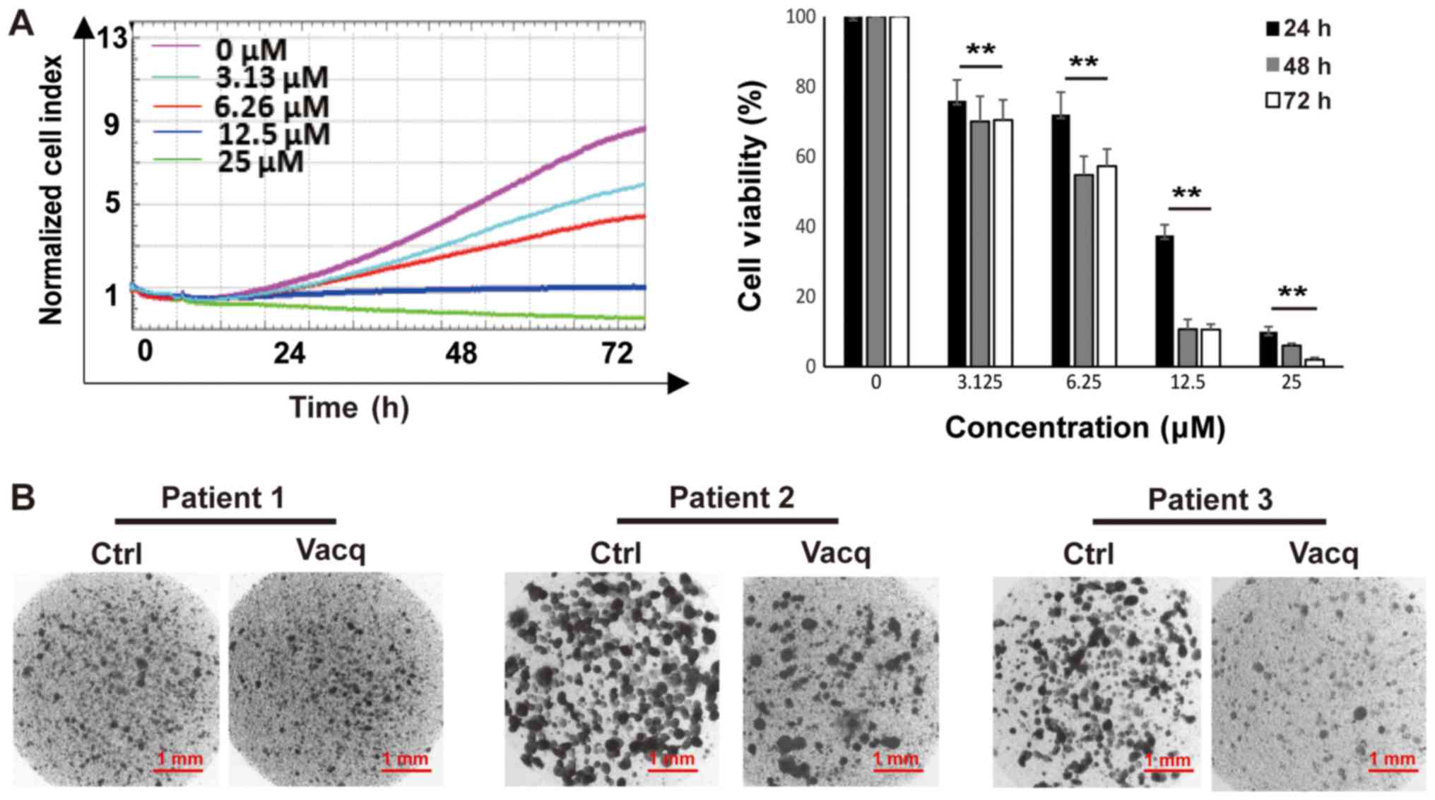
Vacquinol‑1 (Vacq), a quinolone derivative, has recently been reported to display potent antitumor effects in glioblastomas by inducing cellular massive vacuolization and cell death. However, whether Vacq induces cytotoxicities in other types of cancers, and the potential underlying mechanism, remain to be investigated. In the present study, it was revealed that Vacq suppressed cell growth and colony formation in the hepatocellular carcinoma (HCC) cell lines BEL7402 and Huh7. In addition, treatment with Vacq increased the number of early and late apoptotic cells as assessed by flow cytometry with fluorescein isothiocyanate‑conjugated Annexin V and propidium iodide double staining. Notably, the effect by Vacq in the tested cells could be inhibited by pretreatment with a broad specificity caspase inhibitor Z‑VAD‑FMK, suggesting that Vacq may induce apoptosis in HCC cells. Morphologically, exposure to Vacq resulted in nuclear fragmentation and the apoptotic body formation in HCC cells. Furthermore, Vacq treatment increased the cleavage of caspase‑3, caspase‑9 and poly(adenosine diphosphate‑ribose) polymerase‑1. Mechanistic analysis revealed that Vacq upregulated the expressions of pro‑apoptotic proteins [B‑cell lymphoma 2 (bcl‑2)‑associated X protein (Bax) and Bcl‑2‑like protein 11] and downregulated the pro‑survival protein, Bcl‑2, expression in HCC cells. Furthermore, Vacq induced Bax translocation. Of note, Vacq displayed inhibitory effects on patient‑derived HCC cells in two‑dimensional (2D) and three-dimensional (3D) cultures. Taken together, the data suggested that Vacq induced intrinsic apoptosis and may be utilized as an effective reagent for HCC treatment.