Introduction
Significant improvements have been made in the
chemotherapy of unresectable or recurrent colorectal carcinomas.
These improvements are due to the development of combination
chemotherapies, including fluorouracil, irinotecan or oxaliplatin,
in combination with molecular-targeting agents, including
bevacizumab and cetuximab. The use of a totally implantable venous
access device (TIVAD) is recommended for administration of these
chemotherapeutic agents.
TIVADs are generally placed in position by the
percutaneous subclavian vein approach. This approach causes
infrequent intraoperative or postoperative complications, including
pneumothorax, arterial puncture, hemothorax, injury to brachial
plexus (1–5) and pinch-off syndrome (6).
Venous access via the cephalic vein cut-down (CVCD)
method has been widely described as a safe and rapid approach
(7–10). However, this technique is not
currently widely used for the placement of TIVADs. Since the
cephalic vein shows few anatomical anomalies (11), this approach is suitable for the
placement of TIVADs.
This study examined the outcome of the venous access
via the CVCD method for the placement of TIVADs.
Materials and methods
A total of 79 consecutive patients with unresectable
or recurrent colorectal carcinomas underwent TIVAD placement
surgeries between June 2007 and October 2008.
The patients were brought to the operating room and
placed in a supine position. The operation was performed using
local anesthesia (0.5% of xylocaine; AstraZeneca, UK). Patients who
felt uneasy during the operation were administered an intravenous
injection of 15 mg of pentazocine (Astellas, Japan). Surgery was
performed under maximal barrier precaution. The TIVAD placements
were performed by two surgeons. Each stage of the surgical
procedure of this study was supervised by one of the two
surgeons.
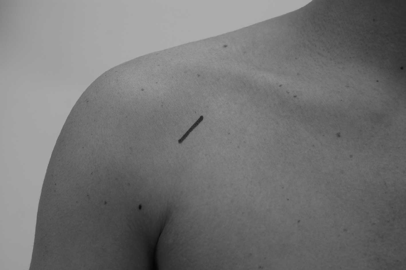
The cephalic vein passes through the clavipectoral
(deltopectoral) triangle to join the axillary vein. A 3-cm wide
skin incision was made in the infraclavicular region between the
pectoralis major muscle and the triangular muscle (Fig. 1). The cephalic vein was identified
in the adipose tissue along the deltopectoral groove. An incision
of 3 mm in length was made on the surface of the vein. A Groshong
catheter was inserted via the cephalic vein and connected to the
port (BardPort X-port isp; Bard Access Systems Inc., Salt Lake
City, UT, USA). The port was implanted in the subcutaneous space.
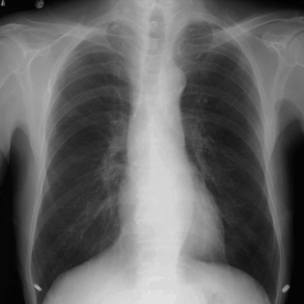
The position of the catheter tip and the shape of the catheter
lumen were confirmed by X-ray.
If the TIVAD placement by the CVCD approach was
unsuccessful, the procedure was converted to the conventional
percutaneous puncture approach of the subclavian vein. The
operation time, and any intraoperative and post-operative
complications were recorded.
Results
A total of 79 TIVAD placements were performed for 79
consecutive patients. TIVAD placement via the CVCD approach was
completed in 74 patients (93.7%). The CVCD approach was not
successful in the remaining 5 patients (6.3%). Subsequently, the 5
patients required conversion to a percutaneous puncture approach.
The mean operation time was 34.7 min (median 30, range 17–103 min).
The cephalic vein was not detected in 2 cases, and in 1 case the
cephalic vein was too narrow to insert the catheter. In 2 cases
where the cephalic vein merged to the axillary vein, the tip of the
catheter was not able to reach the superior vena cava. When the
cephalic vein merged to the axillary vein vertically, the catheter
tip reached only as far as the distal side of the axillary vein. In
such cases, we attempted to change the merging angle of the
cephalic and axillary veins to a more acute angle by leaning the
head of the patient to the opposite side or moving the arm and
shoulder. Manipulation of the angle normally allows the tip of the
catheter to reach the superior vena cava when the merging point was
vertical. However, in the 2 cases noted above, this manipulation
was not successful.
No intraoperative or immediate postoperative
complications were observed. A chest radiograph was obtained
following the surgery to detect the position of the catheter tip
and the distortion of the catheter lumen (Fig. 2). A distortion in the catheter may
indicate pinch-off syndrome. In this study, no catheter distortion
was detected.
Discussion
TIVADs are normally placed using the percutaneous
subclavian vein approach. The percutaneous approach has
occasionally caused intraoperative and postoperative complications,
such as pneumothorax, arterial puncture, hemothorax or injury to
the brachial plexus (1–5). The intraoperative complication rate of
the percutaneous approach was reported to be 3–7% (1–5). The
technique used for TIVAD implantation is now considered to be safer
due to the use of image-guided navigation techniques, such as
venous ultrasonography or venography. However, the risk of
complications remains when the percutaneous puncture approach is
used.
CVCD does not require risking a puncture and is
associated with a very low rate of complications. A number of
studies have compared the cut-down and percutaneous approaches and
reported the superiority and safety of the cut-down approach
compared to the conventional percutaneous method (9,10).
Pinch-off syndrome has been reported as a
complication of TIVAD (12,13). This syndrome is thought to be caused
by the compression of the catheter by the clavicle and the first
rib (12,13). Catheter compression may lead to
obstruction followed by fracture of the catheter. Of note is that
the pinch-off syndrome was not observed in a number of studies
using the cut-down technique (9,10,12).
Moreover, in the cardiovascular field, pinch-off
syndrome is known as subclavian crush syndrome, which has been
reported to occur with pacemaker leads implanted via the subclavian
puncture technique. In these cases, conductor fracture and
insulation breaches develop via compression of a lead that passes
between the first rib and the clavicle. In this study, it was
suggested that strong consideration should be given to obtain
venous access primarily via the cephalic cut-down technique, due to
the possibility of complications of subclavian crush syndrome with
the percutaneous puncture approach.
The CVCD approach requires a surgical technique.
Given the increase in the number of colorectal carcinoma patients
worldwide, this technique may be of practical value. In this study,
TIVAD placements were performed by the CVCD method and the outcome
was evaluated.
The cephalic vein passes through the clavipectoral
(deltopectoral) triangle to merge with the axillary vein. Since the
cephalic vein shows few anatomical anomalies (11), it is suitable for cut-down and the
placement of TIVAD.
TIVAD placement by CVCD was performed in 79 patients
and completed in 74 patients. The remaining 5 patients required
conversion to a percutaneous approach. The reasons for the
conversion were due to abnormalities of the cephalic vein,
including failure to detect the cephalic vein, a narrow cephalic
vein and abnormal merging of the cephalic and axillary veins. In
this study, the failure rate was found to be 6%, which is lower
than that in previous studies where a range of 8–30% was reported
(7,9,10).
Currently, the depth of the deltopectoral groove,
location of cephalic vein and the margins of the axillary vein are
routinely checked by ultrasonography prior to surgery. This
increases the success rate of completing a CVCD method. We are
therefore developing safer, more rapid and more practical placement
methods for TIVADs.
In conclusion, this study showed that the CVCD
technique is a safe and feasible approach for TIVAD placement.
Additionally, this technique is associated with a lower rate of
severe complications, reported to be up to 10%, compared to the
percutaneous method (9,10).
References
|
1
|
Biffi R, de Braud F, Orsi F, et al:
Totally implantable central venous access ports for long-term
chemotherapy. A prospective study analyzing complications and costs
of 333 devices with a minimum follow-up of 180 days. Ann Oncol.
9:767–773. 1998. View Article : Google Scholar
|
|
2
|
Kincaid EH, Davis PW, Chang MC,
Fenstermaker JM and Pennell TC: ‘Blind’ placement of long-term
central venous access devices: report of 589 consecutive
procedures. Am Surg. 65:520–524. 1999.
|
|
3
|
Poorter RL, Lauw FN, Bemelman WA, Bakker
PJ, Taat CW and Veenhof CH: Complications of an implantable venous
access device (Port-a-Cath) during intermittent continuous infusion
of chemotherapy. Eur J Cancer. 32A:2262–2266. 1996. View Article : Google Scholar : PubMed/NCBI
|
|
4
|
Nightingale CE, Norman A, Cunningham D,
Young J, Webb A and Filshie J: A prospective analysis of 949
long-term central venous access catheters for ambulatory
chemotherapy in patients with gastrointestinal malignancy. Eur J
Cancer. 33:398–403. 1997. View Article : Google Scholar : PubMed/NCBI
|
|
5
|
Di Carlo I, Barbagallo F, Toro A, Sofia M,
Lombardo R and Cordio S: External jugular vein cutdown approach, as
a useful alternative, supports the choice of the cephalic vein for
totally implantable access device placement. Ann Surg Oncol.
12:1–4. 2005.PubMed/NCBI
|
|
6
|
Di Carlo I, Fisichella P, Russello D,
Puleo S and Latteri F: Catheter fracture and cardiac migration: a
rare complication of totally implantable venous devices. J Surg
Oncol. 73:172–173. 2000.PubMed/NCBI
|
|
7
|
Povoski SP: A prospective analysis of the
cephalic vein cutdown approach for chronic indwelling central
venous access in 100 consecutive cancer patients. Ann Surg Oncol.
7:496–502. 2000. View Article : Google Scholar : PubMed/NCBI
|
|
8
|
Di Carlo I, Cordio S, La Greca G, et al:
Totally implantable venous access devices implanted surgically: a
retrospective study on early and late complications. Arch Surg.
136:1050–1053. 2001.PubMed/NCBI
|
|
9
|
Sarzo G, Finco C, Parise P, et al:
Insertion of prolonged venous access device: a comparison between
surgical cutdown and percutaneous techniques. Chir Ital.
56:437–442. 2004.PubMed/NCBI
|
|
10
|
Chang HM, Hsieh CB, Hsieh HF, et al: An
alternative technique for totally implantable central venous access
devices. A retrospective study of 1311 cases. Eur J Surg Oncol.
32:90–93. 2006. View Article : Google Scholar : PubMed/NCBI
|
|
11
|
Le Saout J, Vallee B, Person H, Doutriaux
M, Blanc J and Nguyen H: Anatomical basis for the surgical use of
the cephalic vein (V. Cephalica). 74 anatomical dissections
189 surgical dissections. J Chir. 120:131–134. 1983.PubMed/NCBI
|
|
12
|
Andris DA, Krzywda EA, Schulte W, Ausman R
and Quebbeman EJ: Pinch-off syndrome: a rare etiology for central
venous catheter occlusion. J Parenter Enteral Nutr. 18:531–533.
1994. View Article : Google Scholar : PubMed/NCBI
|
|
13
|
Hinke DH, Zandt-Stastny DA, Goodman LR,
Quebbeman EJ, Krzywda EA and Andris DA: Pinch-off syndrome: a
complication of implantable subclavian venous access devices.
Radiology. 177:353–356. 1990. View Article : Google Scholar : PubMed/NCBI
|
|
14
|
Roelke M, O'Nunain SS, Osswald S, Garan H,
Harthorne JW and Ruskin JN: Subclavian crush syndrome complicating
transvenous cardioverter defibrillator systems. Pace. 18:973–979.
1995. View Article : Google Scholar : PubMed/NCBI
|


