Introduction
Selenium is a trace element for which no direct sign
of its being essential in human nutrition was found until 1979. In
the same year, the existence of a correlation between the low
concentration of selenium in the geographical area of Keshan in
China and the pathology, known as Keshan disease, was identified by
a research group in this country (1). Selenium is stored in human tissues in
varying amounts: 30% of tissue selenium is in the liver, 15% in the
kidney, 30% in muscle, 10% in the plasma and the remaining 15%
throughout the other organs. Selenium is found in trace quantities
in a number of dietary agents such as dairy products, meat
products, poultry, fish, fruits, vegetables and cereals, rendering
it part of the food chain.
Selenium is considered to be a significant
antioxidant, a precursor of the antioxidant enzyme known as
‘glutathione peroxidase’ (GPX), which protects cells from free
radical damage in a number of neuronal and neuromuscular disorders,
such as stroke and cerebrovascular disease, Alzheimer’s disease,
Parkinson’s disease, familial amyotrophic lateral sclerosis and
Duchenne muscular dystrophy. The proposed mechanisms mainly invoke
the function of GPX as well as that of selenoprotein P (2). The evidence linking a lack of selenium
with cancer is found in epidemiological and clinical studies. Low
dietary selenium levels became an accurate way of predicting future
cancer rates. A previous study showed that selenium supplementation
led to a 50% reduction in cancer mortality (3). In general, the mean content of
selenium in serum from patients with various types of cancer was
lower than that of the control groups. Therefore, the correlation
between decreased levels of selenium and increased DNA damage and
increased oxidative stress further indicates the significance of
this trace element (4).
A number of studies, including geographic,
pre-clinical animal and prospective, as well as intervention
studies, have shown selenium to be involved in a number of types of
gastrointestinal cancer, such as esophageal, colorectal and
prostate, as well as liver cancer (5–6), and
have suggested its putative role in cancer, as well as progression,
and, subsequently, metastasis prevention (7). A significant positive correlation
between plasma selenium levels and liver cancer was also found. A
number of epidemiological studies performed on humans showed
significantly lower serum, plasma and liver selenium levels in
patients with liver diseases, such as chronic hepatitis and
cirrhosis, and different grades of hepatocellular injury, than
those measured in the healthy control groups (8–13).
Hepatocellular carcinoma (HCC) develops in the liver with severe
impairment of cellular antioxidant systems, since, in patients with
liver metastases from various types of cancer, cellular scavenger
enzymes exhibited normal activities, despite low selenium
concentrations (14).
In particular, HCC is a major health problem
worldwide, being the fifth most common malignancy in males and the
eighth in females and the third most common cause of cancer-related
mortality in the world. The incidence of HCC is on the increase,
with marked variations among geographic regions and racial and
ethnic groups, relative to the exposure to documented environmental
risk factors (15). In particular,
Southern Italy has the highest rates of HCC in Europe (16). Previously, we evaluated the
expression of the human selenium binding protein-1 (SELENBP1) in
tissue samples of HCC patients. SELENBP1 is a protein that
incorporates exogenously administered radioactive (75Se) sodium
selenite in the liver in vivo (17–18).
These studies provide evidence that this protein is down-regulated
in the liver tissue of HCC patients and that its gradual loss is
associated with an increased malignant grade (19).
In the present study, liver selenium levels in
tissue samples of Italian HCC patients were measured by atomic
absorption spectrometry in order to investigate whether there was a
correlation between selenium and SELENBP1 concentrations. To the
best of our knowledge, this is the first study that has examined
this type of correlation in human liver tissue.
Materials and methods
Tissue sample
Paraffin-embedded specimens of liver tissue from 20
HCC patients (5 females, 15 males) were analyzed. The
clinicopathological characteristics of these patients are shown in
Table I. The study included 5 HCC
patients with grade I tumors, 9 HCC patients with grade II and 6
HCC patients with grade III tumors. These patients had underlying
hepatitis C virus (HCV)-related cirrhosis and their age ranged
between 57 and 82 years. All 20 patients in this study provided
informed consent. Liver tissue samples were obtained from patients
recruited as part of an ongoing study to investigate associations
between cytokine dynamics and liver injury progression. Following
standard diagnostic procedures, biopsies were obtained from the
liver and were collected for research purposes.
 | Table IClinicopathological characteristics of
HCC patients subdivided according to grading.a |
Table I
Clinicopathological characteristics of
HCC patients subdivided according to grading.a
| Grading | Age | Gender | Tumor invasion | Tumor size (cm) |
|---|
|
|
|
|
|
|---|
| >70 | <70 | M | F | T1 | T2 | T3 | <4 | >4 |
|---|
| I | 1 | 4 | 4 | 1 | 3 | 2 | 0 | 2 | 3 |
| II | 7 | 2 | 7 | 2 | 1 | 3 | 5 | 3 | 6 |
| III | 3 | 3 | 4 | 2 | 2 | 2 | 2 | 3 | 3 |
Atomic absorption spectrometer
studies
Nitric acid, 65%; xylene anhydrous, 99%;
formaldehyde and ultra pure water were from Sigma-Aldrich
(Steinheim, Germany). Hydrogen peroxide, 30% and selenium standard
1000 ppm were from Carlo Erba Reagents (Milan, Italy). Each liver
tissue was fixed in formaldehyde and embedded in paraffin as part
of routine histological processing. For this study, each
paraffin-embedded tissue was dissected from the paraffin block to
obtain 20-μm slices. The preparation of samples was performed
according to a previous study (20). The wax was removed by washing (2×30
min) in xylene. The deparaffinized specimens were dried at 80°C for
24 h and digested with a mixture of HNO3 and
H2O2 at a ratio of 1:2 (v/v) at a temperature
of 100°C in a bath for 15–20 min.
The selenium concentration was determined by
graphite furnace atomic absorption spectroscopy on a Varian
SpectrAA 200 (Victoria, Australia) spectrometer with Zeeman
background correction. The quantitative determinations were carried
out by a calibration curve using selenium standard solutions (5–20
ppb). Digested samples were diluted with ultra pure water to yield
the selenium concentration within the calibration range.
The furnace settings were as follows: for drying,
ramp to 85°C (5 sec), ramp to 95°C (40 sec), and ramp to 120°C (10
sec); for ashing, ramp to 1000°C (5 sec) and hold at 1000°C (3
sec); for atomization, ramp to 2600°C (0.8 sec, read signal), hold
at 2600°C (2 sec, read signal), and hold at 2600°C (2 sec, tube
clean). The absorbance was determined at 196.0 nm, and the slit was
1.0 nm.
Data analysis
Selenium concentrations (expressed in μg/g) are
reported as the arithmetic means ± standard deviation for each
group of patients. The non-parametric Mann-Whitney U test was used
to evaluate differences between selenium concentrations from
patients with grades I-III HCC. The test was capable of
distinguishing values where p<0.05 with one asterisk
(*), with two asterisks (**) where values
were p<0.01 and with three asterisks (***) where
values were p<0.0001. The correlations between the selenium
levels and clinical data or immunohistochemical scores related to
SELENBP1 (19) were determined
using the Pearson’s correlation coefficient. P<0.05 was
considered to be statistically significant. The statistical program
Prism 4 (GraphPad Software, San Diego, CA, USA) was employed.
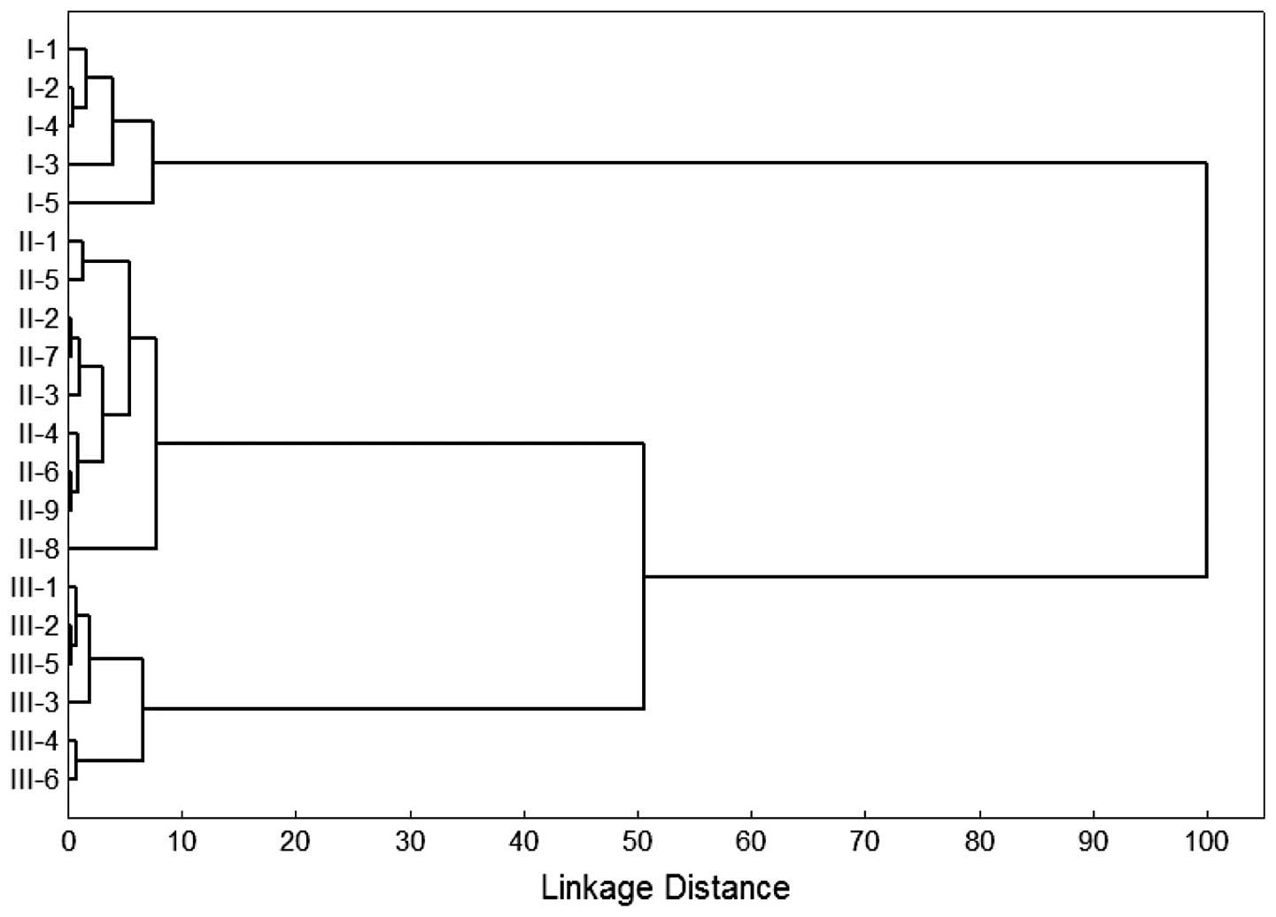
The relationship degrees between selenium levels and
patients were studied by means of the hierarchical agglo-merative
clustering method. Cluster analyses were performed using the
STATISTICA 8.0 statistical package (Statsoft Inc., Tulsa, OK, USA).
In particular, the patients were assigned to clusters step-by-step.
Following each step, clusters were joined to form new clusters. The
course of this process of increasing the degree of clustering was
exhibited in a dendrogram. At any distance from the start of the
clustering process, the number of clusters formed at that point are
shown in the graph (Fig. 1). The
basis for a cluster analysis was formed by calculating the degree
of similarity among the relevant variables of the different
patients to be clustered. In this study, the Euclidean distance
between patients was selected as the measure of similarity
(21–22). Although this study included a
relatively small number of patients, the approach of selecting
these patients for high, medium and low malignant grade is a way in
which to increase the statistical power.
Results
In a recent study, we found that the human SELENBP1
is also down-regulated in HCC (19). The present study correlates the
amount of selenium in HCC patients with the liver content of this
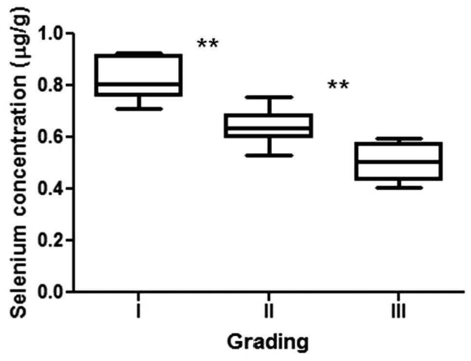
protein. The concentration of selenium in HCC patients subdivided
into three groups based on malignant grades (I–III) has been
evaluated by atomic absorption spectrometry (Fig. 1). The mean concentrations found in
the liver were 0.823±0.083 μg/g of selenium for HCC patients with
grade I, 0.638±0.066 μg/g for those with grade II and 0.505±0.076
μg/g for those with grade III. These values are lower than those
found in the livers of healthy individuals (1.9±0.6 μg/g) (23). The results are also in agreement
with scattered data reported in other studies (8–13).
We compared the selenium levels in three groups
using the Mann-Whitney U test. The differences between the patients
with grade I and II or with grade II and III are statistically
significant with p=0.0017 and p=0.0018, respectively. This finding
is also confirmed by the hierarchical clustering analysis of
patients based on the selenium concentrations. The analysis
provides evidence that the patients were placed into three groups,
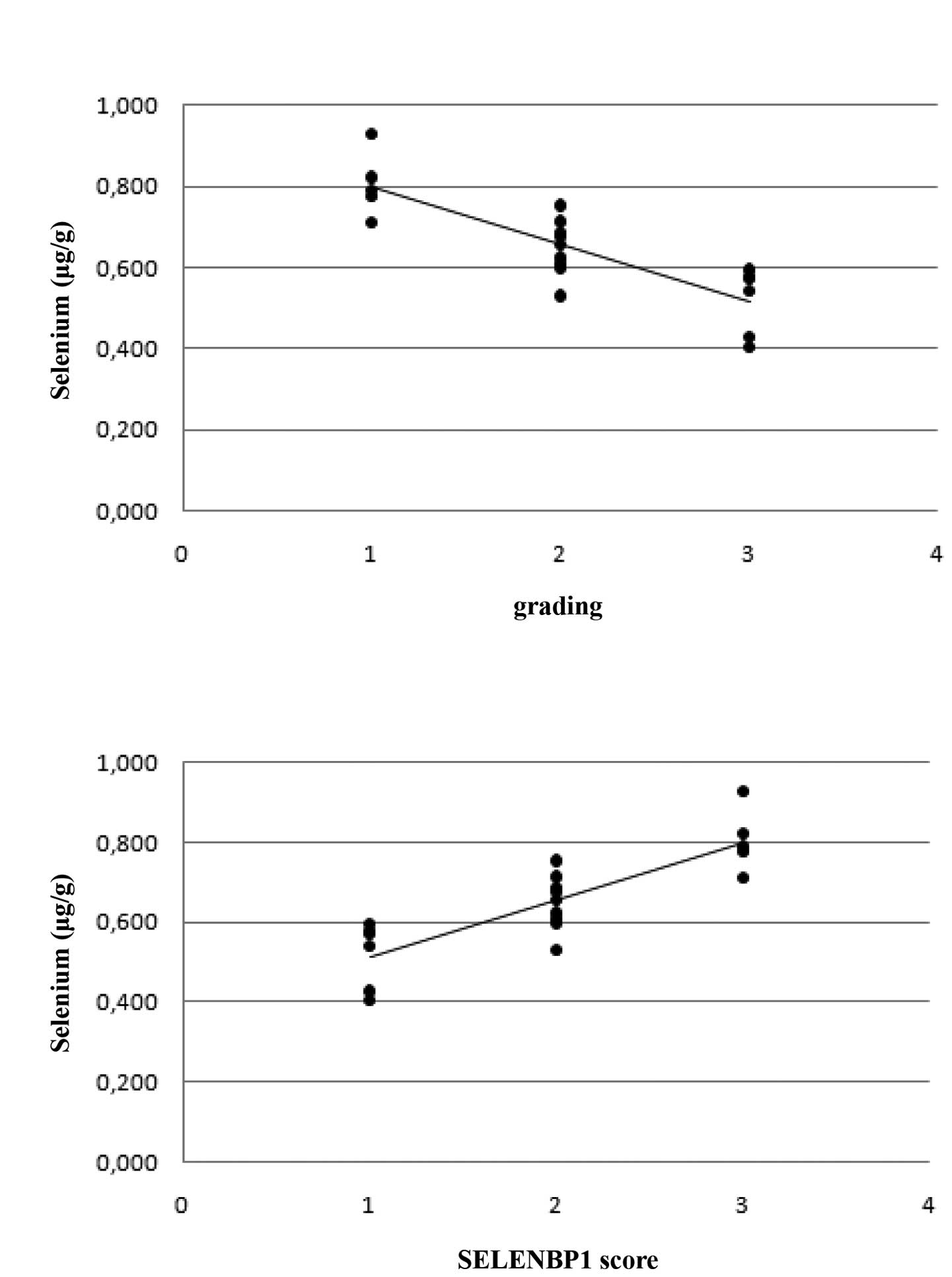
corresponding to the three different malignant grades (Fig. 2). Moreover, the data highlight that
selenium concentrations decrease when the malignant grade increases
with a correlation coefficient equal to −0.86 (Fig. 3A), whereas no correlation has been
observed between selenium levels and patient characteristics such
as age/gender, tumor invasion and tumor size. Finally, we have
correlated the selenium concentrations of patients with grades
I–III HCC and the expression of SELENBP1, which was recently
evaluated in the same patients, by immunohistochemical studies
(19). The rationale of this
evaluation is based on the fact that this protein incorporates
exogenously administered sodium selenite in the liver. Our studies
provided evidence that SELENBP1 was also down-regulated in the
liver tissues of HCC patients and its gradual loss was associated
with an increased malignant grade (19). As reported in Fig. 3B, a significant correlation between
SELENBP1 expression and selenium levels with a correlation
coefficient equal to 0.83 was detected. Thus, a decrease in
selenium concentration in the liver corresponds to lower values of
SELENBP1. This is a notable result since we have shown a clear
correlation with SELENBP1 in liver cancer, while data published in
a recent study (24) report no
correlation between serum or prostate tissue selenium and GPX
activity in prostate cancer patients. It is noteworthy that
prostate cancer also shows SELENBP1 down-regulation (25). Therefore, in general, these data
appear to suggest that the selenoenzymes containing endogenous
selenium as selenocysteine are not involved in those mechanisms
related to the low selenium levels in HCC tissues.
Discussion
Therapy for HCC remains poor; therefore, it is
essential to identify novel HCC markers for early detection of this
disease, and tumor-specific proteins as potential therapeutic
targets. Despite the various studies conducted to understand the
clinicopathological characteristics of the disease and to improve
patient treatment, conventional clinicopathological characteristics
have a limited predictive capacity. In 1960, it was suggested that
α fetoprotein (AFP) may be a useful tumor marker for HCC (26). Although AFP is widely used as a
marker, its drawbacks include a limited sensitivity and the fact
that AFP is not specific to HCC (27). Thus, the diagnosis of HCC cannot
depend mainly on the measurement of AFP. Consequently, molecular
biomarkers associated with HCC should be identified. Different
proteomics have been applied to the study of HCC, resulting in
panels of altered proteins that are proposed as markers with key
roles in the progression of the disease in certain cases (28–35).
However, the protein repertoires reported in these studies are
barely coincident and, consequently, their value as biomarkers for
HCC is limited. At present, only five biomarkers for HCC are
available for clinical use, from which only AFP partially fulfills
the requirements of an ideal tumor marker (36).
Selenium is capable of being tightly bound by
proteins, known as selenium-binding proteins, to distinguish them
from real selenoproteins (37). The
exact role of selenium-binding proteins is unknown, although
certain evidence suggests that these proteins are involved in
intra-Golgi protein transport. A homolog in Lotus japonicus
has been found to be involved in the modulation of the
oxidation/reduction status of target proteins in vesicular Golgi
transport (38–39). Moreover, selenium has been found to
be involved in the protection from ROS-induced cell damage, which
is evident in a number of diseases, and its serum and tissue levels
have been found to be significantly lower in patients with certain
types of cancer in respect to healthy controls (5–6).
Therefore, a functional correlation may be suspected between the
antique protein of Lotus and that in humans. The Golgi apparatus
has the crucial cellular function of protein release following
protein biosynthesis on ribosomes; therefore, a functional
protection against oxidative phenomena always present in the
eukaryotic cell may be necessary.
In this study, we have evaluated liver selenium
levels in tissue samples of HCC patients subdivided on the basis of
grading by atomic absorption spectrometry. The mean selenium
concentrations in the liver were lower in patients with the highest
malignant grade in the following order: grade I> grade
II>grade III. Moreover, these values correlated with the
immunohistochemical scores related to SELENBP1 evaluated in a
recent study (19). Therefore, the
results suggest that i) the evaluation of selenium and SELENBP1
concentrations are particularly useful for prognosis improvement
when clinical considerations suggest a hepatic biopsy in cirrhotic
patients, and ii) the intake of selenium and its derivatives in
cirrhotic patients should be considered as potential drugs against
HCC, which is in agreement with a number of studies on human
hepatoma cells and on animal models of liver cancer that showed the
cytotoxic and antioxidant effects of selenium (40). To the best of our knowledge, no
study has investigated the correlation between selenium and
SELENBP1 concentrations in the liver tissue of patients affected by
HCC, being a potentially significant target tissue for a
chemopreventive effect of selenium. Although there is no other
human tissue study, a number of studies investigated circulating
concentrations of selenium, which were used as a surrogate for
tissue measurements. Our findings are specific for HCC, however we
suggest that this correlation should be confirmed for other types
of cancer in which down-regulation of SELENBP1 has been found.
References
|
1
|
Keshan Disease Research Group.
Epidemiologic studies on the ethiologic relationship of selenium
and Keshan disease. Chi Med. 92:477–482. 1979.PubMed/NCBI
|
|
2
|
Sagara Y, Tan S, Maher P and Schubert D:
Mechanisms of resistance to oxidative stress in Alzheimer’s disease
brain. Neurotoxicology. 19:339–345. 1998.
|
|
3
|
Clark LC, Combs GF Jr, Turnbull BW, et al:
Effects of Selenium supplementation for cancer prevention in
patients with carcinoma of the skin. JAMA. 276:1957–63. 1996.
View Article : Google Scholar
|
|
4
|
Rayman MP: Selenium in cancer prevention:
a review of the evidence and mechanism of action. Proc Nutr Soc.
64:527–542. 2005. View Article : Google Scholar : PubMed/NCBI
|
|
5
|
Peters U and Takata Y: Selenium and the
prevention of prostate and colorectal cancer. Mol Nutr Food Res.
52:1261–1262. 2008. View Article : Google Scholar : PubMed/NCBI
|
|
6
|
Trottier G, Boström PJ, Lawrentschuk N and
Fleshner NE: Nutraceuticals and prostate cancer prevention: a
current review. Nat Rev Urol. 7:21–30. 2009. View Article : Google Scholar : PubMed/NCBI
|
|
7
|
Yoo MH, Xu XM, Carlson BA, Patterson VN,
Gladyshev AD and Hatfield DL: Targeting thioredoxin reductase 1
reduction in cancer cells inhibits self-sufficient growth and DNA
replication. PLoS One. 2:11122007. View Article : Google Scholar : PubMed/NCBI
|
|
8
|
Lecomte E, Herberth B, Pirrollet P, et al:
Effect of alcohol comsumption on blood antioxidant nutrients and
oxidative stress indicators. Am J Clin Nutr. 60:255–261.
1994.PubMed/NCBI
|
|
9
|
Loguercio C, De Girolamo V, Federico A, et
al: Trace elements and chronic liver diseases. J Trace Elem Med
Biol. 11:158–161. 1997. View Article : Google Scholar : PubMed/NCBI
|
|
10
|
Guarini P, Stanzial AM, Olivieri O, et al:
Erythrocyte membrane lipids and serum selenium in post-viral and
alcoholic cirrhosis. Clin Chim Acta. 270:139–150. 1998. View Article : Google Scholar : PubMed/NCBI
|
|
11
|
Navarro-Alarcòn M and Lòpez-Martìnez MC:
Essentiality of selenium in the human body: relationship with
different diseases. Sci Total Environ. 249:347–371. 2000.PubMed/NCBI
|
|
12
|
Navarro-Alarcòn M, Lòpez-G de la Serrana
H, Pèrez-Valero V and Lòpez-Martìnez MC: Selenium concentrations in
serum of individuals with liver diseases (cirrhosis or hepatitis):
relationships with some nutritional and biochemical markers. The
Science of the total environment. 291:135–141. 2002.
|
|
13
|
Tashiro H, Kawamoto T, Okubo T and Koide
O: Variation in the distribution of trace elements in hepatoma.
Biol Trace Element Research. 95:49–63. 2003. View Article : Google Scholar : PubMed/NCBI
|
|
14
|
Casaril M, Corso F, Bassi A, et al:
Decreased activity of scavenger enzymes in human hepatocellular
carcinoma, but not in liver metastases. International Journal of
Clinical and Laboratory Research. 24:94–97. 2006. View Article : Google Scholar
|
|
15
|
Castello G, Scala S, Palmieri G, Curley SA
and Izzo F: HCV-related hepatocellular carcinoma: from chronic
inflammation to cancer. Clin Immunol. 134:237–50. 2010. View Article : Google Scholar : PubMed/NCBI
|
|
16
|
Fusco M, Girardi E, Piselli P, et al:
Epidemiology of viral hepatitis infections in an area of southern
Italy with high incidence rates of liver cancer. Eur J Cancer.
44:847–53. 2008. View Article : Google Scholar : PubMed/NCBI
|
|
17
|
Bansal MP, Mukhopadhyay T, Scott J, Cook
RG, Mukhopadhyay R and Medina D: DNA sequencing of a mouse liver
protein that binds selenium: implications for selenium’s mechanism
of action in cancer prevention. Carcinogenesis. 11:2071–2073.
1990.PubMed/NCBI
|
|
18
|
Chang PW, Tsui SK, Liew C, Lee CC, Waye MM
and Fung KP: Isolation, characterization, and chromosomal mapping
of a novel cDNA clone encoding human selenium binding protein. J
Cell Biochem. 64:217–224. 1997. View Article : Google Scholar
|
|
19
|
Raucci R, Colonna G, Guerriero E, et al:
Structural and functional studies of the human selenium binding
protein-1 and its involvment in hepatocellular carcinoma. Biochimi
et Biophys Acta. 1814:513–522. 2011. View Article : Google Scholar : PubMed/NCBI
|
|
20
|
Beilby JP, Prins AW and Swanson NR:
Determination of hepatic iron concentration in fresh and
paraffin-embedded tissue. Clinical Chemistry. 45:573–574.
1999.PubMed/NCBI
|
|
21
|
Everitt BS, Landau S and Leese M: Cluster
analysis. New York: Oxford Univ Press; 2001
|
|
22
|
Hartigan JA: Clustering algorithms. New
York: John Wiley and Sons Inc; 1975
|
|
23
|
Oldereid NB, Thomassen Y and Purvis K:
Selenium in human male reproductive organs. Human Reproduction.
13:2172–2176. 1998. View Article : Google Scholar : PubMed/NCBI
|
|
24
|
Takata Y, Morris JS, King IB, Kristal AR,
Lin DW and Peters U: Correlation between selenium concentrations
and glutathione peroxidise activity in serum and human prostate
tissue. The Prostate. 69:1635–1642. 2009. View Article : Google Scholar
|
|
25
|
Jeong JY, Wang Y and Sytkowski AJ: Human
selenium binding protein-1 (hSP56) interacts with VDU1 in a
selenium-dependent manner. BBRC. 379:583–588. 2009.PubMed/NCBI
|
|
26
|
Johnson P: Role of α-fetoprotein in the
diagnosis and management of hepatocellular carcinoma. Gastroenterol
and Hepatol. 14:S32–S36. 1999.
|
|
27
|
Nguyen M, Garcia R, Simpson P, Wright T
and Keeffe B: Racial differences in effectiveness of α-fetoprotein
for diagnosis of hepatocellular carcinoma in hepatitis C virus
cirrhosis. Hepatology. 6:410–417. 2002.
|
|
28
|
Kim W, Oe Lim S, Kim JS, et al: Comparison
of proteome between hepatitis B virus- and hepatitis C
virus-associated hepatocellular carcinoma. Clin Cancer Res.
9:5493–5500. 2003.PubMed/NCBI
|
|
29
|
Kuramitsu Y and Nakamura K: Current
progress in proteomic study of hepatitis C virus-related human
hepatocellular carcinoma. Expert Rev Proteomics. 2:589–601. 2005.
View Article : Google Scholar : PubMed/NCBI
|
|
30
|
Li C, Hong Y, Tan YX, et al: Accurate
qualitative and quantitative proteomic analysis of clinical
hepatocellular carcinoma using laser capture microdissection
coupled with isotope-coded affinity tag and two-dimensional liquid
chromatography mass spectrometry. Mol Cell Proteomics. 3:399–409.
2004. View Article : Google Scholar
|
|
31
|
Liang CR, Leow CK, Neo JC, et al: Proteome
analysis of human hepatocellular carcinoma tissues by
two-dimensional difference gel electrophoresis and mass
spectrometry. Proteomics. 5:2258–2271. 2005. View Article : Google Scholar
|
|
32
|
Lim SO, Park SJ, Kim W, et al: Proteome
analysis of hepatocellular carcinoma. BBRC. 291:1031–1037.
2002.PubMed/NCBI
|
|
33
|
Blanc JF, Lalanne C, Plomion C, et al:
Proteomic analysis of differentially expressed proteins in
hepatocellular carcinoma developed in patients with chronic viral
hepatitis C. J Proteomics. 5:3778–3789. 2005. View Article : Google Scholar
|
|
34
|
Kim J, Kim SH, Lee SU, et al: Proteome
analysis of human liver tumor tissue by two-dimensional gel
electrophoresis and matrix assisted laser
desorption/ionization-mass spectrometry for identification of
disease-related proteins. Electrophoresis. 23:4142–4156. 2002.
View Article : Google Scholar
|
|
35
|
Paradis V, Degos F, Dargere D, et al:
Identification of a new marker of hepatocellular carcinoma by serum
protein profiling of patients with chronic liver diseases.
Hepatology. 41:40–47. 2005. View Article : Google Scholar : PubMed/NCBI
|
|
36
|
Santamaría E, Muñoz J, Fernández-Irigoyen
J, et al: Molecular profiling of hepatocellular carcinoma in mice
with a chronic deficiency of hepatic S-adenosylmethionine:
relevance in human liver diseases. J Proteome Res. 5:944–953.
2006.
|
|
37
|
Kryukov GV, Castellano S, Novoselov SV, et
al: Characterization of mammalian selenoproteomes. Science.
300:1439–1443. 2003. View Article : Google Scholar : PubMed/NCBI
|
|
38
|
Porat A, Sagiv Y and Elazar Z: A 56-kDa
selenium-binding protein participates in intra-Golgi protein
transport. J Biol Chem. 275:14457–65. 2000. View Article : Google Scholar : PubMed/NCBI
|
|
39
|
Flemetakis E, Agalou A, Kavroulakis N, et
al: Lotus japonicus gene Ljsbp is highly conserved among
plants and animals and encodes a homologue to the mammalian
selenium-binding proteins. Mol Plant Microbe Interact. 15:313–22.
2002. View Article : Google Scholar
|
|
40
|
Darvesh AS and Bishayee A: Selenium in the
prevention and treatment of hepatocellular carcinoma. Anti Canc
Agents Med Chem. 10:338–345. 2010. View Article : Google Scholar : PubMed/NCBI
|


