Introduction
Rhabdomyosarcoma (RMS) is a malignant soft-tissue
sarcoma that is believed to develop from primitive totipotent
embryonic mesenchyme. RMS is a highly aggressive tumor with a
tendency for advanced and disseminated disease early in its course.
The condition is the most common soft tissue sarcoma in children.
However, RMS in adults is an uncommon tumor that arises mainly in
the large skeletal muscles (1–4).
Pleomorphic RMS was first described by Stout in 1946 (5). More recent studies have reported that
pleomorphic RMS is rare and occurs predominantly in adults. The
present study describes a case of pleomorphic RMS in the right
adrenal region of a 61-year-old female and reviews the literature
on this rare disease. The study was approved by the ethics
committee of the First Affiliated Hospital, School of Medicine,
Zhejiang University (Hangzhou, China). Informed consent was
obtained from the patient.
Case report
A 61-year-old female was referred to the urological
ward of The First Affiliated Hospital, College of Medicine,
Zhejiang University for a right adrenal mass that had been detected
incidentally by ultrasound examination two weeks previously. The
patient had no underlying disease and the physical examination was
unremarkable. Abdominal ultrasound revealed a large adrenal tumor.
A computed tomography scan revealed a right adrenal tumor measuring
6.0×4.0 cm (Fig. 1). The patient
had undergone a complete adrenal endocrinological evaluation, which
demonstrated that the lesion was not a secreting tumor.
A pre-operative transcutaneous fine-needle
aspiration biopsy was performed and the cytological diagnosis was
consistent with a malignant neoplasm. Right adrenalectomy was
performed. The tumor was observed to have invaded into the right
lobe of the liver, and was well-demarcated from the hepatic
parenchyma by a thick fibrous capsule. The total operating time was
3 h. The estimated blood loss was 200 ml (calculated and recorded
by the attending anesthetist). The patient tolerated the procedure
well and there were no post-operative complications. The drainage
tube was removed at 48 h following the surgery. The patient was
discharged on the fifth post-operative day, tolerating a regular
diet. Pathological examination of the surgical specimen was
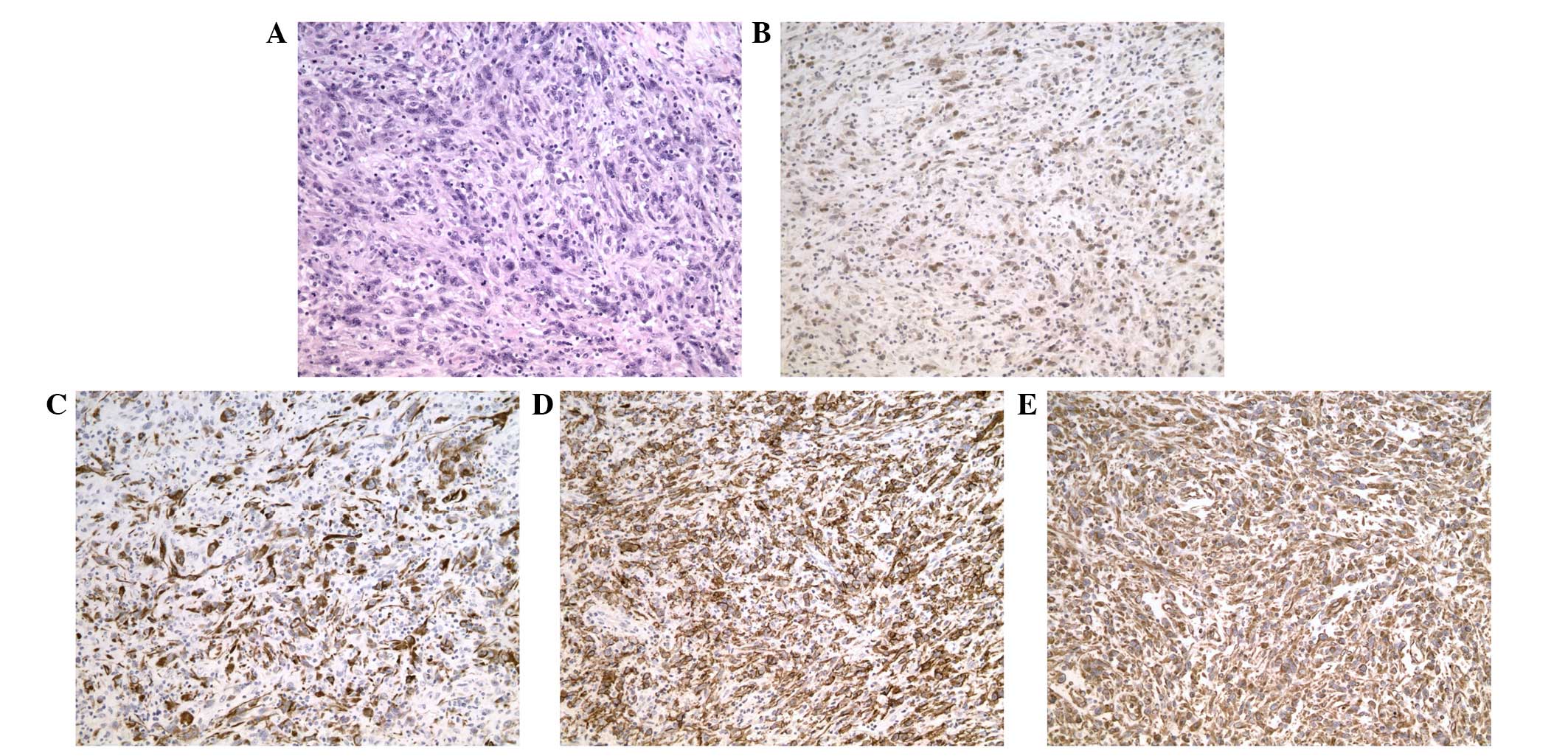
pleomorphic RMS containing spindle cells (Fig. 2A). Immunohistochemistry revealed a
positive stain for MyoD1, desmin, vimentin and CD56 (Fig. 2B–D). No expression of smooth muscle
actin (SMA), SYN or S100 protein was identified in the tumor
tissue.
Discussion
In the current World Health Organization
Classification of Soft Tissue and Bone Neoplasms, RMS is divided
into three distinct subtypes, embryonic, alveolar and pleomorphic
(6). RMS is a rare disease of the
adrenal gland neoplasm, which predominantly occurs in adults.
Charytonowicz et al(7)
suggest that RMS may arise from non-muscle cells, including
mesenchymal stem cells. Theoretically, RMS may affect any body
part, including the adrenal glands, as shown in the present case.
To date, only two RMS cases in the adrenal region have been
described in the English literature. Yi et al(8) reported a case of alveolar RMS in the
right adrenal region of a pediatric patient with a characteristic
history of hypertension and fever. Katayama et al(9) reported a case of RMS in the adrenal
region of an elderly hypertensive patient. However, pleomorphic RMS
of the adrenal gland in an adult has not been previously
reported.
In the present case, light microscopic examination
revealed a malignant pleomorphic mesenchymal neoplasm,
characterized mainly by the proliferation of atypical spindle cells
and few epithelioid cells. Immunohistochemistry revealed positive
staining for MyoD1, desmin, vimentin and CD56. By contrast, no
expression of SMA, SYN or S100 protein was identified in the tumor
tissue. A diagnosis of pleomorphic RMS was confirmed according to
the clinical and pathological findings.
In conclusion, the present study described a rare
case of pleomorphic RMS in the right adrenal region based on the
histopathology and immunohistochemistry results. Due to the small
number of described cases of adrenal gland RMS, inadequate
information is available for evaluating the treatment procedure and
the final prognosis of the patient. An accumulation of such cases
and an improved understanding of the molecular biology driving RMS
tumor behavior are required for further evaluation and research to
identify the histogenesis of the condition. Primary pleomorphic RMS
of the adrenal gland in an adult is a rare condition. To the best
of our knowledge, this is the first case of pleomorphic RMS of the
adrenal gland in an adult diagnosed by light microscopy and
immunohistochemical staining.
References
|
1
|
Furlong MA, Mentzel T and Fanburg-Smith
JC: Pleomorphic rhabdomyosarcoma in adults: a clinicopathologic
study of 38 cases with emphasis on morphologic variants and recent
skeletal muscle-specific markers. Mod Pathol. 14:595–603. 2001.
View Article : Google Scholar
|
|
2
|
Ogilvie CM, Crawford EA, Slotcavage RL, et
al: Treatment of adult rhabdomyosarcoma. Am J Clin Oncol.
33:128–131. 2010.
|
|
3
|
Stock N, Chibon F, Binh MB, et al:
Adult-type rhabdomyosarcoma: analysis of 57 cases with
clinicopathologic description, identification of 3 morphologic
patterns and prognosis. Am J Surg Pathol. 33:1850–1859. 2009.
View Article : Google Scholar
|
|
4
|
Sultan I, Qaddoumi I, Yaser S,
Rodriguez-Galindo C and Ferrari A: Comparing adult and pediatric
rhabdomyosarcoma in the surveillance, epidemiology and end results
program, 1973 to 2005: an analysis of 2,600 patients. J Clin Oncol.
27:3391–3397. 2009. View Article : Google Scholar : PubMed/NCBI
|
|
5
|
Stout AP: Rhabdomyosarcoma of the skeletal
muscles. Ann Surg. 123:447–472. 1946. View Article : Google Scholar
|
|
6
|
Fletcher CDM, Unni KK and Mertens F:
Pathology and genetics of tumors of soft tissue and bone. Soft
Tissue Tumors. IARC Press; Lyon: pp. 146–153. 2002
|
|
7
|
Charytonowicz E, Cordon-Cardo C,
Matushansky I and Ziman M: Alveolar rhabdomyosarcoma: is the cell
of origin a mesenchymal stem cell? Cancer Lett. 279:126–136. 2009.
View Article : Google Scholar : PubMed/NCBI
|
|
8
|
Yi X, Long X, Xiao D, Zai H and Li Y:
Rhabdomyosarcoma in adrenal region of a child with hypertension and
fever: A case report and literature review. J Pediatr Surg.
48:e5–e8. 2013. View Article : Google Scholar : PubMed/NCBI
|
|
9
|
Katayama A, Otsuka F, Takeda M, et al:
Rhabdomyosarcoma discovered in the adrenal region of an elderly
hypertensive patient. Hypertens Res. 34:784–786. 2011. View Article : Google Scholar : PubMed/NCBI
|


