Introduction
Studies worldwide have reported that patients with
medium or advanced cancer had a higher risk for bone metastasis,
accounting for 50–85% (1). Bone
metastasis in the spine is a relatively common site in clinic. The
occurrence rate of spinal metastatic carcinoma is higher than that
of primary malignant tumors. Previous findings showed that the
growth and metastasis of tumor cells are inseparable from
neovascularization, which is a necessary condition in the promotion
of tumor growth in cells (2,3). The principal factor that affects the
generation of neovascularization is vascular endothelial cell
growth factor (VEGF) (4). VEGF-A and
VEGF-C are important members of the VEGF family and can promote
endothelial cell migration and the construction of
neovascularization.
The regenerating gene family member 4 (RegIV) is a
new cancer gene. The expression of RegIV in carcinoma tissues,
including gastric and colorectal cancers has been reported
(5–8).
Nevertheless, the studies on the expression of spinal metastatic
carcinoma are not sufficient. Moreover, to the best of our
knowledge, there is no study on the relationship of RegIV and the
VEGF-A and VEGF-C correlation and microvessel density (MVD) of
patients with spinal metastatic carcinoma. The current study aimed
to reveal the expression of RegIV in spinal metastatic carcinoma
and its correlation with angiogenesis in order to provide valuable
reference for the studies on pathogenesis of spinal metastatic
carcinoma.
Patients and methods
Patients and study criteria
Fifteen patients with spinal metastatic tumor who
conformed to the conditions of this study and who were admitted to
our hospital from January 2011 to January 2013 were selected and
included in the study. Spinal metastatic tumor tissue was
surgically removed and its corresponding normal tissue distant from
lesion area was pathologically studied. Inclusion criteria were: i)
All patients were diagnosed as spinal metastatic tumor; ii) without
undergoing operation; iii) >18 years of age; iv) found with at
least 1 symptom related to spine and spinal cord injury; and v)
expected survival time was >3 months. Exclusion criteria were:
i) Patients with primary spinal malignant tumor; ii) had been
treated with spinal tumor operation; and iii) patients with poor
physical condition, unable to adapt to the operational treatment
implementation.
All the included cases in this study signed written
informed consent. We obtained the approval of the Ethics Committee
of Tianjin Nankai Hospital, all patients were planned to undergo
operation.
Methods
Main reagents
The antibodies and reagents used were: i) Rabbit
polyclonal RegIV antibody (dilution, 1:100; cat. no. sc-80320;
Santa Cruz Biotechnology, Inc., Santa Cruz, CA, USA); ii) rabbit
polyclonal anti-human VEGF-A antibody (dilution, 1:100; cat. no.
dm-10343; Shanghai Duma Biological Technology Co., Ltd., Shanghai,
China); iii) rabbit polyclonal anti-human VEGF-C antibody
(dilution, 1:100; cat. no. dm-10456; Shanghai Duma Biological
Technology Co., Ltd.); iv) DAB reagent and related
immunohistochemical kit (Shanghai Jingke Chemical Technology Co.,
Ltd., Shanghai, China); and v) rabbit polyclonal anti-human FVIII
antibody (dilution, 1:100; cat. no. BA0046; Wuhan Boster Biological
Technology, Ltd., Wuhan, China).
Detection method
RegIV detection was carried out as follows.
Streptomycin-biotin-peroxidase (SP) immunohistochemical method was
used to measure the RegIV protein expression level,
paraffin-embedded tissues were sectioned, and procedures were in
strict accordance with the instructions. Known positive sections
were regarded as the positive control, PBS instead of primary
antibody as the negative control.
VEGF-A, VEGF-C, FVIII detection was identified using
SP immunohistochemical method to measure VEGF-A, VEGF-C, FVIII
expression on spinal metastatic tumor tissue and para-cancer normal
tissue, and the FVIII-positive MVD was counted. Procedures of
staining were in strict accordance with the instructions of the
VEGF-A, VEGF-C, FVIII kits. Results of the staining were
evaluated.
RegIV expression level was detected using RT-qPCR.
Briefly, specimens were treated by liquid nitrogen, and total RNA
was extracted by TRIzol reagent. A260/A280 values of total RNA
detected by UV spectrophotometry were 1.8–2.0. RT-qPCR experiment
was carried out according to the kit instructions. Reverse
transcription system was 20 µl, RT was conducted in accordance with
the following conditions: 37°C for 60 min, and 95°C for 5 min. The
reaction system of PCR used 20 µl, and the reaction was carried out
on the ABI7700 quantitative PCR in accordance with the following
conditions: Initial activation of 95°C for 15 min; 3-step cycle: at
94°C for 15 sec; at 55°C for 30 sec; at 70°C for 30 sec, 30 cycles
in total. RNA U6 served as the internal reference, with reaction
conditions are the same as above. The experiment was repeated 3
times, obtained data were analyzed by RQ=2∆∆Cq.
Observation index
RegIV expression level in spinal metastatic tumor
tissue and paracancer normal tissue was observed, and the
expression level of RegIV, VEGF-A, VEGF-C in spinal metastatic
tumor tissue and paracancer normal tissue was contrasted. The
effect of different pathological parameters on RegIV, VEGF-A,
VEGA-C positive expression, as well as the correlation between
RegIV and VEGF-A, VEGF-C expression were analyzed.
Evaluation criteria
i) We employed the semi-quantitative scoring method
combined with Berry grading method (9): Positive expression of RegIV, VEGF-A,
VEGF-C was stained with cytoplasm, scored, respectively, according
to the positive cell rate and positive cell staining strength,
expression level was determined by the staining degree: Staining
score was the same with the negative control scored 0 point, pale
yellow scored 1 point, pale brown scored 2 points, brown scored 3
points. It could be divided according to the proportion of positive
cells in the observed cell: Number of positive cells ≤10%: 1 point,
11–50%: 2 points, 51–75%: 3 points, >75%: 4 points. Products of
two scores: 0–3 point was (−), 4–5 points were (+), 6–7 points
(++), >8 points (+++), ≤3 points negative, >3 points
positive.
ii) MVD count was based on Weidner and other methods
(10): FVIII-labeled vascular
endothelial cells with brownish yellow staining was positive
standard, the lumen and vascular remodeling formation of positive
endothelial cell clusters was regarded as a single microvessel
number, and for the lumen area with diameter of >8 red cells, or
vessels with a thicker layer, or single positive cell, the counts
were not carried out. Three microvascular distribution areas with
the highest density were selected, MVD count was carried out under
high-power field of vision (x400), its average value was taken as
the MVD value of this case.
Statistical analysis
SPSS 21.0 software (Chicago, IL, USA)was used for
statistical analysis. Positive rates of various groups were tested
by χ2, comparisons among groups were tested by t-test,
correlation analysis was tested by Spearman's rank correlation.
Relationship between the expression and prognosis of RegIV, VEGF-A,
VEGF-C were measured by Cox regression analysis. P <0.05 was
considered statistically significant.
Results
RegIV expression level in spinal
metastatic tumor tissue and paracancer normal tissue
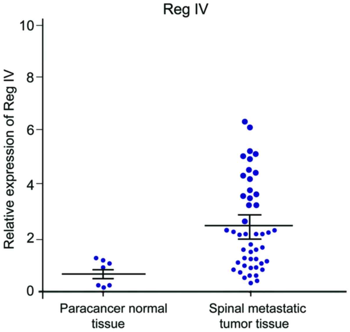
RT-qPCR results showed that the expression level of
RegIV in paracancer normal tissue and spinal metastatic tumor
tissue increased successively, and differences were statistically
significant (P<0.05) (Fig. 1).
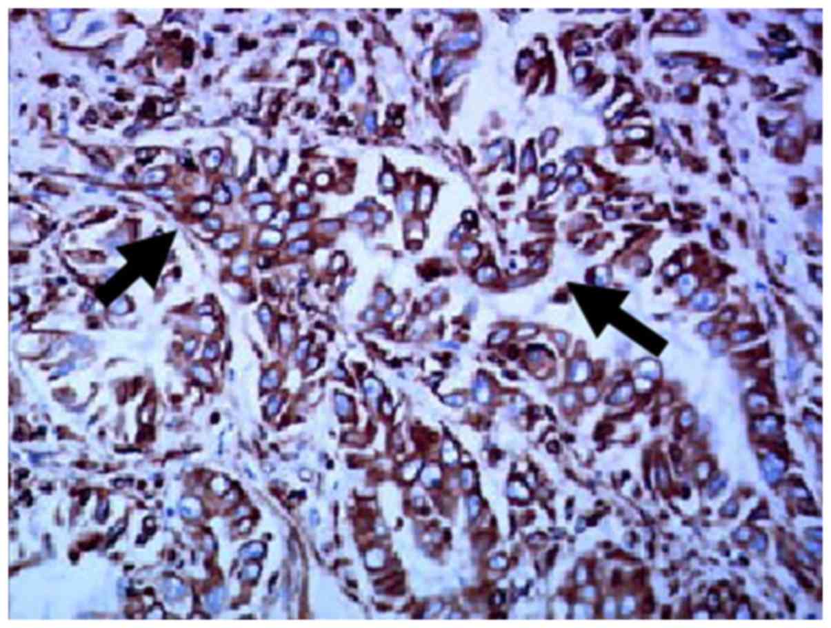
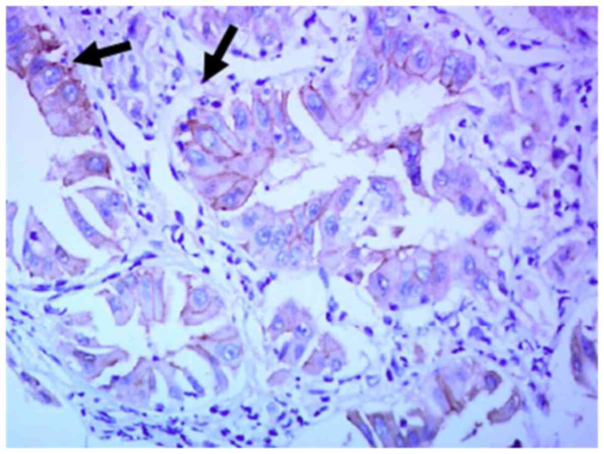
Expression of RegIV, VEGF-A, VEGF-C in
spinal metastatic tumor tissue and paracancer normal tissue
RegIV, VEGF-A, VEGF-C positive expression product
presented brownish yellow particles (Figs. 2–4).
RegIV expression positive rate of cancer tissue of patients with
spinal metastatic tumor was 53.33%, which was significantly higher
than the RegIV expression positive rate of 6.67% of paracancer
normal tissue (P<0.05) (Table I).
VEGF-A expression positive rate of cancer tissue of patients with
spinal metastatic tumor was 60%, which was significantly higher
than the VEGF-A expression positive rate of 6.67% of paracancer
tissue (P<0.05) (Table II). The
VEGF-C expression positive rate of cancer tissue of patients with
spinal metastatic tumor was 66.7%, which was significantly higher
than VEGF-A expression positive rate of 6.67% of paracancer tissue
(P<0.05) (Table III).
 | Table I.RegIV expression in spinal metastatic
tumor and paracancer normal tissue (case). |
Table I.
RegIV expression in spinal metastatic
tumor and paracancer normal tissue (case).
| Group | n | + | ++ | +++ | Positive rate
(%) | − |
|---|
| Spinal metastatic
tumor tissue | 15 | 2 | 3 | 3 | 53.33 | 7 |
| Paracancer normal
tissue | 15 | 1 | 0 | 0 | 6.67 | 14 |
| P-value |
|
|
|
|
| <0.05 |
| Table II.VEGF-A expression in spinal metastatic
tumor and paracancer normal tissue (case). |
Table II.
VEGF-A expression in spinal metastatic
tumor and paracancer normal tissue (case).
| Group | n | + | ++ | +++ | Positive rate
(%) | − |
|---|
| Spinal metastatic
tumor tissue | 15 | 3 | 3 | 3 | 60.0 | 6 |
| Paracancer normal
tissue | 15 | 1 | 0 | 0 | 6.67 | 14 |
| P-value |
|
|
|
|
| <0.05 |
| Table III.VEGF-C expression in spinal metastatic
tumor and paracancer normal tissue (case). |
Table III.
VEGF-C expression in spinal metastatic
tumor and paracancer normal tissue (case).
| Group | n | + | ++ | +++ | Positive rate
(%) | − |
|---|
| Spinal metastatic
tumor tissue | 15 | 3 | 4 | 3 | 66.67 | 5 |
| Paracancer normal
tissue | 15 | 1 | 0 | 0 | 6.67 | 14 |
| P-value |
|
|
|
|
| <0.05 |
Relationship between RegIV expression
and VEGF-A, VEGF-C
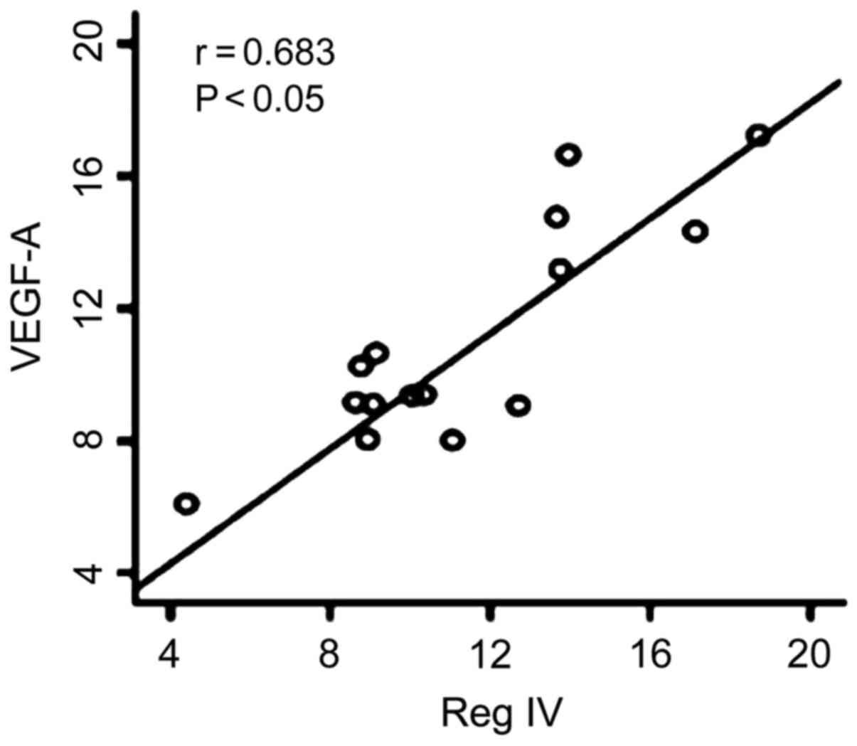
The Spearman's correlation analysis revealed that,
RegIV expression of spinal metastatic tumor tissue was positively
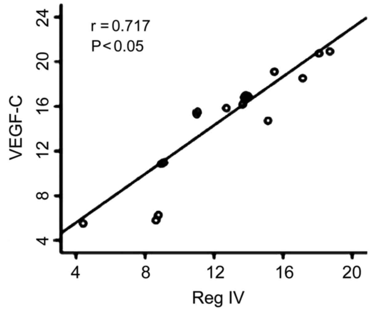
correlated with VEGF-A expression (r=0.683, P<0.05) (Fig. 5). RegIV expression of spinal
metastatic tumor tissue was positively correlated with VEGF-C
expression (r=0.71, P<0.05) (Fig.
6).
MVD comparison of tumor tissue of
RegIV-positive expression cases and paracancer normal tissue
MVD value of RegIV expression cases of spinal
metastatic tumor tissues was 57.67±4.43, which was significantly
higher than that of paracancer normal tissue (40.53±2.71)
(P<0.05) (Fig. 7).
Follow-up result
The average follow-up time was 18.4 months (4.5–36
months), there was no lost follow-up. Survival rate 1 year after
operation was 65.42%, 2 years was 19.35%, and 3 years was
7.26%.
Cox regression analysis
RegIV, VEGF-A, VEGF-C positive expression value and
MVD count were analyzed by Cox proportional hazard model. Results
showed that, RegIV, VEGF-A, VEGF-C expression and MVD count were
the factors that affected prognosis of spinal metastatic tumors
(P<0.05), RegIV positive expression had the maximum relative
risk (RR) to the patient survival (RR) (Table IV).
| Table IV.Cox regression analysis of RegIV,
VEGF-A, VEGF-C expression and MVD count. |
Table IV.
Cox regression analysis of RegIV,
VEGF-A, VEGF-C expression and MVD count.
| Index | U | s | Wald | P-value | RR |
|---|
| RegIV | 0.67 | 0.28 | 5.89 | 0.02 | 1.93 |
| VEGF-A | 0.21 | 0.17 | 1.66 | 0.03 | 1.33 |
| VEGF-C | 0.55 | 0.26 | 4.72 | 0.04 | 1.74 |
| MVD | 0.43 | 0.22 | 3.15 | 0.02 | 1.54 |
Discussion
In the skeletal system of human body, spine is the
tumor location easily transferred to and invaded. Autopsy for
cancer patients found that over 90% of patients had spinal
metastasis (11). Studies showed that
the growth and metastasis of tumor cells were closely related to
the generation of neovascularization (11–13).
VEGF-A and VEGF-C adjusted angiogenesis mainly through the
following two ways (14–16): i) Increasing the permeability of
microvessel; ii) promoting the growth, proliferation and migration
of endothelial cells through acting on the specific receptors on
endothelial cells; the upregulated expression of VEGF-A and VEGF-C
was closely related to hypoxia. Previous findings showed that
inhibiting the expression of VEGF-A and VEGF-C of tumor tissues
successfully inhibited the metastatic phenotypes of various tumors,
including breast, gastric cancer and colon cancers, and even
reversed the malignant phenotype of tumors (17).
Regenerating gene (Reg) family is a micromolecular
and multifunctional secreted protein. These family members are
similar in gene structure and can be divided into four subtypes (I,
II, III and IV). Their functional characteristics are equivalent to
that of acute phase protein and defenders against apoptotic death,
and they all positively take part in the occurrence and development
process of tissue injuries, tumor and inflammation. A previous
study (18) showed that Reg family
mainly had a low expression level in gastrointestinal tract, but
in situ or atopic high expression was evident in case of
tissue injuries. However, RegIV, as the newest member of Reg
family, was first separated and obtained from patients with
inflammatory bowel disease and it was hoped to become a biomarker
to mark highly malignant potential (19). At present, the correlation between the
expression of RegIV in patients with spinal metastatic carcinoma
and angiogenesis remains unknown.
In this study, the positive expression rates of
RegIV, VEGF-A and VEGF-C in spinal metastatic carcinoma tissues
were 53.33, 60 and 66.67%, respectively, which were significantly
higher than the positive expression rate in paracancer normal
tissue (P<0.05). This indicated that RegIV, VEGF-A and VEGF-C
may play an important role in the occurrence and development of
spinal metastatic carcinoma.
According to relevant analysis of Spearman, the
expression of RegIV in spinal metastatic carcinoma tissues was
positively correlated with the expression of VEGF-A (r=0.683,
P<0.05), the expression of RegIV in spinal metastatic carcinoma
tissues was positively correlated with the expression of VEGF-C
(r=0.717, P<0.05). The MVD value of RegIV expression cases in
spinal metastatic carcinoma tissues was 57.67±4.43, which was
significantly higher than the MVD value in paracancer normal tissue
(40.53±2.71) (P<0.05). This result suggested that RegIV was an
important positive regulator for angiogenesis and could promote the
generation of neovascularization in spinal metastatic carcinoma
tissues by activating the expression of VEGF-A and VEGF-C as well
as upregulated VEGF-A and VEGF-C. Its mechanism may be that RegIV
could carry out repeated degradation for vascular basement membrane
and extracellular matrix, promote the migration of vascular
endothelial cells and trigger the formation of neovascularization;
while VEGF-A and VEGF-C changed the active form of endothelial
cells and promoted tumor cells to pass through matrix, thus to
induce the occurrence of tumor infiltration and metastasis.
In conclusion, high expression of RegIV in spinal
metastatic carcinoma may increase microvessel density and promote
angiogenesis by promoting the expression of VEGF-A and VEGF-C,
thereby accelerating the occurrence and progression of spinal
metastatic carcinoma. Therefore, in clinical treatment for patients
with spinal metastatic carcinoma, the method of targeted therapy
can be taken into consideration to inhibit the expression of RegIV,
VEGF-A and VEGF-C so as to block tumor activity, adjusting the
pathway of tumor angiogenesis, markedly reducing malignant degree
of tumor and improving prognosis of patients.
References
|
1
|
Van Cutsem E, Köhne CH, Hitre E, Zaluski
J, Chien Chang CR, Makhson A, D'Haens G, Pintér T, Lim R, Bodoky G,
et al: Cetuximab and chemotherapy as initial treatment for
metastatic colorectal cancer. N Engl J Med. 360:1408–1417. 2009.
View Article : Google Scholar : PubMed/NCBI
|
|
2
|
Strube A, Hoffmann J, Stepina E, Hauff P,
Klar U and Käkönen SM: Sagopilone inhibits breast cancer bone
metastasis and bone destruction due to simultaneous inhibition of
both tumor growth and bone resorption. Clin Cancer Res.
15:3751–3759. 2009. View Article : Google Scholar : PubMed/NCBI
|
|
3
|
Murakami H, Kawahara N, Demura S, Kato S,
Yoshioka K and Tomita K: Total en bloc spondylectomy for lung
cancer metastasis to the spine. J Neurosurg Spine. 13:414–417.
2010. View Article : Google Scholar : PubMed/NCBI
|
|
4
|
Eastley N, Newey M and Ashford RU:
Skeletal metastases - the role of the orthopaedic and spinal
surgeon. Surg Oncol. 21:216–222. 2012. View Article : Google Scholar : PubMed/NCBI
|
|
5
|
Patchell RA, Tibbs PA, Regine WF, Payne R,
Saris S, Kryscio RJ, Mohiuddin M and Young B: Direct decompressive
surgical resection in the treatment of spinal cord compression
caused by metastatic cancer: a randomised trial. Lancet.
366:643–648. 2005. View Article : Google Scholar : PubMed/NCBI
|
|
6
|
Inoue T, Oh RJ and Shiomi H: New approach
for treatment of vertebral metastases using intensity-modulated
radiotherapy. Strahlenther Onkol. 187:108–113. 2011. View Article : Google Scholar : PubMed/NCBI
|
|
7
|
Arrigo RT, Kalanithi P, Cheng I, Alamin T,
Carragee EJ, Mindea SA, Park J and Boakye M: Predictors of survival
after surgical treatment of spinal metastasis. Neurosurgery.
68:674–681; discussion 681. 2011. View Article : Google Scholar : PubMed/NCBI
|
|
8
|
Cole JS and Patchell RA: Metastatic
epidural spinal cord compression. Lancet Neurol. 7:459–466. 2008.
View Article : Google Scholar : PubMed/NCBI
|
|
9
|
Stieler F, Wolff D, Bauer L, Wertz HJ,
Wenz F and Lohr F: Reirradiation of spinal column metastases:
comparison of several treatment techniques and dosimetric
validation for the use of VMAT. Strahlenther Onkol. 187:406–415.
2011. View Article : Google Scholar : PubMed/NCBI
|
|
10
|
Tokuhashi Y, Matsuzaki H, Oda H, Oshima M
and Ryu J: A revised scoring system for preoperative evaluation of
metastatic spine tumor prognosis. Spine. 30:2186–2191. 2005.
View Article : Google Scholar : PubMed/NCBI
|
|
11
|
Lee BH, Kim TH, Chong HS, Moon ES, Park
JO, Kim HS, Kim SH, Lee HM, Cho YJ, Kim KN, et al: Prognostic
factor analysis in patients with metastatic spine disease depending
on surgery and conservative treatment: review of 577 cases. Ann
Surg Oncol. 20:40–46. 2013. View Article : Google Scholar : PubMed/NCBI
|
|
12
|
Tabouret E, Cauvin C, Fuentes S, Esterni
B, Adetchessi T, Salem N, Madroszyk A, Gonçalves A, Casalonga F and
Gravis G: Reassessment of scoring systems and prognostic factors
for metastatic spinal cord compression. Spine J. 15:944–950. 2015.
View Article : Google Scholar : PubMed/NCBI
|
|
13
|
Zhang D, Xu W, Liu T, Yin H, Yang X, Wu Z
and Xiao J: Surgery and prognostic factors of patients with
epidural spinal cord compression caused by hepatocellular carcinoma
metastases: retrospective study of 36 patients in a single center.
Spine (Phila Pa 1976). 38:E1090–E1095. 2013. View Article : Google Scholar : PubMed/NCBI
|
|
14
|
Santini D, Tampellini M, Vincenzi B,
Ibrahim T, Ortega C, Virzi V, Silvestris N, Berardi R, Masini C,
Calipari N, et al: Natural history of bone metastasis in colorectal
cancer: final results of a large Italian bone metastases study. Ann
Oncol. 23:2072–2077. 2012. View Article : Google Scholar : PubMed/NCBI
|
|
15
|
Scagliotti GV, Hirsh V, Siena S, Henry DH,
Woll PJ, Manegold C, Solal-Celigny P, Rodriguez G, Krzakowski M,
Mehta ND, et al: Overall survival improvement in patients with lung
cancer and bone metastases treated with denosumab versus zoledronic
acid: Subgroup analysis from a randomized phase 3 study. J Thorac
Oncol. 7:1823–1829. 2012. View Article : Google Scholar : PubMed/NCBI
|
|
16
|
Escudier B, Szczylik C, Porta C and Gore
M: Treatment selection in metastatic renal cell carcinoma: expert
consensus. Nat Rev Clin Oncol. 9:327–337. 2012. View Article : Google Scholar : PubMed/NCBI
|
|
17
|
Shostak K and Chariot A: EGFR and NF-κB:
partners in cancer. Trends Mol Med. 21:385–393. 2015. View Article : Google Scholar : PubMed/NCBI
|
|
18
|
Crnalic S, Hildingsson C, Wikström P,
Bergh A, Löfvenberg R and Widmark A: Outcome after surgery for
metastatic spinal cord compression in 54 patients with prostate
cancer. Acta Orthop. 83:80–86. 2012. View Article : Google Scholar : PubMed/NCBI
|
|
19
|
Tatsui CE, Suki D, Rao G, Kim SS, Salaskar
A, Hatiboglu MA, Gokaslan ZL, McCutcheon IE and Rhines LD: Factors
affecting survival in 267 consecutive patients undergoing surgery
for spinal metastasis from renal cell carcinoma. J Neurosurg Spine.
20:108–116. 2014. View Article : Google Scholar : PubMed/NCBI
|