Introduction
Malignant lymphoma of the uterine cervix and corpus
is relatively rare and accounts for only 0.5% of extranodal
lymphomas. The overall incidence of malignant lymphomas of the
uterine cervix is less than 1% among all cervical malignancies
(1), and only a few cases have been
reported to date (2–4). It is difficult to diagnose malignant
lymphomas of the uterine cervix by cervical cytology because
lymphomas at this site are rare and can be misdiagnosed as other
clinical conditions such as chronic cervicitis and epithelial
malignancies (e.g., small cell carcinoma and poorly differentiated
adenocarcinoma) (1). As a result,
there are many cases where malignant lymphoma was diagnosed after
invasive surgery.
Here, we present a case of malignant lymphoma of the
uterine cervix presumptively diagnosed by the Papanicolaou (Pap)
smear. We also discuss the clinicopathological features and
therapeutic management of this rare tumor.
Case report
A 74-year-old woman (gravida 6, para 3) with no
clinical symptoms had a medical checkup. Her past medical history
was not significant. The result from the uterine cancer test was
‘possible non-epithelial malignant tumor, including malignant
lymphoma’. A detailed examination was conducted at our
hospital.
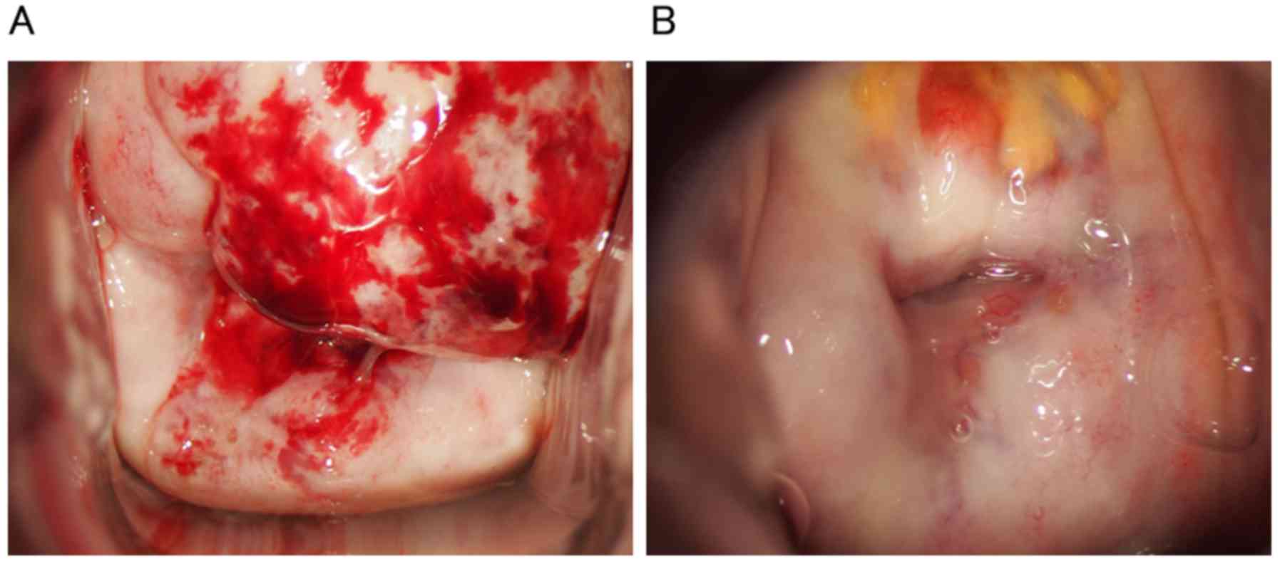
Vaginal examination revealed a macroscopic, whitish
hemorrhagic tumor (Fig. 1) occupying
the anterior lip of the uterine cervix (Fig. 1A). Pelvic ultrasound showed an
irregular round hypoechoic mass approximately 4 cm in diameter.
Positron emission tomography (PET) scan demonstrated a uterine
cervical mass with high PET avidity, and another swollen lymph node
was detected near the gastric cardia. No notable abnormalities,
including the levels of serum LDH and soluble IL 2 receptor, were
found in a blood test.
We obtained written informed consent, and patient
anonymity has been preserved.
Pathological findings
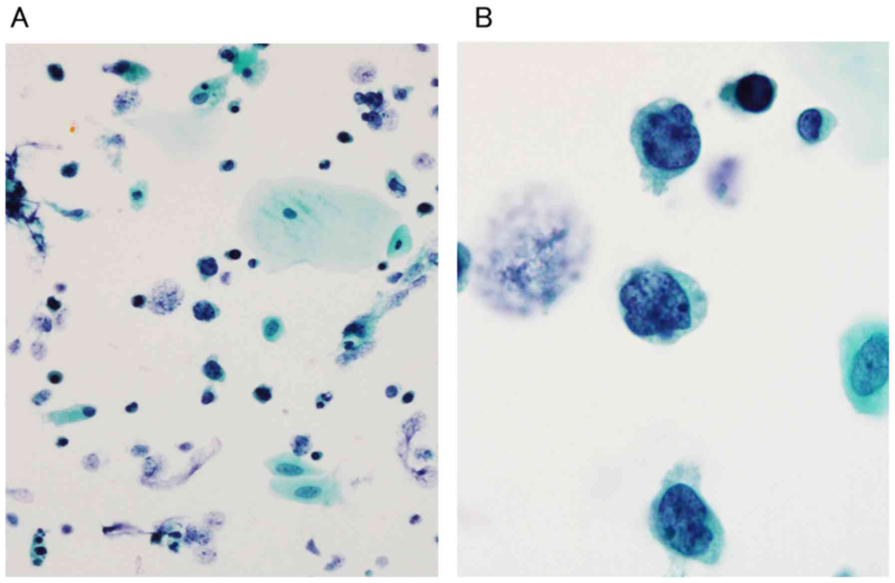
Cervical cytology was performed by liquid-based
cytology (LBC) using the Sure Path method. The LBC slide
demonstrated a necrotic background and scattered atypical cells
with high nuclear/cytoplasmic ratios (Fig. 2A). Approximately 80% of the cells
observed in the slide were atypical cells. These cells had almost
bare and coarse, hyperchromatic, focally cleaved nuclei with
prominent nucleoli using a high-power field (Fig. 2B). Since it lacked adepithelial
construction, it was suspected to be a non-epithelial malignant
tumor, including malignant lymphoma, by cervical cytology. It was
classified as ‘other malignancy’ according to the Bethesda System
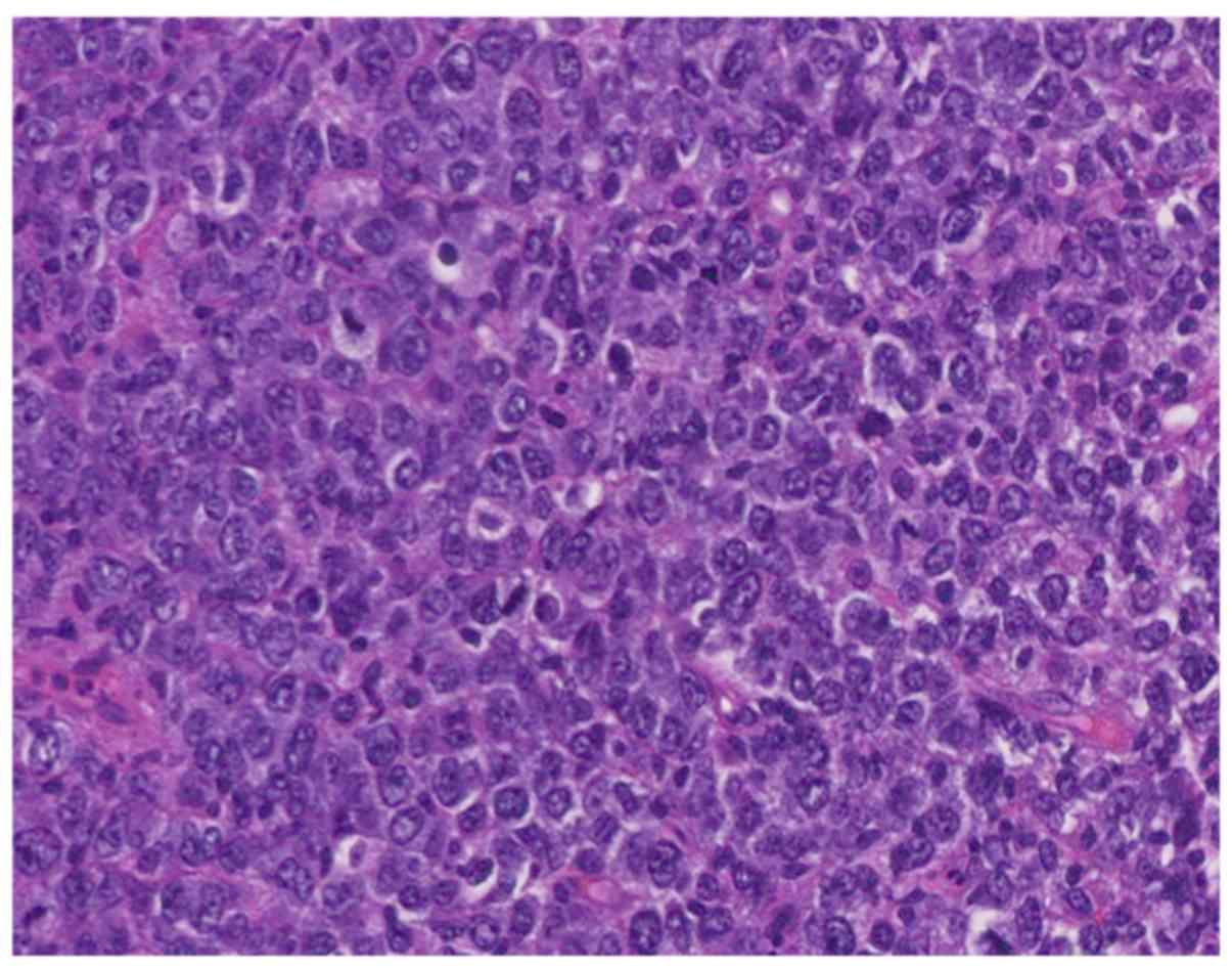
2001. A cervical biopsy was concurrently conducted, and the
histological examination revealed diffuse large atypical lymphocyte
infiltration with prominent nucleoli and mitosis (Fig. 3). Immunohistochemical analysis showed
strong positivity for CD45 and CD20 while CD3 was negative. Further
results from the immunohistochemical analysis were as follows:
AE1/3 (−), p40 (−), CEA (−), S-100 (−), CD10 (−), BCL-2 (−).
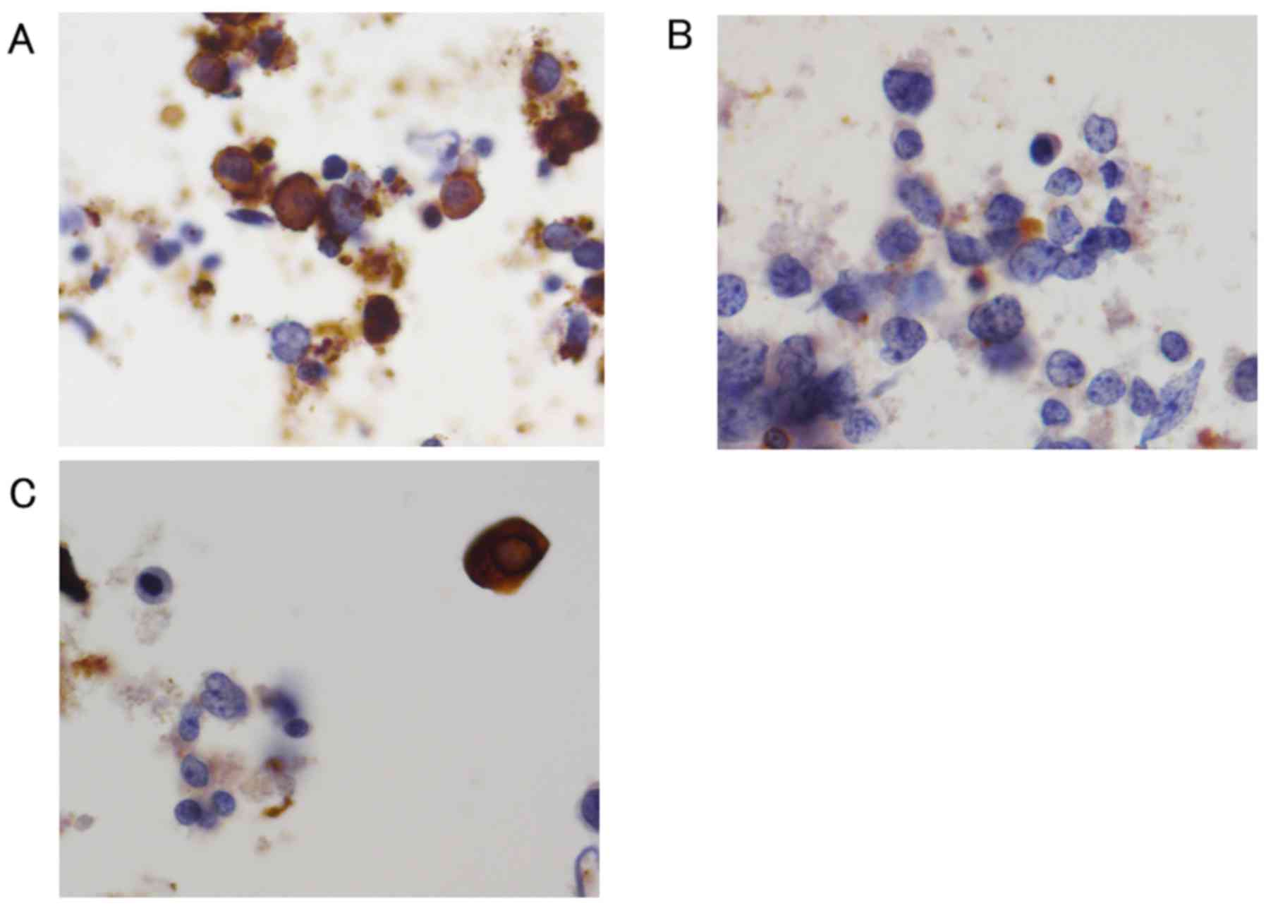
Immunocytochemical staining demonstrated similar findings to the
immunohistochemical analysis (Fig.
4). A bone marrow aspiration biopsy revealed normocellular
marrow, and no lymphoma cell invasion was observed.
Immunophenotyping of the bone marrow aspirate by flow cytometry
showed no aberrant antigen expression or monotypic B cells.
Based on these results, the final diagnosis was
diffuse large B cell lymphoma (DLBCL) of the uterine cervix, stage
IIEA (Ann Arbor classification). The patient received 6 courses of
R-CHOP chemotherapy (rituximab, 375 mg/m2 at day 1;
cyclophosphamide, 750 mg/m2 at day 1; doxorubicin, 50
mg/m2 at day 1; vincristine, 1.4 mg/m2 at day
1; prednisolone 100 mg/body at day 1–5) and achieved complete
remission. The cervical tumor disappeared grossly (Fig. 1B), and atypical cells were not present
in the Pap smear and biopsy specimens. Complete remission was also
confirmed by PET/CT scan and no residual or relapse tumor was found
(data not shown).
Discussion
We encountered a case of malignant lymphoma of the
uterine cervix that was presumptively diagnosed by a Pap smear
screening.
Due to its rarity, it is usually difficult to
diagnose malignant lymphoma of the uterine cervix by Pap smears. In
cervical cytology, the positivity for malignant lymphoma has been
reported to be 30–40% (5–7). This may be due to the fact that while
lymphoma cells often infiltrate the cervical stroma, epithelial
lining tissues remain preserved (5).
In Pap screenings, the classical cytological features of malignant
lymphoma are as follows: The presence of a dispersed monomorphic
cell population, high nuclear/cytoplasmic ratios, coarse granular
nuclear chromatin, focally cleaved nuclei, and the presence of
prominent nucleoli (8). The
differential diagnosis of malignant lymphoma of the uterine cervix
includes chronic cervicitis, lymphoma-like lesions (LLLs),
carcinoma, carcinosarcoma, endometrial stromal sarcoma, melanoma
(small cell variant), and primitive neuroectodermal tumor (PNET).
However, meticulous morphological observations and
immunohistochemical analysis can efficiently distinguish malignant
lymphoma from these other lesions (9). In our case, the tumor cells were easily
obtained due to its extroverted growth, and typical cytological
features of malignant lymphoma were observed in cervical cytology.
Although approximately 80% of the cells were atypical cells in the
Pap staining of our case, precise percentage for the lymphoma cells
in the slide has not yet been elucidated to date. Further
cytological study seems to be required. In general, diagnosis of B
cell lymphoma is based on morphology (H&E staining) and CD20
positive and CD3 negative, additional immunohistochemical analyses
including CD10, BCL-2, and c-Myc may be useful to distinguish
histological subtypes.
The clinical diagnosis of cervical lymphoma may be
difficult due to the lack of specific symptoms. The most common
clinical symptoms have been reported as vaginal bleeding or
discharge, and abdominal or pelvic pain. However, some patients
were reported to be asymptomatic (9).
The correct diagnosis is often delayed and, as a result, the
disease is diagnosed at an advanced stage. Our patient was also
asymptomatic; however, we were able to detect malignant lymphoma at
an early stage through cervical cytology. Therefore, Pap test
screenings may be useful for the early diagnosis of malignant
lymphoma of the uterine cervix. We did not measure the serum level
of such parameters as EBV-DNA and CMV-DNA in the present case.
Other hematological malignancies including virus associated ones
should be considered when diagnosing malignant lymphoma.
Standard therapy for DLBCL is R-CHOP chemotherapy
with or without radiotherapy. In a previous review article, they
reported that 30–50% of patients underwent surgery, which included
invasive radical hysterectomy with pelvic lymphadenectomy. Since
R-CHOP chemotherapy is the most effective treatment, invasive
surgery should be avoided (10).
Despite its rarity, the possibility of malignant
lymphoma should be considered when screening for cervical cancers
by Pap tests. In addition to conventional morphological
observations, it is important to perform immunohistochemical
analyses. A rapid and accurate diagnosis can lead to immediate
treatment without requiring surgery. The accumulation of more
clinical cases is important for obtaining a clearer understanding
of the clinicopathological features of this rare tumor.
Acknowledgements
We thank Daisuke Kawashima and Kanako Inoue for
their technical assistance.
Funding
No funding was received.
Availability of data and materials
The datasets used and/or analyzed during the current
study are available from the corresponding author on reasonable
request.
Authors' contributions
TK, AT and HT diagnosed, investigated and managed
the patient. TK, HK and HT determined the medical significance of
this case and wrote the manuscript. MA, SI, MI, HY, SN, YT, TF, MG
and FE provided advice in managing the patient's treatment and
preparing the manuscript.
Ethics approval and consent to
participate
Not applicable.
Consent for publication
The patient provided written informed consent for
the publication of their data.
Competing interests
The authors confirm that they have no competing
interests.
References
|
1
|
Calli AO, Rezanko T, Yigit S and Payzin B:
Lymphoma of the cervix: A diagnostic pitfall on cervicovaginal
smear. J Cytol. 29:213–215. 2012. View Article : Google Scholar : PubMed/NCBI
|
|
2
|
Bellevicine C, Zabatta A, Malapelle U,
Vetrani A and Troncone G: Diffuse large B-cell extranodal lymphoma
of the uterine cervix: An incidental pap smear finding with
histological and immunohistochemical correlation. Diagn Cytopathol.
42:644–646. 2014. View
Article : Google Scholar : PubMed/NCBI
|
|
3
|
Hanley KZ, Tadros TS, Briones AJ, Birdsong
GG and Mosunjac MB: Hematologic malignancies of the female genital
tract diagnosed on liquid-based Pap test: Cytomorphologic features
and review of differential diagnoses. Diagn Cytopathol. 37:61–67.
2009. View
Article : Google Scholar : PubMed/NCBI
|
|
4
|
Chan JK, Loizzi V, Magistris A, Hunter MI,
Rutgers J, DiSaia PJ and Berman ML: Clinicopathologic features of
six cases of primary cervical lymphoma. Am J Obstet Gynecol.
193:866–872. 2005. View Article : Google Scholar : PubMed/NCBI
|
|
5
|
Whitaker D: The role of cytology in the
detection of malignant lymphoma of the uterine cervix. Acta Cytol.
20:510–513. 1976.PubMed/NCBI
|
|
6
|
Komaki R, Cox JD, Hansen RM, Gunn WG and
Greenberg M: Malignant lymphoma of the uterine cervix. Cancer.
54:1699–1704. 1984. View Article : Google Scholar : PubMed/NCBI
|
|
7
|
Cardillo MR and Forte F: The diagnostic
value of cytology in a case of lymphoma of the uterine cervix. Eur
J Gynaecol Oncol. 8:597–602. 1987.PubMed/NCBI
|
|
8
|
Cahill LA, Stastny JF and Frable WJ:
Primary lymphoma of the endometrium. A report of two cases
diagnosed on cervicovaginal smears. Acta Cytol. 41:533–538. 1997.
View Article : Google Scholar : PubMed/NCBI
|
|
9
|
Lagoo AS and Robboy SJ: Lymphoma of the
female genital tract: Current status. Int J Gynecol Pathol.
25:1–21. 2006. View Article : Google Scholar : PubMed/NCBI
|
|
10
|
Mandato VD, Palermo R, Falbo A, Capodanno
I, Capodanno F, Gelli MC, Aguzzoli L, Abrate M and La Sala GB:
Primary diffuse large B-cell lymphoma of the uterus: Case report
and review. Anticancer Res. 34:4377–4390. 2014.PubMed/NCBI
|