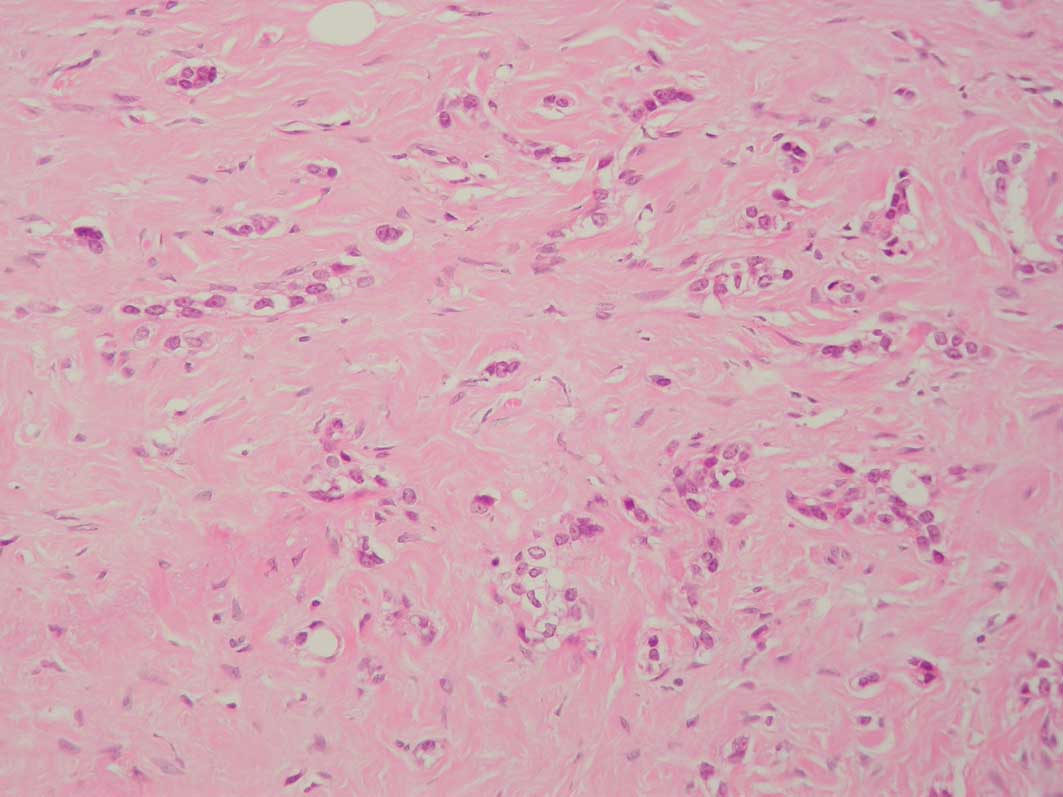
Pseudoangiomatous stromal hyperplasia: An observation on its microscopic involvement in breast carcinoma and the presence of lymph node metastases
- Authors:
View Affiliations
Affiliations: Department of Pathology, Royal Liverpool Hospital, Duncan Building, Liverpool L7 8XP, UK. johnnycoyne@doctors.org.uk
- Published online on: September 1, 2010 https://doi.org/10.3892/ol_00000141
-
Pages:
805-807
Metrics: Total
Views: 0 (Spandidos Publications: | PMC Statistics: )
Metrics: Total PDF Downloads: 0 (Spandidos Publications: | PMC Statistics: )
This article is mentioned in:
Abstract
The spaces of pseudoangiomatous stromal hyperplasia (PASH) are postulated to be important in the intramammary spread of breast carcinoma. The present study aimed to note the prevalence of inconspicuous, microscopic foci of PASH (identified as CD34+ve, CD31-ve and D2-40-ve spaces containing tumour emboli) involved in breast carcinoma and to establish the significance of its relationship to lymph node metastases. A total of 80 cases of breast carcinoma were examined for microscopic foci of PASH permeated by carcinoma and, of the four cases found to demonstrate such involvement, three had lymph node metastases.
View References
1
|
Damiani S, Peterse JL and Eusebi V:
Malignant neoplasms infiltrating ‘pseudoangiomatous’ stromal
hyperplasia of the breast: an unrecognised pathway of tumour
spread. Histopathology. 41:208–215. 2002.
|
2
|
Asioli S, Eusebi V, Gaetano L, Losi L and
Bussolati G: The pre-lymphatic pathway, the roots of the lymphatic
system in breast tissue: a 3D study. Virchows Arch. 453:401–406.
2008. View Article : Google Scholar : PubMed/NCBI
|
3
|
Vuitch MF, Rosen PP and Erlandson RA:
Pseudoangiomatous hyperplasia of mammary stroma. Hum Pathol.
17:185–191. 1986. View Article : Google Scholar
|
4
|
Hartveit F: Attenuated cells in breast
stroma; the missing lymphatic system of the breast. Histopathology.
16:533–543. 1990. View Article : Google Scholar : PubMed/NCBI
|
5
|
Hartveit F: Pericytes in human breast
stroma and the cells to which they relate. Eur J Morphol.
30:289–296. 1992.PubMed/NCBI
|
6
|
Fisher CJ, Hanby AM, Robinson L and Mills
RR: Mammary hamartoma – a review of thirty-five cases.
Histopathology. 20:99–106. 1992.
|
7
|
Tavassoli FA and Eusebi V: Tumors of the
Mammary Gland. AFIP Atlas of Tumor Pathology, 4th Series, Fasicle
10. ARP Press; Maryland: pp. 269–274. 2009
|
8
|
Cserni G: Evaluation of sentinel lymph
nodes in breast cancer. Histopathology. 46:697–702. 2005.
View Article : Google Scholar : PubMed/NCBI
|
9
|
Agarmal B, Saxena R, Morimiya A, Mehrotra
S and Badve S: Lymphoangiogenesis does not occur in breast cancer.
Am J Surg Pathol. 29:1449–1455. 2005. View Article : Google Scholar : PubMed/NCBI
|
10
|
Barsky SH, Ye Y and Karlin NJ: ‘Separation
Artefact’ V lymphovascular invasion: are mimics only mimics? Mod
Pathol. 86(Suppl 1): 289A2006.
|
11
|
Acs G, Dumoff KL, Solin LJ, Pasha T, Xu
Xiaowei and Zhang PJ: Extensive retraction artifact correlates with
lymphatic and nodal metastasis and predicts poor outcome in early
stage breast carcinoma. Am J Surg Pathol. 31:129–140. 2007.
View Article : Google Scholar : PubMed/NCBI
|
12
|
Acs G, Paragh G, Shang-Tian C, Laronga C
and Zhang PJ: The presence of micropapillary features and
retraction artifact in core needle biopsy material predict lymph
node metastasis in breast carcinoma. Am J Surg Pathol. 33:202–210.
2009. View Article : Google Scholar : PubMed/NCBI
|