Introduction
Breast cancer is the most common cancer affecting
women and is one of the leading causes of cancer-related deaths in
women (1). Although the mortality
of breast cancer has decreased with the advancement of early
detection and treatment (2),
thousands of women die from this disease each year and the
prognosis of patients with distant metastasis remains poor
(2,3). Therefore, the development of new
therapeutic strategies is a matter of pressing concern.
The PVRL4 gene encodes nectin-4, one of the
nectin and nectin-like family calcium-independent cell adhesion
molecules (4). This family consists
of two subgroups, one containing nectin-1 to −4 that associate with
afadin, a PDZ domain-containing cytoplasmic adaptor protein, and
another containing nectin-like cell adhesion molecules,
nectin-like-1 to −5 (5). Nectin-1
to −4 have an extracellular region containing a distal IgV-like
domain, two IgC-like domains, a single transmembrane region and a
cytoplasmic region with a C-terminal PDZ binding motif (6). Nectins are involved in cell adhesion
by interacting with each other and/or other adhesion molecules,
including cadherins, through their extracellular regions, and their
cytoplasmic regions function as an anchor to the cellular
cytoskeleton by binding with adaptor proteins such as afadin, PAR3
and band 4.1B (7). In addition,
nectin-1 has been shown to act as a viral entry receptor for the
human herpes simplex virus (8), and
nectin-4 as a receptor for the measles virus (MV) (9). Although nectin-1, −2 and −3 are widely
expressed in adult tissues, nectin-4 expression is restricted to
fetal tissues and adult organs such as the throat and salivary
gland (ducts), mammary gland and the skin (epidermis and sweat
glands) (6). Notably, PVRL4
expression is elevated in a wide range of human cancer types such
as breast, lung and ovarian cancer (10–13).
Elevated PVRL4 expression confers the anchorage-independent
proliferation of breast cancer cells, induction of integrin β4
signaling and subsequent Src family kinase activation in a matrix
attachment-independent manner (14). In addition, nectin-4 overexpression
in MDA-MB-231 cells, a nectin-4 null breast cancer cell line,
induced epithelial-mesenchymal transition and metastasis, and
upregulated the WNT/β-catenin signaling (15). Furthermore, PVRL4 could serve as a
useful prognostic predictor of breast, lung, esophageal and
high-grade T1 bladder cancer (12,16,17).
Notably, recent studies revealed that PVRL4 is a
promising molecular target for the treatment of cancer (18–20).
The antibody-drug conjugate, enfortumab vedotin, interacts with
PVRL4 and is administered for the treatment of urothelial bladder
cancer and other PVRL4+ solid tumors. The proliferation
of human breast, bladder, pancreatic and lung cancer cells was
significantly suppressed by treatment with enfortumab vedotin in
mice xenograft models (21). Since
PVRL4 is one of the known entry receptors for the MV, the
application of oncolytic viruses may become another strategy for
treatments that target PVRL4. Previously, Sugiyama et al
(20) generated a recombinant MV
HL-strain (rMV-SLAMblind) that carried a mutation in the region
responsible for the interaction with signaling lymphocytic
activation molecule (SLAM), another entry receptor for the MV. This
genetically engineered virus efficiently infected breast cancer
cells in a PVRL4-dependent fashion and decreased the viability of
the cancer cells in vitro and in vivo, suggesting a
therapeutic potential of rMV-SLAMblind as an oncolytic virus
against human cancer expressing PVRL4.
Although expression of PVRL4 is elevated in a
number of cancer types, including breast cancer (12,13,21),
the mechanism of its induction in cancer cells remains to be
elucidated. In addition, the global gene expression profile
associated with PVRL4 has not yet, to the best of our knowledge,
been clarified. Therefore, the aim of the present study was to
identify the transcriptional regulator(s) of PVRL4 and to
disclose the genes and pathways enhanced by PVRL4 overexpression in
cancer cells. For this, regulatory regions within the PVRL4
gene were searched for using Assay for Transposase-Accessible
Chromatin-sequencing (ATAC-seq) and chromatin
immunoprecipitation-sequencing (ChIP-seq) data in combination with
a reporter assay. In addition, candidate transcription factors
whose binding motifs are localized in an enhancer region identified
in the present study were further investigated. Furthermore,
RNA-seq and subsequent pathway analyses were conducted using
PVRL4-small interfering RNA (siRNA) in breast cancer cells
expressing abundant PVRL4 to disclose the characteristics
associated with its expression.
Materials and methods
Cell lines
Human breast cancer cell lines, SKBR3, T47D and
MCF7, and human colorectal cancer cell lines, DLD1, LS174T, HCT116
and RKO, were obtained from the American Type Culture Collection.
T47D and DLD1 cells were cultured in RPMI medium (Gibco; Thermo
Fisher Scientific, Inc.), MCF7 and LST174T cells in EMEM (Gibco;
Thermo Fisher Scientific, Inc.), SKBR3 and HCT116 cells in McCoy's
5A (Gibco; Thermo Fisher Scientific, Inc.) and RKO cells in DMEM
medium (Gibco; Thermo Fisher Scientific, Inc.), all containing 10%
fetal bovine serum (Biosera, Ltd.) and antibiotic/antimycotic
solution. The cells were incubated at 37°C under a humidified
atmosphere containing 5% CO2. Mycoplasma contamination
was tested using MycoStrip (Thermo Fisher Scientific, Inc.), which
indicated that all cell lines were free of mycoplasma.
Reverse transcription-quantitative PCR
(RT-qPCR)
Total cellular RNA was extracted from the cultured
cells using a RNeasy Mini Kit (Qiagen, Inc.). cDNA was synthesized
from 1 µg of total RNA with ReverTra Ace (Toyobo Life Science)
according to the manufacturer's protocol. qPCR was performed using
KAPA SYBR FAST qPCR Master Mix (2X) Kit (Sigma-Aldrich; Merck KGaA)
with sets of primers for JUNB, JUN, FOS, JUND, activating
transcription factor 3 (ATF3), ATF7, Jun dimerization
protein 2 (JDP2), CAMP responsive element binding protein 1
(CREB1), CREB3L4, PVRL4 and hypoxanthine
phosphoribosyltransferase 1 (HPRT1) (Table SI) and a StepOnePlus system (Thermo
Fisher Scientific, Inc.). The thermocycler conditions were as
follows: Denaturation at 95°C for 20 sec, followed by annealing and
extension at 60°C for 20 sec, for a total of 40 cycles. Quantities
of transcripts were determined by relative standard curves, and
HPRT1 was used as the internal control. The quantification
of the JUNB, JUN, FOS, JUND, ATF3, ATF7, JDP2, CREB1, CREB3L4,
PVRL4 and HPRT1 transcripts were calculated from
five-point standard curves prepared by amplifying the pooled
control cDNA derived from SKBR3 cells according to Getting Started
Guide of Applied Biosystems StepOne™ and StepOnePlus™ Real-Time PCR
Systems Standard Curve Experiments (PN: 4376784; Thermo Fisher
Scientific, Inc.). The standard curves were automatically generated
by the StepOne Software v2.2.3. Baseline cycles and thresholds were
set manually, and the quantification cycles were calculated
automatically by the software using an in-built algorithm.
Gene silencing
Pools of human specific siRNAs were obtained from
Merck KGaA (Table SII).
ON-TARGETplus Non-targeting Pool (cat. no. D-001810-10; GE
Healthcare Dharmacon, Inc.) was used as the control. All gene
silencing experiments were performed in SKBR3 cells excepting
si-FOS, where T47D cells were also used. SKBR3 and T47D cells were
seeded 1 day before siRNA treatment. Cells were transfected with 10
nM siRNA using Lipofectamine RNAiMAX (Thermo Fisher Scientific,
Inc.). After 48 h of incubation, total RNA was isolated from the
cells using an RNeasy Mini Kit according to the manufacturer's
instruction. The silencing effect was evaluated by quantitative
RT-qPCR, as aforementioned.
Preparation of plasmids
Putative promoter regions in the 5′-flanking
sequence, intron, and 3′-flanking sequence of PVRL4 were
amplified by PCR with specific primer sets (Table SIII and IV) containing recognition sites for the
following restriction enzymes: XhoI, BglII,
KpnI or HindIII. PCR products were digested with
restriction enzymes and cloned into the pGL4.23 vector (Promega
Corporation). Mutant versions of PVRL4 reporter plasmids were
generated by site-directed mutagenesis. Briefly, PCR was performed
using KOD Plus NEO enzyme (Toyobo Life Science), 100 ng of the
pGL4.23 -PVRL4#10-III plasmid containing the enhancer region as a
template and a set of primers for Mut-1 and −2 (Table SV), according to the manufacturer's
protocol. The PCR products were digested with Dpn1
restriction enzyme for 2 h at 37°C and were transformed into
Escherichia coli DH5α cells.
To generate plasmids expressing FOS, the entire
coding sequence of FOS was amplified by PCR using KOD ONE (Toyobo
Life Science) with a set of primers (Table SVI) containing EcoRI or
XhoI restriction sites, according to the manufacturer's
protocol. The PCR product was cloned into a pCMV-myc vector
(Promega Corporation). Sanger sequencing [using Applied Biosystems
3500 (Thermo Fisher Scientific, Inc.) according to the
manufacturer's protocol] confirmed the full-length cDNA sequence of
FOS was inserted into the plasmid.
Luciferase assay
Cells seeded in 12-well plates were transfected with
0.3 µg of pGL4.23 reporter plasmid and 0.05 µg of pRL-TK plasmid
(Promega Corporation) by FuGENE 6 reagent (Promega Corporation).
After 48 h of transfection, a PicaGene Dual Sea Pansy Luminescence
Kit (TOYOB-Net) was utilized to measure the activities of firefly
and Renilla luciferase according to the supplier's protocol.
The Renilla activity was normalized to the firefly
activity.
Putative transcription factor binding
site
To identify DNA sequences for putative transcription
factor binding sites, ChIP-seq data from the ENCODE project
[http://www.genome.ucsc.edu; The University of California Santa
Cruz Genome Browser Database (UCSC); accession nos. GSM733646,
GSM733674 and GSM733771] and JASPER (http://jaspar.genereg.net/) were used. In the present
study, a JASPAR score >15.5 was deemed a putative-binding
motif.
ATAC-Seq
ATAC-Seq of breast and colorectal cancer cells
expressing either high or low levels of PVRL4 was performed using
an ATAC-Seq Kit (Active Motif, Inc.), according to the
manufacturer's instructions. Briefly, the cells were resuspended in
100 µl of ATAC lysis buffer to extract the nuclei. The nuclei were
collected by centrifugation at 500 × g for 5 min at 4°C,
resuspended in 50 µl of Tagmentation Master Mix (including
assembled transposomes) and then incubated at 37°C for 30 min with
shaking. The tagmented DNA was purified using 250 µl of DNA
Purification Binding Buffer and 5 µl of 3 M sodium acetate. The DNA
library was collected using SPRI beads, which binds to a magnet,
and eluted with 20 µl of DNA Purification Elution Buffer. The
quality of library was verified using a bioanalyzer (Agilent
Technologies, Inc.). The libraries were sequenced (primers listed
in Table SVII) with HiSeq Rapid
SBS Kit v2 (Illumina, Inc) and HiSeq Rapid SR Cluster Kit v2
(Illumina, Inc) using 60 bp single-end reads on the HiSeq2500
platform (cat. no. SY-401-2501; Illumina, Inc). The raw sequencing
reads were analyzed for quality using FastQC and then aligned to
the human genome (hg19) using Bowtie2 (v2.4.1; http://bowtie-bio.sourceforge.net/bowtie2/index.shtml).
Integrative Genomics Viewer was used to visualize the sequencing
data (https://igv.org/doc/desktop/).
Chromatin conformation capture (3C)
assay
The 3C assay was performed as previously described
(22,23). Briefly, SKBR3 cells were
cross-linked with 1% formaldehyde for 10 min at room temperature
and then quenched with 125 mM glycine. The cross-linked chromatin
was digested at 37°C overnight with 400 U BgIII (Takara Bio,
Inc.), which was then heat-inactivated for 25 min at 65°C with 1.6%
SDS. Subsequently, DNA fragments were ligated with T4 DNA ligase
(New England Biolabs, Inc.) for 8 h at 16°C. Samples were treated
with 300 µg of proteinase K (Merck KGaA) at 37°C overnight to
remove the cross-link and then with 300 µg of RNase A (Merck KGaA).
The ligated DNA was purified by phenol/chloroform extraction and
ethanol precipitation. The first PCR reaction was amplified with
the outer primer sets. The first PCR parameters for relative
quantification were as follows: 2 min at 98°C, followed by 35
cycles at 98°C for 10 sec, 60°C for 5 sec and 68°C for 10 sec.
After the products of the first PCR were purified, nested PCR (KOD
One; Toyobo Life Science) was performed using these products with
the inner primer sets to investigate a possible interaction between
the promoter and enhancer regions of PVRL4. The nested PCR
parameters for relative quantification were as follows: 2 min at
98°C, followed by 30 cycles at 98°C for 10 sec, 62.5°C for 5 sec
and 68°C for 10 sec. The sequences of the PCR primers used are
shown in Table SVIII. The PCR
products were confirmed by gel electrophoresis using 1.5% agarose
gels and visualized with ethidium bromide staining (Sigma-Aldrich;
Merck KGaA).
ChIP
The ChIP and ChIP-qPCR analysis were performed as
previously described (24). A total
of 2×10⁷ SKBR3 cells were cross-linked with 1%
formaldehyde for 10 min, followed by quenching with 125 mM glycine
for 5 min. DNA fragmentation was performed using a UD-201
homogenizer (Tomy Seiko Co., Ltd.) with the following settings:
Output level 5, 50% duty, 15 sec, 3 cycles and on floating ice to
obtain 200–500 bp DNA fragments. The fragmented DNA samples were
confirmed by gel electrophoresis using 1.5% agarose gels and
visualized with ethidium bromide staining (Sigma-Aldrich; Merck
KGaA). An aliquot of the sample was kept as an input and the
remaining sample was used for ChIP analysis. The fragmented samples
from the SKBR cells were incubated with 8 µg of anti-phospho-FOS
antibody (Cell Signaling Technology, Inc.; cat. no. 5348; 1:100) or
8 µg of anti-IgG antibody (normal mouse IgG; Santa Cruz
Biotechnology, Inc.; cat. no. sc-2025; 1:400) and bound to protein
G-sepharose beads (GE Healthcare) at 4°C overnight. The beads were
separated with a column and washed sequentially for 5 min with 1 ml
of the following buffers: 1X low salt wash buffer [1% Triton X-100,
1 mM EDTA, 150 mM NaCl, 20 mM Tris-HCl (pH 8.0)], 1X high salt wash
buffer [0.1% SDS, 1% Triton X-100, 2 mM EDTA, 500 mM NaCl, 20 mM
Tris-HCl (pH 8.0)], 1X LiCl wash buffer [0.25 M LiCl, 0.5% Nonidet
P-40, 0.5% sodium deoxycholate, 1 mM EDTA, 10 mM Tris-HCl (pH 8.0)]
and 2X Tris EDTA buffer [10 mM Tris-HCl (pH 8.0), 1 mM EDTA]. The
beads were then eluted with 200 µl of elution buffer (100 mM
NaHCO3, 1% SDS, 5 mM NaCl) and 2 µl of proteinase K (10
mg/ml; Sigma-Aldrich; Merck KGaA). The immunoprecipitated and input
DNA were purified by using a QIAquick PCR Purification Kit (Qiagen,
Inc.). The concentration of two purified DNA samples was measured
using Qubit dsDNA HS Assay Kit (Thermo Fisher Scientific, Inc.)
with a Qubit fluorometer (Thermo Fisher Scientific, Inc.). qPCR was
performed using KAPA SYBR FAST qPCR Master Mix (2X) Kit
(Sigma-Aldrich; Merck KGaA) with primers shown in Table SIX. Amplification of the
glyceraldehyde-3-phosphate dehydrogenase gene was used as a
negative control, and relative quantification was performed using
StepOne Software v2.2.3. The PCR parameters for relative
quantification were as follows: 2 min at 98°C, followed by 40
cycles at 95°C for 1 sec and 60°C for 20 sec.
RNA-seq and Gene Ontology (GO)
analysis
To identify genes regulated by PVRL4, RNA-seq
analysis was performed using an Ion Proton system™ (Thermo Fisher
Scientific, Inc.). Total RNA was extracted from SKBR3 cells treated
with siPVRL4#1, #2 or control siRNA using a RNeasy Mini Kit
(Qiagen, Inc.), and the quality of RNA was assessed using an
Agilent bioanalyzer device (Agilent Technologies, Inc). Libraries
were prepared using all samples that had a RNA integrity number
>7.0. RNA-seq libraries were prepared with 100 ng total RNA
using the Ion AmpliSeq Transcriptome Human GeneExpression Kit
(Thermo Fisher Scientific, Inc.) according to the manufacturer's
instructions. The libraries were sequenced using an Ion Proton
system with an Ion PI Hi-Q Sequencing 200 kit and Ion PI Chip v3
(Thermo Fisher Scientific, Inc.). The sequencing reads were aligned
to hg19_AmpliSeq_Transcriptome_ERCC_v1 using the Torrent Mapping
Alignment Program (https://github.com/iontorrent/TMAP), and the raw count
data were generated using the AmpliSeqRNA plug-in (v5.2.0.3), both
from the Torrent Suite Software (v5.2.2; Thermo Fisher Scientific,
Inc.). The DESeq2 package (v1.26.0; http://bioconductor.org/packages/release/bioc/html/DESeq2.html)
was used to normalize the read count data and test for differential
gene expression. False discovery rate-adjusted P-values (q-values)
<0.5 were considered to indicate a statistically significant
difference. Functional enrichment analysis was performed using
Metascape (https://metascape.org/gp/index.html#/main/step1)
(25). Metascape pathway enrichment
analysis uses GO, Kyoto Encyclopedia of Genes and Genomes
(https://www.genome.jp/kegg/kegg_ja.html), Reactome
(https://reactome.org/) and MSigDB (https://www.gsea-msigdb.org/gsea/msigdb). In brief,
pairwise similarities between two enriched terms were calculated
based on a κ test score. Then the similarity matrix was clustered
hierarchically, and a similarity threshold score of 0.3-κ test
score was applied to trim the result into separate clusters.
Statistical analysis
Unpaired two-tailed Student's t-test and one-way
ANOVA followed by Dunnett's test were applied for statistical
analyses. P<0.05 was considered to indicate a statistically
significant difference. Data obtained from three independent
experiments are presented as the mean ± SD.
Results
Candidate regulatory regions of
PVRL4
It was reported that PVRL4 expression was elevated
in ~61% of ductal carcinomas of the breast (11). However, genetic amplification of the
PVRL4 gene was observed in ~10% of breast invasive carcinoma
according to The Cancer Genome Atlas Pan Cancer Atlas data
(https://gdc.cancer.gov/about-data/publications/pancanatlas),
suggesting that different mechanisms may play a role in the
elevated PVRL4 expression in breast cancer tissues.
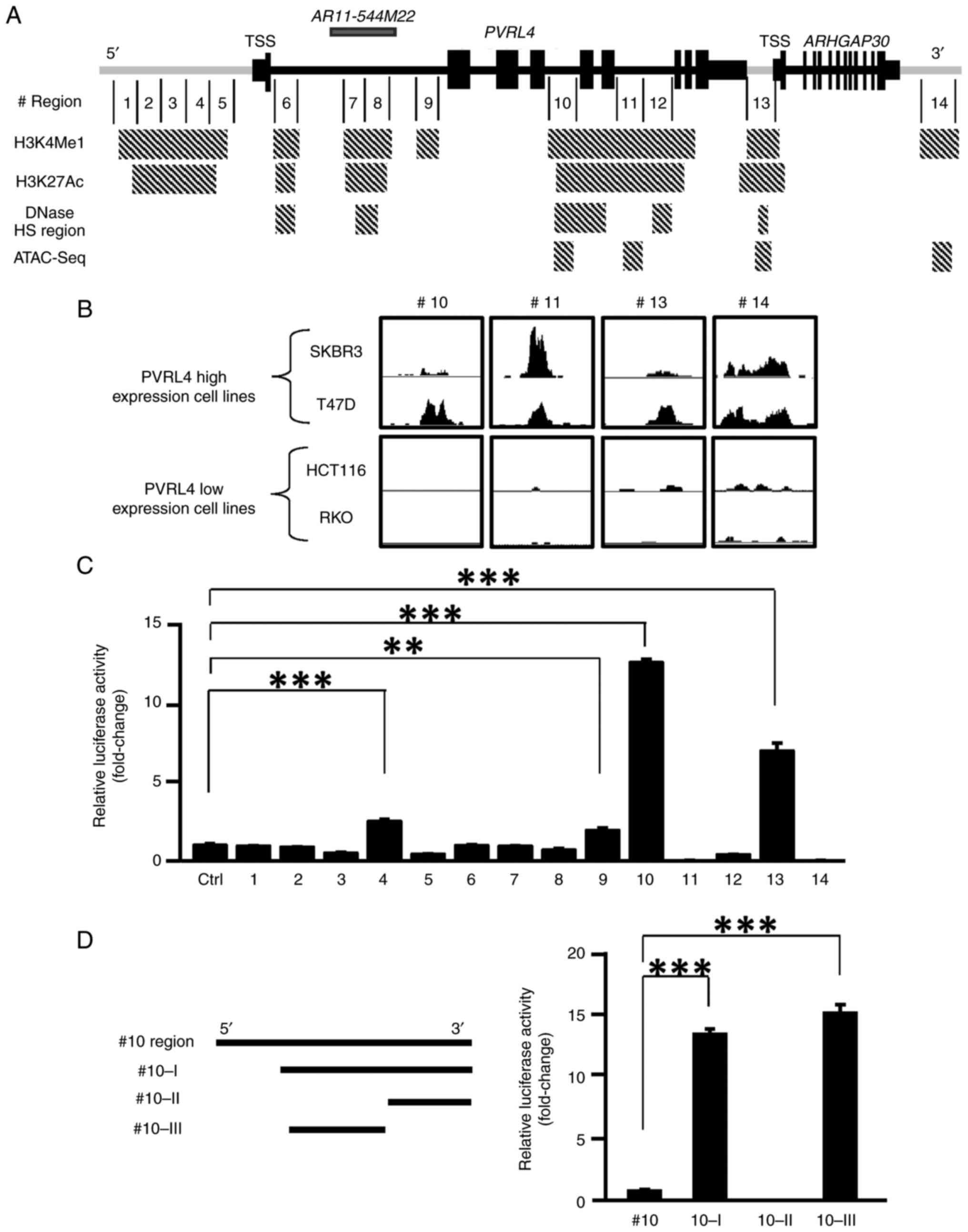
To clarify the regulatory mechanisms of its
expression in cancer cells, transcriptional regulatory elements in
the PVRL4 genomic region were searched for. Analysis of the
histone modifications, H3K4me1 and H3K27ac, in the UCSC genome
database identified seven regions high in H3K4me1 and five regions
high in H3K27ac, and the five high H3K27ac-regions overlapped with
the high H3K4me1-regions (Figs. 1A
and S1A). A further search of the
database identified five DNase high sensitivity regions within the
overlapped high H3K4me1 and high H3K27ac regions. To identify open
chromatin regions, ATAC-seq was performed using breast cancer cells
expressing high levels of PVRL4 (SKBR3 and T47D) and colon
cancer cells expressing low levels of PVRL4 (HCT116 and RKO)
(Fig. S1B). Subsequently, four
putative open chromatin regions were identified with high ATAC-seq
peaks in SKBR3 and T47D cells but not in HCT116 and RKO cells
(Fig. 1B). Of these four regions,
two were located in intron 4 and intron 6 and the remaining two
were in the 3′-flanking region. Notably, three of the four regions
were localized in the overlapped high H3K4me1 and high H3K27ac
regions.
Identification of a transcriptional
regulatory region in PVRL4
To identify the enhancer region within PVRL4,
14 regions were selected from the high H3K4me1 regions (Fig. 1A) and cloned into the pGL4.23
reporter plasmid. Transcriptional regulation is restricted by the
physical constraints imposed by topologically associating domain
(TAD). It is of note that these 14 regions were localized within
the same TAD of the PVRL4- transcription start site according to
the Topologically Associating Domain Knowledge Base (http://dna.cs.miami.edu/TADKB/), suggesting that
the candidate regions may physically associate with the PVRL4
promoter region (Fig. S2). To
examine the transcriptional activity of the candidate regions, a
dual-luciferase assay using the 14 reporter plasmids was conducted
in SKBR3 and T47D cells. The reporter plasmids containing regions
#4, #9, #10 or #13 and those containing region #10 or #13 showed
significantly higher luciferase activities compared with the
control (mock) plasmids in SKBR3 and T47D cells, respectively
(P<0.01; Figs. 1C and S3A). Since region #13 was localized in
intron 1 of the Rho GTPase activating protein 30 (ARHGAP30)
gene, this region was excluded from further analysis. Region #10
became the focus of further study as it demonstrated the highest
transcriptional activity among the candidate regions. To elucidate
the important region within region #10, three deletion mutants of
#10 reporter plasmids (#10-I, #10-II and #10-III) were prepared and
their reporter activities were compared with that of wild-type #10
reporter plasmids (Figs. 1D and
S3B). Notably, deletion of the
5′-region (#10-I) increased the reporter activity in both SKBR3 and
T47D cells. Furthermore, reporter plasmids containing #10-III also
had a higher activity than the wild-type #10 plasmid in both cell
lines, suggesting that this region may include transcriptionally
important elements. Since the #10-III region was within an
overlapping region (with high H3K4me1, high H3K27ac, high DNase
sensitivity and open chromatin), it may play a role in the
transcription of PVRL4 as a distant enhancer region.
Involvement of two putative
FOS-binding motifs in the enhancer region
Using the ChIP-seq data from the ENCODE project,
transcription factors that may interact with the candidate enhancer
region (#10-III) and putative-binding motifs were searched for
using the JASPER database. Subsequently, 11 candidate
binding-motifs of transcription factors were identified (Table I). Since FOSB and FOS-like 2 (FOSL2)
act as heterodimers with JUN, knockdown of JUN by siRNA will
decrease the activity of the FOSB-JUN and FOSL2-JUN complexes
(26). However, FOS is known to
function both as a homodimer and a heterodimer with JUN (27). Therefore, FOSB and FOSL2 were
excluded from the subsequent knockdown experiments and the effect
of the other nine transcription factors (JUN, JUNB, JUND, JDP2, FOS
ATF3, ATF7, CREB1 and CREB3L4) were examined. siRNAs targeting the
nine transcription factors were generated and the knockdown effect
of each siRNA on the expression of its target gene was confirmed by
qPCR analysis (Fig. S4). The
effect of each siRNA on the reporter activity of plasmids
containing #10-III was then investigated in SKBR3 cells. FOS siRNA
significantly reduced the reporter activity (P<0.005), but the
remaining eight siRNAs did not have a significant effect (Fig. 2A). Since the #10-III region
contained a putative FOS binding motif (TGACGTCA), mutant reporter
plasmids carrying nucleotide substitutions in the binding motif
(mut1, CAACGTCA;
mut2, TGACGCAA;
mut1+2, CAACGCAA) were constructed (Fig. 2B). As expected, these substitutions
significantly reduced the reporter activity compared with the
wild-type plasmid (pGL4.23-PVRL4#10-III) in SKBR3 cells (Figs. 2B and S5A).
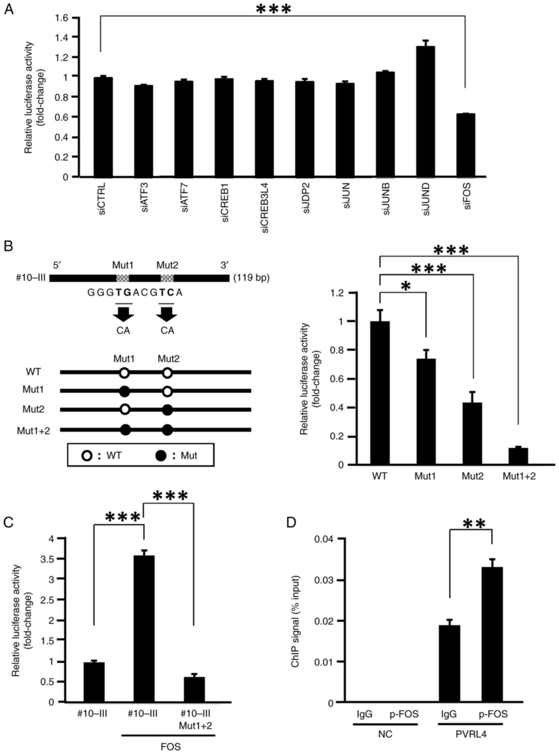 | Figure 2.Analysis of transcriptional factors
associated with the enhancer activity in region #10-III. (A) Effect
of the siRNA for nine candidate transcription factors and the CTRL
on the reporter activity of wild type #10-III plasmids in SKBR3
cells. ***P<0.005 vs. control. (B) Reporter activities of
#10-III mutant plasmids containing different substitutions in the
putative activator protein-1 binding motif. Schematic presentation
of mutant reporter plasmids containing different substitutions in
the motif (left panel). Reporter activities of WT (#10-III) and
three mutant plasmids (Mut1, Mut2, and Mut1 + 2) were analyzed in
SKBR3 cells (right panel). *P<0.05, ***P<0.005 vs. WT. (C)
Effect of exogenous FOS overexpression on the reporter activity of
the WT and mutant plasmids (#10-III and Mut1 + 2, respectively) in
SKBR3 cells. ***P<0.005 vs. WT. (D) ChIP-quantitative PCR
analysis with anti-p-FOS disclosed an interaction between FOS and
the enhancer region (#10-III). Mouse IgG was used as the NC.
**P<0.01 vs. IgG. ATF, activating transcription factor; CREB,
CAMP responsive element binding protein; CTRL, control; ChIP,
chromatin immunoprecipitation; JDP2, Jun dimerization protein 2;
Mut, mutant; NC, negative control; p-FOS, phosphorylated-FOS;
siRNA, small interfering RNA; WT, wild-type. |
 | Table I.JASPAR scores and binding motifs of
transcription factor candidates for PVRL4. |
Table I.
JASPAR scores and binding motifs of
transcription factor candidates for PVRL4.
| Transcription
factor | Sequence
(5′-3′) | JASPAR score | Strand |
|---|
| JDP2 | ATGACGTCA | 19.28 | - |
| JUNB | ATGACGTCAT | 18.18 | - |
| ATF3 | ATGACGTCAT | 18.06 | + |
| ATF7 | ATGACGTCAT | 17.63 | - |
| FOS | GATGACGTCAT | 17.30 | + |
| JUN | GATGACGTCAT | 16.70 | + |
| FOSL2 (JUN
dimer) | GATGACGTCAT | 16.67 | + |
| FOSB (JUN
dimer) | GATGACGTCATCG | 16.55 | + |
| JUND | GATGACGTCAT | 16.08 | + |
| CREB3L4 | GGTGACGTCACC | 15.80 | + |
| CREB1 | TGACGTCA | 15.79 | + |
To confirm that FOS transcriptionally regulates the
expression of PVRL4, the reporter activity of
pGL4.23-PVRL4#10-III was analyzed in the presence and absence of a
plasmid expressing wild-type FOS (pCMV-FOS; efficiency of the
overexpression of FOS is shown in Fig.
S5B) in SKBR3 cells. As shown in Fig. 2C, the reporter activity increased
3.58-fold in the presence of pCMV-FOS. This increased reporter
activity by pCMV-FOS was not observed with a mutant reporter
plasmid containing two substitutions in the FOS-binding motifs
(Mut1 + 2; Fig. 2C and S5C). Furthermore, the ChIP-qPCR assay
demonstrated that the immunoprecipitation with p-FOS antibody
enriched the DNA of region #10-III compared with the control IgG
antibody (Fig. 2D), which is in
agreement with the ENCODE results. These results suggested that FOS
transcriptionally upregulated PVRL4 through its interaction
with two FOS-binding motifs in intron 4.
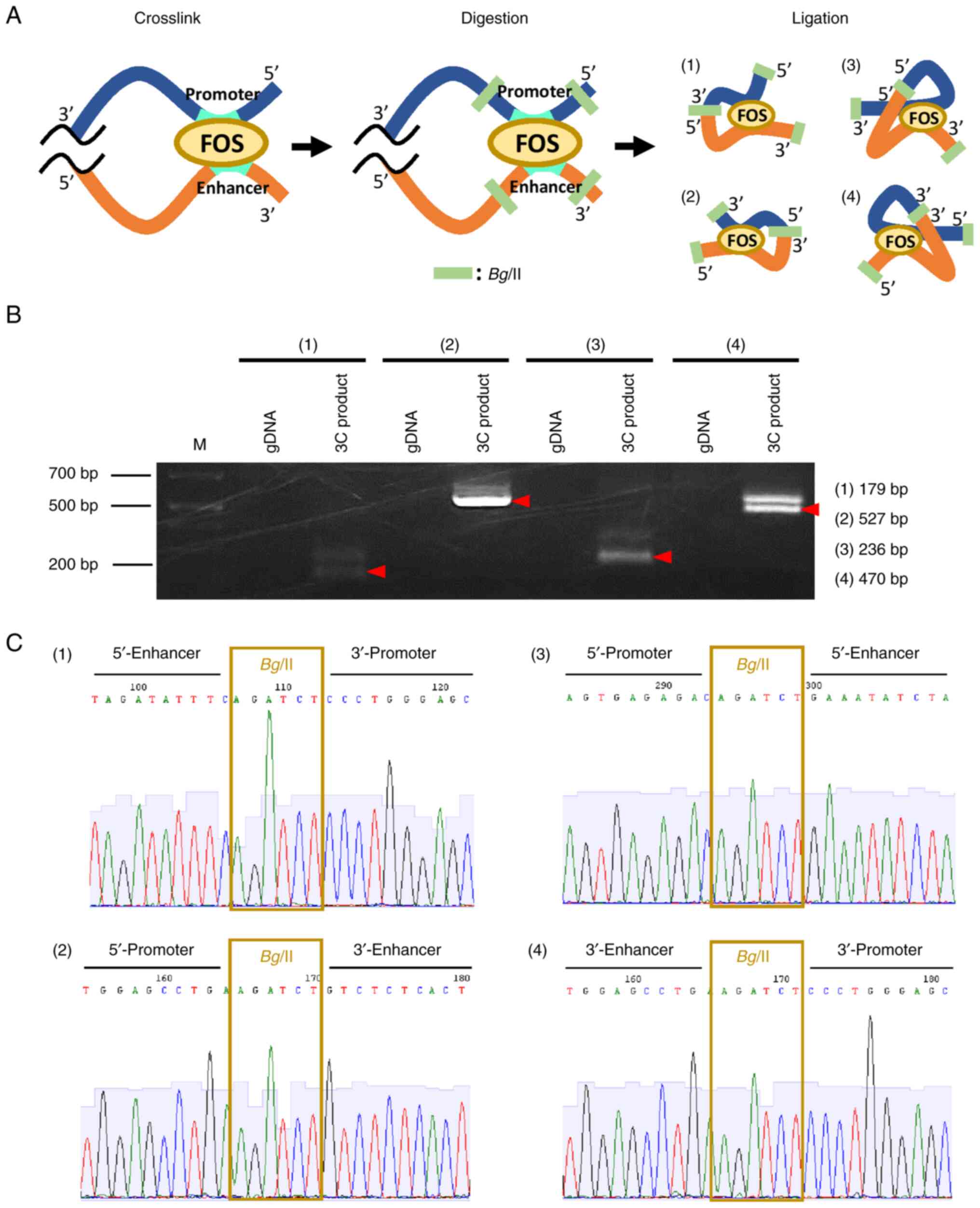
Interaction of the enhancer region
with the promoter of PVRL4
To examine whether the enhancer region interacts
with the PVRL4 promoter, a 3C assay was conducted. DNA from
SKBR3 cells was cross-linked with formaldehyde and subsequently
digested with a restriction enzyme, Bglll. Self-ligation of
the DNA was expected to produce chromatin loops between the
enhancer and promoter regions when the two were closely associated
(Fig. 3A). In total, four sets of
first and nested PCR primers were designed that could detect
associations between the two regions (Table SVII). Subsequently, amplification
of the 3C DNA with the four primer sets produced PCR products with
the expected sizes, but the amplification of control SKBR3 DNA
failed to produce PCR products (Fig.
3B). Additionally, sequence analysis of the nested PCR products
was conducted with primers in Table
SVIII, which confirmed a ligated DNA sequence of the enhancer
region (#10-III) and the promoter region (Fig. 3C). These data suggested that the
enhancer interacted with the promoter region through the formation
of a chromatin loop.
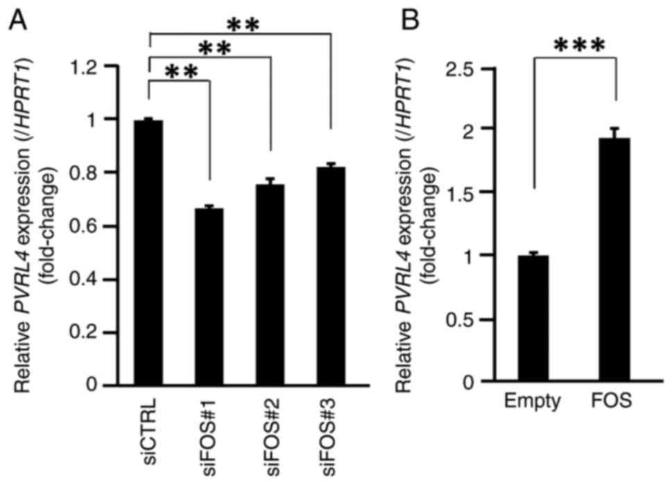
FOS is involved in PVRL4
expression
To investigate the involvement of FOS in the
regulation of PVRL4, the effect of FOS knockdown on
PVRL4 expression in SKBR3 and T47D cells was analyzed by
qPCR. The efficiency of FOS knockdown by FOS siRNA
transfection is shown in Fig. S4I and
J. The expression of PVRL4 was significantly decreased
by transfection with the three different FOS siRNAs (siFOS#1, #2
and #3; P<0.01; Figs. 4A and
S6).
To confirm the effect of FOS on PVRL4
expression, MCF7 cells were transfected with pCMV-FOS and the
expression of PVRL4 was analyzed by qPCR. Consistent with
the reporter assay, PVRL4 expression was significantly
enhanced by the overexpression of FOS (P<0.005; Fig. 4B).
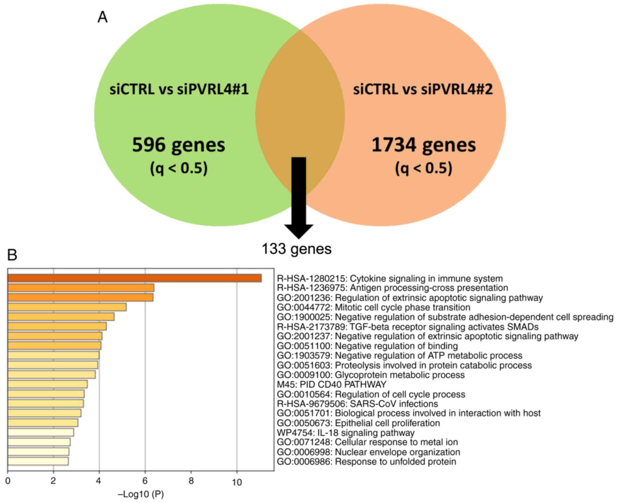
Expression analysis suggested a link
between PVRL4 with the immune system and apoptosis
To clarify the function of PVRL4 in breast cancer
cells, RNA-seq analysis using SKBR3 cells treated with control or
PVRL4 siRNA (siPVRL4#1 and siPVRL4#2) was performed. The efficiency
of PVRL4 knockdown following transfection with these siRNA is shown
in Fig. S7A. A total of 596 and
1734 genes were identified whose expression levels were
significantly altered by siPVRL4#1 and siPVRL4#2, respectively,
compared with control siRNA (q<0.5; Figs. 5A and S7B). The number of genes was low since
PVRL4 is a cell membrane receptor (4). The effect of these two siRNA on
expression may be small compared with the siRNA of transcription
factors. Since the inclusion of unmerged genes may have increased
the likelihood of detecting GO of off-target effects, 133 genes
that were commonly altered by the two siRNAs were used. GO analysis
with the 133 genes found three ontology terms, ‘Cytokine Signaling
in Immune System’, ‘Antigen processing-Cross presentation’ and
‘Regulation of Extrinsic Apoptotic Signaling Pathway’ (Fig. 5B). In both siPVRL4 treatments, the
expression levels of IFIT1, IFI44, IFI44L, MX1, XAF1 and
OAS2, related to ‘Cytokine Signaling in Immune System’, were
upregulated 11.4–2.9-fold, compared with the control siRNA
(Table SX). These six,
IFIT1 (28), IFI44
(29), IFI44L (29), MX1 (30,31),
XAF1 (30,32) and OAS2 (30,33),
genes are known for their involvement in the defense against virus
infection. Since PVRL4 is a member of the nectin family, altered
expression of the genes involved in ‘Negative Regulation of
Binding’ may suggest a decrease in cell adhesion. These results
suggested that PVRL4 was involved in the cytokine response and
immune system.
Discussion
In the present study, a distant enhancer region of
PVRL4 in intron 4 was identified, and it was clarified that
FOS was involved in the transcriptional regulation of PVRL4
through interaction with this enhancer region. Additionally, it was
demonstrated that PVRL4 may downregulate the expression of genes
associated with cytokine signaling and the immune system. In
general, gene expression is regulated by several regions and a
number of factors. Therefore, expression of PVRL4 may be
regulated not only by the enhancer region but also by other
regions. Moreover, the expression of PVRL4 may be controlled
partially by FOS and other undetermined transcription factors.
A previous study revealed that c-FOS protooncogene
expression was induced by estrogen in MCF7 breast cancer cells
(34). Recently, Binato et
al (35) reported that c-FOS
and c-JUN proteins are induced in luminal A-type breast cancer
cells, and that nuclear receptor-interacting protein 1 (NRIP1) was
consequently augmented by the complex. Furthermore, it was found
that expression levels of the progesterone receptor, estrogen
receptor 1 and cyclin D1 were upregulated by NRIP1, suggesting a
link between c-FOS and the proliferation of breast cancer cells
(35). In addition, c-FOS is
transcriptionally induced by ETS Transcription Factor ELK1 in
bladder cancer (36). However, it
remains to be clarified how frequently transcriptional activity of
c-FOS is enhanced in different types of cancer cells. To
transactivate downstream genes, FOS forms a dimeric complex with
various dimer partners, such as JUN family proteins (c-JUN, JUNB
and JUND), and the complex binds to the so-called TPA-responsive
element (TGAC/GTCA) in the downstream genes through its leucine
zipper structure (37). In addition
to this heterodimerization, the activity of FOS is modulated
through its phosphorylation by kinases, including ERK1/2 (38) and RSK1/2 (39). Although it was shown in the present
study that FOS plays a crucial role in the expression of
PVRL4, the involvement of dimer partner proteins in the
induction of expression remains to be clarified. Since the
regulatory mechanism of FOS-mediated transcriptional activity is
complicated, further investigation is necessary.
A recent study reported that estrogen-related
receptor-α (ESRRA) transcriptionally upregulates PVRL4
expression through an interaction with estrogen responsive elements
in its promoter region (40).
Although enhanced FOS expression is not frequently observed
in breast cancer cells (26), the
activity of FOS-heterodimers may be enhanced by its partner
proteins or by post-transcriptional modifications of FOS protein.
In addition, PVRL4 may be transcriptionally regulated by
ESRRA and c-FOS in breast cancer cells, but this requires further
experimental validation.
In the present study, RNA-seq and subsequent pathway
enrichment analyses revealed that PVRL4 expression was
associated with cytokine responses, antigen processing-cross
presentation and the immune system. These results were in agreement
with the report that PVRL4 functions as a ligand of TIGIT, the
inhibitory receptor T-cell immunoreceptor with Ig and ITIM domains,
and that PVRL4 inhibits the activity of natural killer cells
(41). In addition, the cytoplasmic
region of PVRL4 is involved in the interaction with the actin
cytoskeleton through afadin. It was also reported that PVRL4
activates the JAK-STAT signaling pathway through association with
suppressor of cytokine signaling 1 (SOCS1) (42,43).
Therefore, in addition to TIGIT-mediated escape from the immune
checkpoint, PVRL4 expression may mitigate cytokine signaling
through the recruitment of SOCS1 and facilitate cells in
suppressing immune responses. If these hypotheses are correct,
decreased expression of PVRL4 and/or inhibition of
PVRL4-mediated immune suppression may enhance the efficacy of
immune checkpoint inhibitors. It is of note that IFIT1, IFI44,
IFI44L, MX1, XAF1 and OAS2, the six genes upregulated by
the knockdown of PVRL4, are expected to be downregulated in
cells expressing PVRL4. Since these proteins are known to exhibit
antiviral activity through inhibition of viral replication and the
stabilization of antiviral immunity, PVRL4 (the MV receptor)
expression may serve not only in the entry of the MV but also
provide a suitable environment for their replication by suppressing
antiviral reactions. Therefore, the development of new therapeutic
modalities to suppress the expression of PVRL4 may contribute to
efficient treatment for neoplasms expressing abundant PVRL4 as well
as the symptoms caused by the infection of MV.
The limitations of the present study include the
absence of tissue-specific control of PVRL4. Since breast
cancer cell lines were used in the present study, PVRL4
regulatory mechanisms in other tissues may have been missed. As
such, future studies may elucidate tissue-specific regulatory
mechanisms of PVRL4 expression. In addition, the identified
enhancer region, #10-III, may affect the expression of
ARHGAP30 or other genes (44), which should be independently
determined in future studies.
In conclusion, in the present study, it was
determined that FOS directly regulated the transcriptional activity
of PVRL4 in breast cancer cell lines. These results may
assist with understanding the regulatory mechanism of PVRL4
and may contribute to the development of new strategies for cancer
treatment and measles infection.
Supplementary Material
Supporting Data
Supporting Data
Acknowledgements
We are grateful to research assistants: Ms. Seira
Hatakeyama, Ms. Rika Koubo, and Ms. Yumiko Isobe (Division of
Clinical Genome Research, The Institute of Medical Science, The
University of Tokyo) for their technical assistance.
Funding
This study was supported in part by Health and Labour Sciences
Research Grants of Japan (grant no. 15ck0106001h0003) and Japan
Agency for Medical Research and Development (grant no.
19ck0106281h0003).
Availability of data and materials
The datasets generated or analyzed during the
current study are available in the NCBI Gene Expression Omnibus,
https://www.ncbi.nlm.nih.gov/geo/query/acc.cgi?acc=GSE236275
and https://www.ncbi.nlm.nih.gov/geo/query/acc.cgi?acc=GSE240039.
Authors' contributions
TN and YF designed the studies, and TN, KT, KY, MA
and AS performed the experiments. TN and KT confirm the
authenticity of all the raw data. TN, KY and TI provided analysis
and interpretation of data. TN, KT, TF and YF wrote the manuscript.
YO, TF, MY and CK contributed to data collection and
interpretation, and critically reviewed the manuscript. All authors
approved the final version of the manuscript.
Ethics approval and consent to
participate
Not applicable.
Patient consent for publication
Not applicable.
Competing interests
The authors declare that they have no competing
interests.
References
|
1
|
Siegel RL, Miller KD and Jemal A: Cancer
statistics, 2018. CA Cancer J Clin. 68:7–30. 2018. View Article : Google Scholar : PubMed/NCBI
|
|
2
|
Narod SA, Iqbal J, Giannakeas V, Sopik V
and Sun P: Breast cancer mortality after a diagnosis of ductal
carcinoma in situ. JAMA Oncol. 1:888–896. 2015. View Article : Google Scholar : PubMed/NCBI
|
|
3
|
Chung CT and Carlson RW: Goals and
objectives in the management of metastatic breast cancer.
Oncologist. 8:514–520. 2003. View Article : Google Scholar : PubMed/NCBI
|
|
4
|
Duraivelan K and Samanta D: Emerging roles
of the nectin family of cell adhesion molecules in
tumour-associated pathways. Biochim Biophys Acta Rev Cancer.
1876:1885892021. View Article : Google Scholar : PubMed/NCBI
|
|
5
|
Samanta D and Almo SC: Nectin family of
cell-adhesion molecules: Structural and molecular aspects of
function and specificity. Cell Mol Life Sci. 72:645–658. 2015.
View Article : Google Scholar : PubMed/NCBI
|
|
6
|
Chatterjee S, Sinha S and Kundu CN: Nectin
cell adhesion molecule-4 (NECTIN-4): A potential target for cancer
therapy. Eur J Pharmacol. 15:1745162021. View Article : Google Scholar : PubMed/NCBI
|
|
7
|
Ooshio T, Fujita N, Yamada A, Sato T,
Kitagawa Y, Okamoto R, Nakata S, Miki A, Irie K and Takai Y:
Cooperative roles of Par-3 and afadin in the formation of adherens
and tight junctions. J Cell Sci. 120:2352–2365. 2007. View Article : Google Scholar : PubMed/NCBI
|
|
8
|
Ishino R, Kawase Y, Kitawaki T, Sugimoto
N, Oku M, Uchida S, Imataki O, Matsuoka A, Taoka T, Kawakami K, et
al: Oncolytic virus therapy with HSV-1 for hematological
malignancies. Mol Ther. 29:762–774. 2021. View Article : Google Scholar : PubMed/NCBI
|
|
9
|
Mühlebach MD, Mateo M, Sinn PL, Prüfer S,
Uhlig KM, Leonard VH, Navaratnarajah CK, Frenzke M, Wong XX,
Sawatsky B, et al: Adherens junction protein nectin-4 is the
epithelial receptor for measles virus. Nature. 480:530–533. 2011.
View Article : Google Scholar : PubMed/NCBI
|
|
10
|
Athanassiadou AM, Patsouris E, Tsipis A,
Gonidi M and Athanassiadou P: The significance of survivin and
nectin-4 expression in the prognosis of breast carcinoma. Folia
Histochem Cytobiol. 49:26–33. 2011. View Article : Google Scholar : PubMed/NCBI
|
|
11
|
Fabre-Lafay S, Monville F, Garrido-Urbani
S, Berruyer-Pouyet C, Ginestier C, Reymond N, Finetti P, Sauvan R,
Adélaïde J, Geneix J, et al: Nectin-4 is a new histological and
serological tumour associated marker for breast cancer. BMC Cancer.
7:732007. View Article : Google Scholar : PubMed/NCBI
|
|
12
|
Takano A, Ishikawa N, Nishino R, Masuda K,
Yasui W, Inai K, Nishimura H, Ito H, Nakayama H, Miyagi Y, et al:
Identification of nectin-4 oncoprotein as a diagnostic and
therapeutic target for lung cancer. Cancer Res. 69:6694–6703. 2009.
View Article : Google Scholar : PubMed/NCBI
|
|
13
|
Derycke MS, Pambuccian SE, Gilks CB,
Kalloger SE, Ghidouche A, Lopez M, Bliss RL, Geller MA, Argenta PA,
Harrington KM and Skubitz AP: Nectin 4 overexpression in ovarian
cancer tissues and serum: Potential role as a serum biomarker. Am J
Clin Pathol. 134:835–845. 2010. View Article : Google Scholar : PubMed/NCBI
|
|
14
|
Pavlova NN, Pallasch C, Elia AE, Braun CJ,
Westbrook TF, Hemann M and Elledge SJ: A role for PVRL4-driven
cell-cell interactions in tumorigenesis. Elife. 30:e003582013.
View Article : Google Scholar : PubMed/NCBI
|
|
15
|
Siddharth S, Goutam K, Das S, Nayak A,
Nayak D, Sethy C, Wyatt MD and Kundu CN: Nectin-4 is a breast
cancer stem cell marker that induces WNT/β-catenin signaling via
Pi3k/Akt axis. Int J Biochem Cell Biol. 89:85–94. 2017. View Article : Google Scholar : PubMed/NCBI
|
|
16
|
Deng H, Shi H, Chen L, Zhou Y and Jiang J:
Over-expression of Nectin-4 promotes progression of esophageal
cancer and correlates with poor prognosis of the patients. Cancer
Cell Int. 19:1062019. View Article : Google Scholar : PubMed/NCBI
|
|
17
|
Bellmunt J, Kim J, Reardon B, Perera-Bel
J, Orsola A, Rodriguez-Vida A, Wankowicz SA, Bowden M, Barletta JA,
Morote J, et al: Genomic predictors of good outcome, recurrence, or
progression in high-grade T1 non-muscle-invasive bladder cancer.
Cancer Res. 80:4476–4486. 2020. View Article : Google Scholar : PubMed/NCBI
|
|
18
|
Bouleftour W, Guillot A and Magne N: The
anti-nectin 4: A promising tumor cells target. A systematic review.
Mol Cancer Ther. 21:493–501. 2022. View Article : Google Scholar : PubMed/NCBI
|
|
19
|
M-Rabet M, Cabaud O, Josselin E, Finetti
P, Castellano R, Farina A, Agavnian-Couquiaud E, Saviane G,
Collette Y, Viens P, et al: Nectin-4: A new prognostic biomarker
for efficient therapeutic targeting of primary and metastatic
triple-negative breast cancer. Ann Oncol. 28:769–776. 2017.
View Article : Google Scholar : PubMed/NCBI
|
|
20
|
Sugiyama T, Yoneda M, Kuraishi T, Hattori
S, Inoue Y, Sato H and Kai C: Measles virus selectively blind to
signaling lymphocyte activation molecule as a novel oncolytic virus
for breast cancer treatment. Gene Ther. 20:338–347. 2013.
View Article : Google Scholar : PubMed/NCBI
|
|
21
|
Challita-Eid PM, Satpayev D, Yang P, An Z,
Morrison K, Shostak Y, Raitano A, Nadell R, Liu W, Lortie DR, et
al: Enfortumab vedotin antibody-drug conjugate targeting nectin-4
is a highly potent therapeutic agent in multiple preclinical cancer
models. Cancer Res. 76:3003–3013. 2016. View Article : Google Scholar : PubMed/NCBI
|
|
22
|
Hagège H, Klous P, Braem C, Splinter E,
Dekker J, Cathala G, de Laat W and Forné T: Quantitative analysis
of chromosome conformation capture assays (3C-qPCR). Nat Protoc.
2:1722–1733. 2007. View Article : Google Scholar : PubMed/NCBI
|
|
23
|
Schilit SLP and Morton CC: 3C-PCR: A novel
proximity ligation-based approach to phase chromosomal
rearrangement breakpoints with distal allelic variants. Hum Genet.
137:55–62. 2018. View Article : Google Scholar : PubMed/NCBI
|
|
24
|
Yamaguchi K, Yamaguchi R, Takahashi N,
Ikenoue T, Fujii T, Shinozaki M, Tsurita G, Hata K, Niida A, Imoto
S, et al: Overexpression of cohesion establishment factor DSCC1
through E2F in colorectal cancer. PLoS One. 9:e857502014.
View Article : Google Scholar : PubMed/NCBI
|
|
25
|
Zhou Y, Zhou B, Pache L, Chang M,
Khodabakhshi AH, Tanaseichuk O, Benner C and Chanda SK: Metascape
provides a biologist-oriented resource for the analysis of
systems-level datasets. Nat Commun. 10:15232019. View Article : Google Scholar : PubMed/NCBI
|
|
26
|
Kharman-Biz A, Gao H, Ghiasvand R, Zhao C,
Zendehdel K and Dahlman-Wright K: Expression of activator protein-1
(AP-1) family members in breast cancer. BMC Cancer. 13:4412013.
View Article : Google Scholar : PubMed/NCBI
|
|
27
|
Szalóki N, Krieger JW, Komáromi I, Tóth K
and Vámosi G: Evidence for homodimerization of the c-Fos
transcription factor in live cells revealed by fluorescence
microscopy and computer modeling. Mol Cell Biol. 35:3785–3798.
2015. View Article : Google Scholar : PubMed/NCBI
|
|
28
|
Young DF, Andrejeva J, Li X,
Inesta-Vaquera F, Dong C, Cowling VH, Goodbourn S and Randall RE:
Human IFIT1 inhibits mRNA translation of rubulaviruses but not
other members of the paramyxoviridae family. J Virol. 90:9446–9456.
2016. View Article : Google Scholar : PubMed/NCBI
|
|
29
|
Busse DC, Habgood-Coote D, Clare S, Brandt
C, Bassano I, Kaforou M, Herberg J, Levin M, Eléouët JF, Kellam P
and Tregoning JS: Interferon-induced protein 44 and
interferon-induced protein 44-like restrict replication of
respiratory syncytial virus. J Virol. 94:e00297–e00320. 2020.
View Article : Google Scholar : PubMed/NCBI
|
|
30
|
Han Y, Bai X, Liu S, Zhu J, Zhang F, Xie
L, Liu G, Jiang X, Zhang M, Huang Y, et al: XAF1 protects host
against emerging RNA viruses by stabilizing IRF1-dependent
antiviral immunity. J Virol. 96:e00774222022. View Article : Google Scholar : PubMed/NCBI
|
|
31
|
Haller O and Kochs G: Mx genes: Host
determinants controlling influenza virus infection and
trans-species transmission. Hum Genet. 139:695–705. 2020.
View Article : Google Scholar : PubMed/NCBI
|
|
32
|
Kuang M, Zhao Y, Yu H, Li S, Liu T, Chen
L, Chen J, Luo Y, Guo X, Wei X, et al: XAF1 promotes anti-RNA virus
immune responses by regulating chromatin accessibility. Sci Adv.
9:eadg52112023. View Article : Google Scholar : PubMed/NCBI
|
|
33
|
Liao X, Xie H, Li S, Ye H, Li S, Ren K, Li
Y, Xu M, Lin W, Duan X, et al: 2′, 5′-oligoadenylate synthetase 2
(OAS2) inhibits zika virus replication through activation of type I
IFN signaling pathway. Viruses. 12:4182020. View Article : Google Scholar : PubMed/NCBI
|
|
34
|
Duan R, Porter W and Safe S:
Estrogen-induced c-fos protooncogene expression in MCF-7 human
breast cancer cells: Role of estrogen receptor Sp1 complex
formation. Endocrinology. 139:1981–1990. 1998. View Article : Google Scholar : PubMed/NCBI
|
|
35
|
Binato R, Corrêa S, Panis C, Ferreira G,
Petrone I, da Costa IR and Abdelhay E: NRIP1 is activated by
C-JUN/C-FOS and activates the expression of PGR, ESR1 and CCND1 in
luminal A breast cancer. Sci Rep. 11:211592021. View Article : Google Scholar : PubMed/NCBI
|
|
36
|
Kawahara T, Shareef HK, Aljarah AK, Ide H,
Li Y, Kashiwagi E, Netto GJ, Zheng Y and Miyamoto H: ELK1 is
up-regulated by androgen in bladder cancer cells and promotes tumor
progression. Oncotarget. 6:29860–29876. 2015. View Article : Google Scholar : PubMed/NCBI
|
|
37
|
Zhou H, Zarubin T, Ji Z, Min Z, Zhu W,
Downey JS, Lin S and Han J: Frequency and distribution of AP-1
sites in the human genome. DNA Res. 12:139–150. 2005. View Article : Google Scholar : PubMed/NCBI
|
|
38
|
Roskoski R Jr: ERK1/2 MAP kinases:
Structure, function, and regulation. Pharmacol Res. 66:105–143.
2012. View Article : Google Scholar : PubMed/NCBI
|
|
39
|
Bakiri L, Reschke MO, Gefroh HA, Idarraga
MH, Polzer K, Zenz R, Schett G and Wagner EF: Functions of Fos
phosphorylation in bone homeostasis, cytokine response and
tumourigenesis. Oncogene. 31:1506–1517. 2011. View Article : Google Scholar : PubMed/NCBI
|
|
40
|
Wang L, Yang M, Guo X, Yang Z, Liu S, Ji Y
and Jin H: Estrogen-related receptor-α promotes gallbladder cancer
development by enhancing the transcription of nectin-4. Cancer Sci.
111:1514–1527. 2020. View Article : Google Scholar : PubMed/NCBI
|
|
41
|
Reches A, Ophir Y, Stein N, Kol I,
Isaacson B, Charpak Amikam Y, Elnekave A, Tsukerman P, Kucan Brlic
P, Lenac T, et al: Nectin4 is a novel TIGIT ligand which combines
checkpoint inhibition and tumor specificity. J Immunother Cancer.
8:e0002662020. View Article : Google Scholar : PubMed/NCBI
|
|
42
|
Maruoka M, Kedashiro S, Ueda Y, Mizutani K
and Takai Y: Nectin-4 co-stimulates the prolactin receptor by
interacting with SOCS1 and inhibiting its activity on the
JAK2-STAT5a signaling pathway. J Biol Chem. 292:6895–6909. 2017.
View Article : Google Scholar : PubMed/NCBI
|
|
43
|
Liau NPD, Laktyushin A, Lucet IS, Murphy
JM, Yao S, Whitlock E, Callaghan K, Nicola NA, Kershaw NJ and Babon
JJ: The molecular basis of JAK/STAT inhibition by SOCS1. Nat
Commun. 9:15582018. View Article : Google Scholar : PubMed/NCBI
|
|
44
|
Fulco CP, Munschauer M, Anyoha R, Munson
G, Grossman SR, Perez EM, Kane M, Cleary B, Lander ES and Engreitz
JM: Systematic mapping of functional enhancer-promoter connections
with CRISPR interference. Science. 354:769–773. 2016. View Article : Google Scholar : PubMed/NCBI
|