Introduction
Atherothrombosis is contributed to the sudden onset
of arterial occlusion induced by platelet activation and
aggregation, which initiates cerebrovascular and cardiovascular
diseases causing high risk for mortality and disability (1,2). Once the
platelets in blood flow are tethered by the injury sites of vessel,
the formation of glycoprotein (GP)Ib/IX/V and von Willebrand factor
complex followed by GPIIb/IIIa activation induces platelet
aggregation (1,2). By contrast, the secretion of
autocrine/paracrine mediators such as ADP, and the release of
thromboxane A2 (TXA2) from platelets are triggered by activation,
which accerelates thrombus formation (1). In addition, the secretion of platelet
granule contents such as platelet-derived growth factor-AB
(PDGF-AB) is elicited by activation, which modifies vascular
endothelial/smooth muscle cell function resulting in the
development of atherosclerosis (1).
In a variety of pathophysiological conditions, shear stress is
recognized to induce platelet activation (3), which depends on the interaction of
GPIb/IX/V and von Willebrand factor (3,4).
Ristocetin is a potent inducer of the interaction as an activator
of GPIb/IX/V (5).
Ristocetin-inducible GPIb activation reportedly elicits the
generation of TXA2 via the activation of phospholipase A2 in human
platelets (4). We previously
indicated that ristocetin stimulates the release of soluble CD40
(sCD40) ligand from human platelets mediated through TXA2
production (6).
Type 2 diabetes mellitus (DM) is a worldwide concern
of public health due to the elevating incidence of cardiovascular
diseases (7). In addition to the
progression of atherosclerotic change, the spontaneous platelet
aggregation develops from early stage of the disease, resulting in
the increased risk of cardiovascular complications (8). Regarding the functional changes of
platelets in type 2 DM patients, we previously showed that
irreversible platelet microaggregation is inducible by a low-dose
(1 µM) of ADP, and that the notable sensitivity of platelets is
mediated through not P2Y1 but P2Y12 receptors (9). We have also demonstrated that the
upregulation of platelet aggregation is closely correlated with the
activation of p44/p42 mitogen-activated protein (MAP) kinase and
p38 MAP kinase stimulated by collagen in the platelets from type 2
DM patients (10). Thus, the adequate
regulation of platelet dysfunctions underlying the pathogenesis may
be a useful strategy for the improvement of prognosis of DM
patients. Anti-platelet agents, such as acetylsalicylic acid (ASA),
are widely accepted therapeutic tools for the prevention of
ischemic cardiovascular diseases in DM patients (11), whereas non-responders known as
‘aspirin resistance’, reduce the effectiveness of ASA (12).
On the other hand, it is recognized that heat shock
proteins (HSPs) induced by various environmental stresses such as
heat and chemicals, intracellularly act as molecular chaperones
protecting the unfolded proteins against aggregation (13). Among HSPs, low-molecular weight HSPs
(HSPB) such as HSP27 (HSPB1) and αB-crystallin (HSPB5),
characterized by the homological sequences of amino acid, termed
‘α-crystallin domain’, possess a variety of pleiotropic functions
including an anti-apoptotic effect (14,15), and
play roles as stabilizer of actin and microtubules of cytoskeleton
and molecular chaperones (13,16).
Regarding the functional modulation of HSPBs, it is known that
post-translational modification, such as phosphorylation, changes
their functions (13). In human
HSP27, the serine residues Ser-15, Ser-78 and Ser-82, are
recognized as potential sites of phosphoryation (13,17). HSP27
at rest exists as an unphosphorylated aggregated form, which is
rapidly dissociated into dimer or monomer due to phosphorylation,
considered as a hinge of its substrate binding and presenting its
characteristic functions (13,18). It is
well known that the members of MAP kinase superfamily such as p38
MAP kinase are involved in the phosphorylation of HSP27 in the
process of human platelet activation (13,19). We
previously reported that Rac, a low-molecular weight GTP-binding
protein, regulates HSP27 phosphorylation stimulated by collagen via
p44/p42 MAP kinase in human platelets (20). Furthermore, we have recently shown
that phosphorylated-HSP27 stimulated by collagen or thrombin
receptor-activating protein (TRAP) in the platelets of type 2 DM
patients is released together with the secretion of PDGF-AB
(21,22). Accumulating evidence indicates that
HSP27, not only intracellularly, but also extracellularly plays a
role in and modulates inflammation (23). However, the clinical relevance of
HSP27 phosphorylation and the release in human platelets especially
in the diabetic conditions is not precisely understood.
In the present study, we investigated the effect of
ristocetin on the release of HSP27 in platelets in type 2 DM
patients, and the effect of anti-platelet agents on this relase.
Our present findings strongly suggest that anti-platelet agents
inhibit the HSP27 release from platelets stimulated by ristocetin
but not the aggregation in type 2 DM patients.
Materials and methods
Materials
Ristocetin and ADP were purchased from Sigma-Aldrich
(Merck KGaA, Darmstadt, Germany). Phospho-specific HSP27 (Ser-78)
rabbit anti-human polyclonal antibodies (cat. no. SPA-523) were
purchased from Stressgen Biotechnologies (Victoria, BC, Canada).
GAPDH rabbit polyclonal antibodies (cat. no. SC-25778) were
purchased from Santa Cruz Biotechnology, Inc. (Dallas, TX, USA).
The PDGF-AB enzyme-linked immunosorbent assay (ELISA) kit was
purchased from R&D Systems, Inc. (Minneapolis, MN, USA). The
phosphorylated-HSP27 ELISA kit was purchased from Enzo Life
Sciences, Inc. (Farmingdale, NY, USA). Other materials and
chemicals were obtained from commercial sources. Ristocetin was
dissolved in dimethyl sulfoxide. The maximum concentration of
dimethyl sulfoxide was 0.1%, which did not affect platelet
aggregation, western blot analysis or ELISA.
Subjects
The inclusion criteria for the study were the
presence of type 2 DM according to the criteria of the World Health
Organization. We excluded the patients who were complicated with a
malignancy, infectious diseases, including hepatitis B and
hepatitis C, or autoimmune disorders. All the participants were
advised to avoid sleep deprivation or blood donation. The study was
approved by the committee of the conduct of human research at the
National Center for Geriatrics and Gerontology (Obu, Japan) and at
Gifu University Graduate School of Medicine (Gifu, Japan). Written
informed consent was obtained from all the patients.
Blood sampling
Blood (10 ml) was drawn from the vein between 8:00
a.m. and 9:00 a.m. after at least 15 min of bed rest to preserve
steady state conditions. Sodium citrate (14 µM) was added to the
blood immediately as an anticoagulant, and platelet-rich plasma
(PRP) was obtained by centrifugation at 155 × g for 12 min at room
temperature. Platelet-poor plasma (PPP) was prepared from the
residual blood by centrifugation at 1,400 × g for 5 min.
Platelet aggregation
Platelet aggregation was measured using an
aggregometer (PA-200 apparatus; Kowa Co. Ltd., Tokyo, Japan) with a
laser-scattering system, as previously described (9,10). The
system can determine the size of platelet aggregates based on
particle counting (small size, 9–25 µm; medium size, 25–50 µm;
large size, 50–70 µm). Briefly, PRP was preincubated at 37°C for 1
min with a stirring speed of 800 rpm, and then stimulated by 1.5
mg/ml of ristocetin, 1 µM of ADP or vehicle. The percentage of
transmittance of isolated platelets was recorded as 0%, and that of
corresponding PPP (blank) was recorded as 100%. Platelet
aggregation was then terminated by adding ice-cold EDTA (10 mM).
The conditioned mixture was collected and centrifuged at 10,000 × g
at 4°C for 2 min. The supernatant was immediately stored at −80°C
until the determination of PDGF-AB and phosphorylated-HSP27 levels.
The pellet was washed twice with PBS, and then lysed immediately by
boiling in a lysis buffer containing 62.5 mM Tris-HCl, pH 6.8, 2%
sodium dodecyl sulfate (SDS), 50 mM dithiothreitol and 10% glycerol
for a western blot analysis.
Western blot analysis
A western blot analysis was performed as previously
described (24). Briefly,
SDS-polyacrylamide gel electrophoresis (PAGE) was performed by the
method described by Laemmli (25) in
a 12.5% polyacrylamide gel. The proteins fractioned in the gels
were transferred onto polyvinylidene fluoride (PVDF) membranes, and
the membranes were blocked with 5% fat-free dry milk in
phosphate-buffered saline (PBS) with 0.1% Tween-20 (PBS-T; 10 mM
Na2HPO4, 1.8 mM KH2PO4,
pH 7.4, 137 mM NaCl, 0.1% Tween-20) for 2 h before incubation with
the indicated primary antibodies. Peroxidase-labeled antibodies
raised in goat against rabbit IgG (KPL, Gaithersburg, MD, USA, cat.
no. 5220-0336) were used as the secondary antibodies. The primary
and secondary antibodies were diluted to 1:10,000 with 5% fat-free
dry milk in PBS-T. The peroxidase activity on the PVDF membrane was
visualized with X-ray film by means of an ECL Western blotting
detection system (GE Healthcare, Buckinghamshire, UK) according to
the manufacturer's protocol. The bands were analyzed by
densitometry using the ImageJ software program (National Institutes
of Health, Bethesda, MD, USA). The quantitative data of each sample
were obtained as the counts of pixels.
ELISA for PDGF-AB and
phosphorylated-HSP27
The levels of PDGF-AB and phosphorylated-HSP27 in
the supernatant of the conditioned mixture after platelet
aggregation were determined by corresponding ELISA kits, according
to their manufacturer's protocol.
Statistical analysis
The data except representatives were expressed as
the mean ± standard deviation. The statistical significance of the
correlation between two variables, linear regression analysis was
adopted. The comparison of means between the two groups were
performed by one-way ANOVA. Statistical analyses were performed
with SPSS version 19.0 (IBM Japan Ltd., Tokyo, Japan) as a
software. P<0.05 was considered to indicate a statistically
significant difference.
Results
Characterization of the subjects for
western blot analysis and ELISA
The clinical and biochemical characteristics of the
recruited patients with type 2 DM (n=46) are shown in Table I. Of the 46 patients, 15 patients
administered anti-platelet agents such as ASA (n=12), ticlopidine
(n=1) or clopidgrel (n=3), were classified as the anti-platelet
group (one patient was administered both ASA and clopidgrel). The
HbA1c levels of the control and anti-platelet groups, which were
significantly higher than the upper limit of normal range (5.9%),
were 8.4±1.4% and 8.7±1.2%, respectively. No significant difference
was found between the two groups (P<0.05). The anthropometric
indexes were within the normal limits in Japanese patients.
Significant changes of metabolic variables were not observed.
 | Table I.Characteristics of the study
subjects. |
Table I.
Characteristics of the study
subjects.
|
| Group |
|---|
|
|
|
|---|
| Parameters | Control | Anti-platelet |
|---|
| Total number | 31 | 15 |
| Sex (F/M) | 12/19 | 9/6 |
| Age (years) | 70.9±6.9 | 74.3±5.6 |
| DM duration
(years) | 14.0±8.5 | 14.1±5.7 |
| Height (cm) | 159.1±9.0 | 153.7±10.3 |
| Weight (kg) |
62.9±12.6 |
54.1±9.8a |
| BMI | 24.7±3.6 | 22.7±2.4 |
| sBP (mmHg) | 120.3±20.6 | 117.4±15.1 |
| dBP (mmHg) |
68.1±10.2 |
63.8±12.5 |
| HbA1c (%) |
8.4±1.4 |
8.7±1.2 |
| Glu (mg/dl) | 169.9±60.3 | 159.0±48.4 |
| TC (mg/dl) | 185.2±34.3 | 181.6±46.7 |
| TG (mg/dl) | 128.1±57.6 |
140.6±109.8 |
| HDL (mg/dl) |
49.1±14.0 |
48.1±10.6 |
| Plt
(×104/µl) | 21.3±5.3 | 20.0±4.2 |
Comparison of the ristocetin-induced
platelet aggregation between the control and anti-platelet groups
of type 2 DM patients
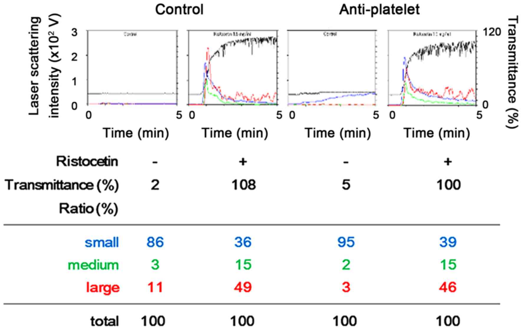
A representative pattern of ristocetin-induced
platelet aggregation in the study groups analyzed by an
aggregometer with a laser scattering system are shown in Fig. 1. Ristocetin (1.5 mg/ml) potently
induced platelet aggregation in the two study groups, and it seems
that there was no difference between the effect observed in the
anti-platelet group and that in the control group. Thus, we
compared the changes induced by restocetin of the area under the
curve (AUC) of light transmittance and the ratio of aggregated
particle sizes of platelets between the two groups. The
ristocetin-induced change of AUC of light transmittance in the
anti-platelet group was not significantly different from that in
the control group (Fig. 2).
Ristocetin significantly induced the changes of the aggregated
particle size ratio, namely, the ratio of large aggregates (50–70
µm) and medium aggregates (25–50 µm) was increased, but that of
small aggregates (9–25 µm) was decreased (Table II). There was little difference
between the two groups in the change induced by ristocetin of the
aggregated particle size ratio (Table
II).
| Table II.Comparison of ristocetin effect on
the ratio of aggregated particle size between the control and
anti-platelet groups in type 2 DM patients. |
Table II.
Comparison of ristocetin effect on
the ratio of aggregated particle size between the control and
anti-platelet groups in type 2 DM patients.
|
|
| Ratio of aggregated
particle size |
|---|
|
|
|
|
|---|
| Group | Ristocetin | Large (%) | Medium (%) | Small (%) |
|---|
| Control | − |
5.4±6.3 |
2.8±2.9 | 91.5±9.0 |
|
| + |
45.5±4.6a |
14.7±1.1a |
39.8±3.9a |
| Anti-platelet | − |
3.7±3.7 |
4.2±5.6 | 91.9±8.4 |
|
| + |
46.6±3.5a |
15.3±2.1a |
37.9±3.3a |
Comparison of the ristocetin-induced
HSP27 phosphorylation between the control and anti-platelet groups
of type 2 DM patients
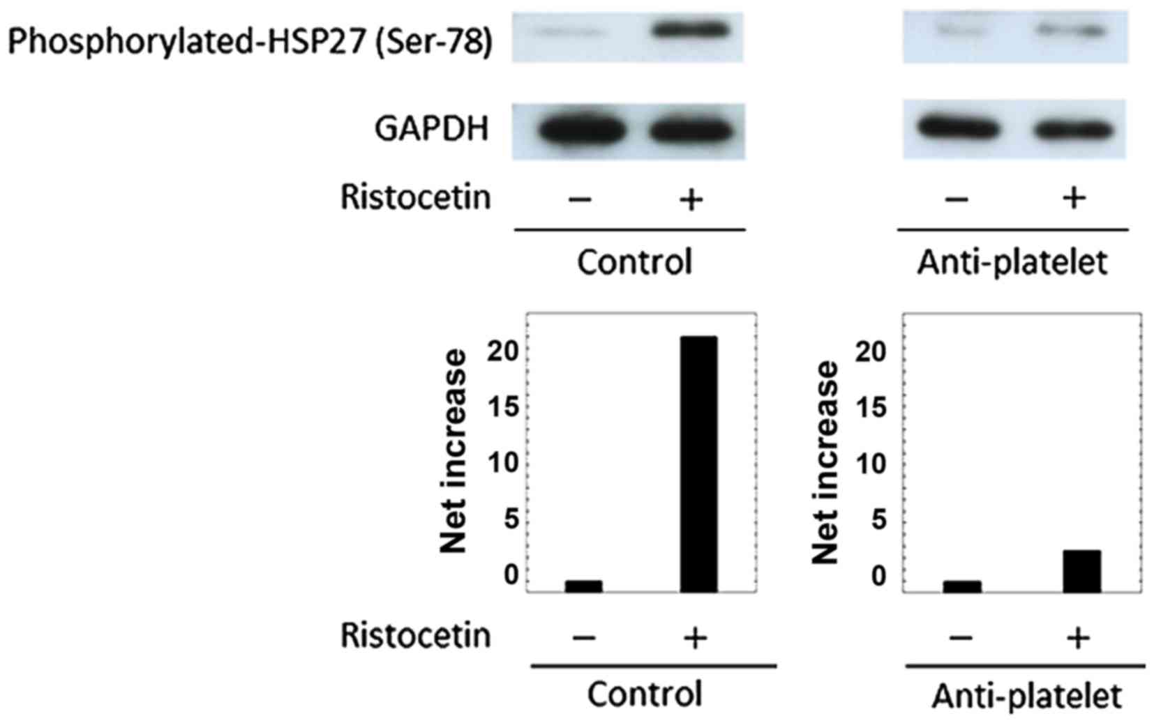
The phosphorylation of HSP27 in human is detected at
three serine residues (Ser-15, Ser-78 and Ser-82) (13,17). In
previous studies (21,22), we demonstrated that the levels of
HSP27 phosphorylation at Ser-78 are significantly correlated with
the levels of released phosphorylated-HSP27 in human platelets
stimulated by collagen or TRAP. Thus, we examined the effect of
ristocetin on the phosphorylation of HSP27 (Ser-78) in the platelet
in the study groups, and compared the phosphorylation levels.
Ristocetin (1.5 mg/ml) elicited the phosphorylation of HSP27
(Ser-78) in the two groups, but the levels observed in a patient of
the anti-platelet group taking ASA were remarkably weaker than
those in a patient of the control group (Fig. 3). Similar results were obtained from
patients taking other types of anti-platelet agents such as
ticlopidine and clopidgrel (data not shown). It is probable that
the administration of anti-platelet agents suppresses the
ristocetin-induced phosphorylation of HSP27 in the platelets of
type 2 DM patients.
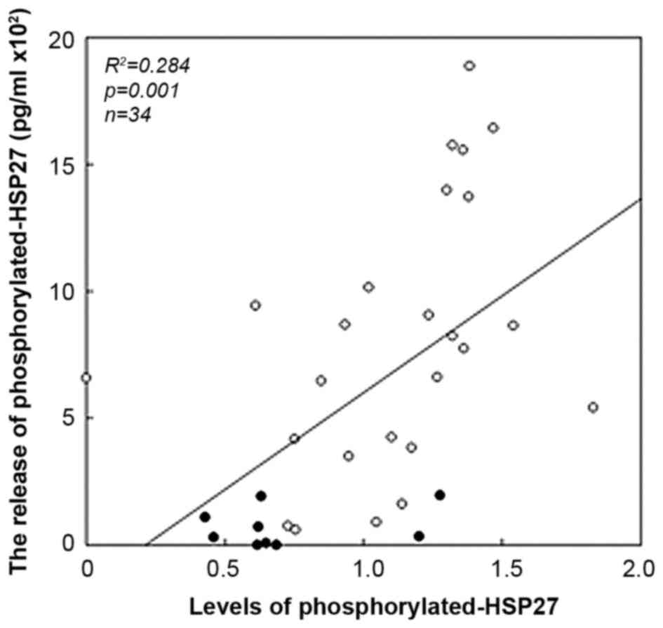
Association between the release of
phosphorylated-HSP27 and the levels of HSP27 phosphorylation
(Ser-78) induced by ristocetin in the platelets from the two
groups-combined type 2 DM patients
Since we found that ristocetin stimulated the
release of phosphorylated-HSP27 from platelets, we next
investigated the relationship between the levels of released
phosphorylated-HSP27 and the levels of HSP27 (Ser-78)
phosphorylation induced by ristocetin (1.5 mg/ml) in the platelets
from two groups-combined type 2 DM patients (n=34; 25 from control
group and 9 from anti-platelet group). The levels were
significantly correlated with the levels of HSP27 (Ser-78)
phosphorylation in the platelets (R2=0.284, P=0.001;
Fig. 4). It is likely that the
ristocetin-induced phosphorylation of HSP27 results in the release
of phosphorylated-HSP27 from the platelets of type 2 DM
patients.
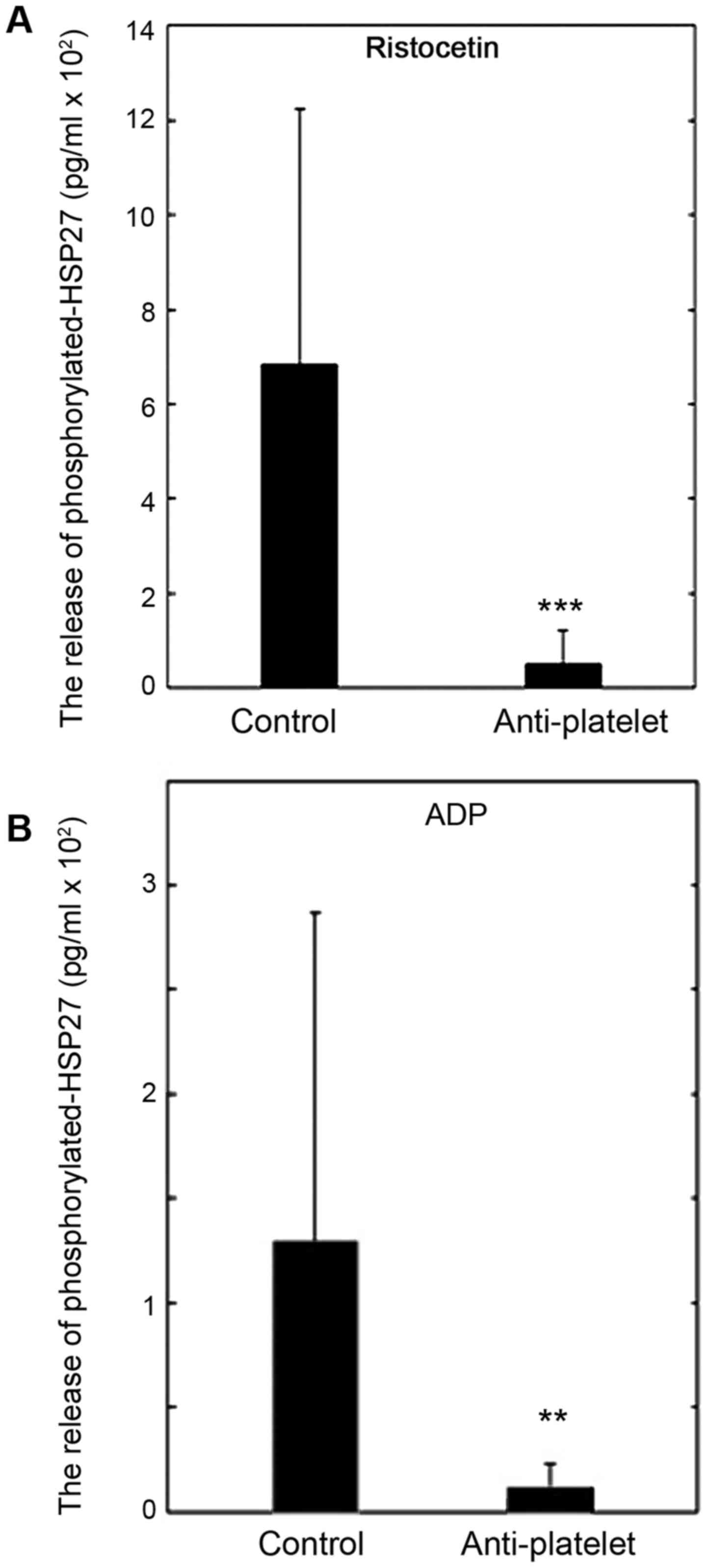
Comparison of the release of
phosphorylated-HSP27 between the control and anti-platelet groups
of type 2 DM patients
We compared the released levels of
phosphorylated-HSP27 induced by ristocetin or ADP between the
control and anti-platelet groups of type 2 DM patients. The levels
of the phosphorylated-HSP27 release induced by 1.5 mg/ml of
ristocetin in the anti-platelet group were significantly lower than
those in the control group (Fig. 5A).
The levels of phosphorylated-HSP27 release induced by 1 µM of ADP
in the anti-platelet group, as well as ristocetin, were
significantly lower than those in the control group (Fig. 5B).
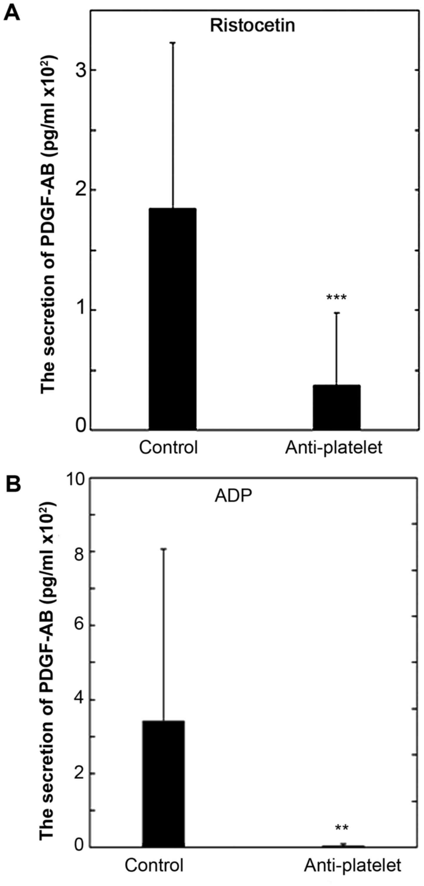
Comparison of the secretion of PDGF-AB
between the control group and the anti-platelet group of type 2 DM
patients
We recently reported that the release of
phosphorylated HSP27 induced by collagen or TRAP is accompanied
with the secretion of PDGF-AB from the platelets of type 2 DM
subjects (21,22). We also compared the secretion of
PDGF-AB induced by ristocetin or ADP between the control and
anti-platelet groups of type 2 DM patients. The levels of the
PDGF-AB secretion induced by 1.5 mg/ml of ristocetin in the
anti-platelet group were significantly lower than those in the
control group (Fig. 6A). In addition,
the levels of PDGF-AB secretion induced by 1 µM of ADP in the
anti-platelet group were significantly lower than those in the
control group (Fig. 6B).
Discussion
In the present study, we investigated the effect of
ristocetin on the release of HSP27 from platelets of type 2 DM
patients, and the effect of anti-platelet agents on the release. At
first, we compared the platelet aggregation induced by ristocetin
between the control and anti-platelet groups of type 2 DM, and
found that there were few differences in the AUC of light
transmittance or the ratio of aggregated particle sizes of
platelets between the two groups. These findings indicate that
anti-platelet agents hardly affect the ristocetin-induced platelet
aggregation in the study subjects. Ristocetin has been established
as a potent inducer of the interaction between GPIb/IX/V and von
Willebrand factor (5), which is a
model of shear stress-activated platelets in vitro (3,4).
Therefore, the present findings strongly suggest that anti-platelet
agents have no effect on the shear stress-induced platelets
aggregation in type 2 DM subjects.
We have recently reported that collagen or TRAP
induces HSP27 phosphorylation in platelets of type 2 DM patients,
and that the phosphorylated HSP27 is released from the platelets,
accompanying the secretion of PDGF-AB (21,22). Thus,
we next examined whether ristocetin induced the phosphorylation and
the release of HSP27, and found that the phosphorylation of HSP27
was truly induced by ristocetin, and that the released levels of
phosphorylated HSP27 was significantly correlated with the
phosphorylated levels of HSP27 stimulated by ristocetin in the
study subjects. Thus, it seems likely that the
ristocetin-stimulated release of phosphorylated HSP27 results from
the phosphorylation of HSP27. This is probably the first report
showing that the ristocetin-induced phosphorylation of HSP27,
resulting in the release of phosphorylated HSP27 from human
platelets, as far as we know. Of note, the levels of phosphorylated
HSP27 release stimulated by ristocetin in the anti-platelet group
were markedly lower than those in the control group, whereas little
difference between the two groups was observed in the
ristocetin-stimulated aggregation. Therefore, it is probable that
anti-platelet agents may successfully reduce the phosphorylation
and release of HSP27 induced by ristocetin without affecting the
aggregation in platelets of type 2 DM patients. In addition, we
investigated the secretion of PDGF-AB induced by ristocetin between
the control and anti-platelet groups. As well as the release of
phosphorylated HSP27, we found that the secretion of PDGF-AB
stimulated by ristocetin in the anti-platelet group was
significantly lower than that in the control group. Therefore, it
is likely that ristocetin-induced PDGF-AB secretion from platelets,
in addition to the release of phosphorylated HSP27, could be
suppressed by anti-platelet agents in type 2 DM subjects.
It has been reported that GPIb/IX/V activation
promotes the generation of TXA2, leading to ADP secretion in rodent
platelets (26). We previously
demonstrated that ristocetin elicits TXA2 synthesis through
cyclooxygenase-1, resulting in the release of soluble CD40 ligand
via the activation of MAP kinases including p38 MAP kinase and
p44/p42 MAP kinase through the binding of TXA2 to TP in human
platelets (6). We have also reported
that ADP induces HSP27 phosphorylation through p38 MAP kinase and
p44/p42 MAP kinase in human platelets (27). Based on these findings, we compared
the effect of ADP on the release of HSP27 between the study groups,
and found that the levels of HSP27 release in the anti-platelet
group were significantly lower than those in the control group. It
seems likely that ADP in fact induces the phosphorylation of HSP27
and its subsequent release from platelets of type 2 DM patients,
which is suppressible by anti-platelet agents. Therefore, these
results suggest that the ristocetin-induced phosphorylation and the
release of HSP27 reducible by anti-platelet agents are dependent on
the downstream events of GPIb/XI/V activation such as TXA2-involved
release of ADP in the platelets of type 2 DM patients.
Additionally, we confirmed that the levels of PDGF-AB secretion
induced by ADP in the anti-platelet group were lower than those in
the control group Taking our findings into account as a whole, it
is most likely that the ristocetin-induced phosphorylation and
release of HSP27 accompanying the secretion of PDGF-AB, are
mediated through the downstream events of GPIb/IX/V activation
including ADP release, which could be suppressed by anti-platelet
agents in type 2 DM patients.
In addition to intracellular functions of HSP27 such
as molecular chaperone, HSP27 released into extracellular space
could affect the functions of adjacent cells (13,23). It
has been reported that HSP27 released from macrophages protectively
acts against the development of atherosclerosis (28). In vascular endothelial cells,
extracellular HSP27 has been shown to increase transcription of
VEGF promoting angiogenesis (29). It
has also been reported that HSP27 diversely upregulates
pro-inflammatory factors including interleukin (IL)-1β in addition
to anti-inflammatory factors including IL-10 in macrophages
(30). Recently, global myocardial
ischemia reportedly causes HSP27 release, which could elicit
pro-inflammatory effect through toll-like receptors in vascular
endothelial cells of coronary arteries (31). It is well recognized that platelets in
blood flow are tethered at the injured sites of vessel through the
formation of GPIb/IX/V and von Willebrand factor complex,
initiating adhesion, first step of the platelet activation
(1,2).
Ristocetin is known to mimic the interaction of GPIb/IX/V and von
Willbrand factor (5). We have
recently reported that collagen and TRAP stimulate the
phosphorylation of HSP27 and the release from the platelets of type
2 DM patients, which is associated with PDGF-AB secretion (21,22). It is
well established that PDGF-AB is involved in the pathological
development of atherosclerosis (1).
Taking account of these findings, it is likely that the pathways of
platelet activation cooperatively upregulates the phosphorylation
of HSP27 leading to the release accompanying PDGF-AB secretion,
which may be involved in the pathogenesis of atherosclerosis, and
inflammation. Therefore, the regulation of HSP27 phosphorylation in
human platelets suggests a novel therapeutic possibility for the
diseases caused by atherosclerosis, major concerns in type 2 DM
patients. Further investigations are required to elucidate the
details underlying the release of phosphorylated-HSP27 from human
platelets.
In conclusion, our present findings strongly suggest
that anti-platelet agents inhibit the HSP27 release from platelets
stimulated by ristocetin but not the aggregation in type 2 DM
patients.
Acknowledgements
Not applicable.
Funding
This study was supported by the Research Funding for
Longevity Sciences (28–9) from National Center for Geriatrics and
Gerontology, Japan.
Availability of data and materials
The datasets used and/or analyzed during the current
study are available from the corresponding author on reasonable
request.
Authors' contributions
HT and OK conceived and designed the experiments.
HT, GK and TO performed the experiments. HT, TD, KK and OK analyzed
the data. HT, YE, HI, TO, SO, TI and OK wrote the paper. All
authors read and approved the final manuscript.
Ethics approval and consent to
participate
The study was approved by the committee of the
conduct of human research at National Center for Geriatrics and
Gerontology, and at Gifu University Graduate School of Medicine.
Written informed consent was obtained from all the patients.
Consent for publication
Not applicable.
Competing interests
The authors declare that they have no competing
interests.
References
|
1
|
Davì G and Patrono C: Platelet activation
and atherothrombosis. N Engl J Med. 357:2482–2494. 2007. View Article : Google Scholar : PubMed/NCBI
|
|
2
|
Furie B and Furie BC: Mechanisms of
thrombus formation. N Engl J Med. 359:938–949. 2008. View Article : Google Scholar : PubMed/NCBI
|
|
3
|
Berndt MC, Shen Y, Dopheide SM, Gardiner
EE and Andrews RK: The vascular biology of the glycoprotein Ib-IX–V
complex. Thromb Haemost. 86:178–188. 2001.PubMed/NCBI
|
|
4
|
Garcia A, Quinton TM, Dorsam RT and
Kunapuli SP: Src family kinase-mediated and Erk-mediated
thromboxane A2 generation are essential for VWF/GPIb-induced
fibrinogen receptor activation in human platelets. Blood.
106:3410–3414. 2005. View Article : Google Scholar : PubMed/NCBI
|
|
5
|
Dong JF, Berndt MC, Schade A, McIntire LV,
Andrews RK and López JA: Ristocetin-dependent, but not
botrocetin-dependent, binding of von Willebrand factor to the
platelet glycoprotein Ib-IX–V complex correlates with
shear-dependent interactions. Blood. 97:162–168. 2001. View Article : Google Scholar : PubMed/NCBI
|
|
6
|
Enomoto Y, Adachi S, Matsushima-Nishiwaki
R, Doi T, Niwa M, Akamatsu S, Tokuda H, Yoshimura S, Iwama T and
Kozawa O: Thromboxane A2 promotes soluble CD40 ligand release from
platelets in atherosclerotic patients. Athelosclerosis.
209:415–421. 2010. View Article : Google Scholar
|
|
7
|
Zimmet P, Alberti KG and Shaw J: Global
and societal implications of the diabetes epidemic. Nature.
414:782–787. 2001. View
Article : Google Scholar : PubMed/NCBI
|
|
8
|
Grundy SM, Benjamin IJ, Burke GL, Chait A,
Eckel RH, Howard BV, Mitch W, Smith SC Jr and Sowers JR: Diabetes
and cardiovascular disease: A statement for healthcare
professionals from the American Heart Association. Circulation.
100:1134–1146. 1999. View Article : Google Scholar : PubMed/NCBI
|
|
9
|
Matsuno H, Tokuda H, Ishisaki A, Zhou Y,
Kitajima Y and Kozawa O: P2Y12 receptors play a significant role in
the development of platelet microaggregation in patients with
diabetes. J Clin Endocrinol Metab. 90:920–927. 2005. View Article : Google Scholar : PubMed/NCBI
|
|
10
|
Hanai Y, Adachi S, Yasuda I, Takai S,
Matsushima-Nishiwaki R, Kato H, Enomoto Y, Akamatsu S, Sakakibara
S, Ogura S, et al: Collagen-induced p38 MAP kinase activation is a
biomarker of platelet hyper-aggregation in patients with diabetes
mellitus. Life Sci. 85:386–394. 2009. View Article : Google Scholar : PubMed/NCBI
|
|
11
|
Colwell JA; American Diabetes Association,
: Aspirin therapy in diabetes. Diabetes Care. 26 Suppl 1:S87–S88.
2003. View Article : Google Scholar : PubMed/NCBI
|
|
12
|
Hankey GJ and Eikelboom JW: Aspirin
resistance. Lancet. 367:606–617. 2006. View Article : Google Scholar : PubMed/NCBI
|
|
13
|
Mymrikov EV, Seit-Nebi AS and Gusev NB:
Large potentials of small heat shock proteins. Physiol Rev.
91:1123–1159. 2011. View Article : Google Scholar : PubMed/NCBI
|
|
14
|
Bruey JM, Ducasse C, Bonniaud P, Ravagnan
L, Susin SA, Diaz-Latoud C, Gurbuxani S, Arrigo AP, Kroemer G,
Solary E, et al: Hsp27 negatively regulates cell death by
interacting with cytochrome c. Nat Cell Biol. 2:645–652. 2000.
View Article : Google Scholar : PubMed/NCBI
|
|
15
|
Arrigo AP: The cellular ‘networking’ of
mammalian Hsp27 and its functions in the control of protein
folding, redox state and apoptosis. Adv Exp Med Biol. 594:14–26.
2007. View Article : Google Scholar : PubMed/NCBI
|
|
16
|
Pockley AG: Heat shock proteins,
inflammation, and cardiovascular disease. Circulation.
105:1012–1017. 2002. View Article : Google Scholar : PubMed/NCBI
|
|
17
|
Landry J, Lambert H, Zhou M, Lavoie JN,
Hickey E, Weber LA and Anderson CW: Human HSP27 is phosphorylated
at serines 78 and 82 by heat shock and mitogen-activated kinases
that recognize the same amino acid motif as S6 kinase II. J Biol
Chem. 267:794–803. 1992.PubMed/NCBI
|
|
18
|
Acunzo J, Katsogiannou M and Rocchi P:
Small heat shock proteins HSP27 (HspB1), αB-crystallin (HspB5) and
HSP22 (HspB8) as regulators of cell death. Int J Biochem Cell Biol.
44:1622–1631. 2012. View Article : Google Scholar : PubMed/NCBI
|
|
19
|
Saklatvala J, Rawlinson L, Waller RJ,
Sarsfield S, Lee JC, Morton LF, Barnes MJ and Farndale RW: Role for
p38 mitogen-activated protein kinase in platelet aggregation caused
by collagen or a thromboxane analogue. J Biol Chem. 271:6586–6589.
1996. View Article : Google Scholar : PubMed/NCBI
|
|
20
|
Kageyama Y, Doi T, Akamatsu S, Kuroyanagi
G, Kondo A, Mizutani J, Otsuka T, Tokuda H, Kozawa O and Ogura S:
Rac regulates collagen-induced HSP27 phosphorylation via p44/p42
MAP kinase in human platelets. Int J Mol Med. 32:813–818. 2013.
View Article : Google Scholar : PubMed/NCBI
|
|
21
|
Tokuda H, Kuroyanagi G, Tsujimoto M,
Enomoto Y, Matsushima-Nishiwaki R, Onuma T, Kojima A, Doi T, Tanabe
K, Akamatsu S, et al: Release of phosphorylated HSP27 (HSPB1) from
platelets is accompanied with the accerelation of aggregation in
diabetic patients. PLoS One. 10:e01289772015. View Article : Google Scholar : PubMed/NCBI
|
|
22
|
Tokuda H, Kuroyanagi G, Tsujimoto M,
Matsushima-Nishiwaki R, Akamatsu S, Enomoto Y, Iida H, Otsuka T,
Ogura S, Iwama T, et al: Thrombin receptor-activating protein
(TRAP)-activated Akt is involved in the release of
phosphorylated-HSP27 (HSPB1) from platelets in DM patients. Int J
Mol Sci. 17(pii): E7372016. View Article : Google Scholar : PubMed/NCBI
|
|
23
|
Reddy VS, Madala SK, Trinath J and Reddy
GB: Extracellular small heat shock proteins: exosomal biogenesis
and function. Cell Stress Chaperones. Oct 30–2017.(Epub ahead of
print). View Article : Google Scholar : PubMed/NCBI
|
|
24
|
Kato K, Ito H, Hasegawa K, Inaguma Y,
Kozawa O and Asano T: Modulation of the stress-induced synthesis of
hsp27 and alpha B-crystallin by cyclic AMP in C6 rat glioma cells.
J Neurochem. 66:946–950. 1996. View Article : Google Scholar : PubMed/NCBI
|
|
25
|
Laemmli UK: Cleavage of structural
proteins during the assembly of the head of bacteriophage T4.
Nature. 227:680–685. 1970. View
Article : Google Scholar : PubMed/NCBI
|
|
26
|
Liu J, Pestina TI, Berndt MC, Steward SA,
Jackson CW and Gartner TK: The roles of ADP and TXA in
botrocetin/VWF-induced aggregation of washed platelets. J Thromb
Haemost. 2:2213–2222. 2004. View Article : Google Scholar : PubMed/NCBI
|
|
27
|
Kato H, Takai S, Matsushima-Nishiwaki R,
Adachi S, Minamitani C, Otsuka T, Tokuda H, Akamatsu S, Doi T,
Ogura S, et al: HSP27 phosphorylation is correlated with
ADP-induced platelet granule secretion. Arch Biochem Biophys.
475:80–86. 2008. View Article : Google Scholar : PubMed/NCBI
|
|
28
|
Rayner K, Chen YX, McNulty M, Simard T,
Zhao X, Wells DJ, de Belleroche J and O'Brien ER: Extracellular
release of the atheroprotective heat shock protein 27 is mediated
by estrogen and competitively inhibits acLDL binding to scavenger
receptor-A. Circ Res. 103:133–141. 2008. View Article : Google Scholar : PubMed/NCBI
|
|
29
|
Thuringer D, Jego G, Wettstein G, Terrier
O, Cronier L, Yousfi N, Hébrard S, Bouchot A, Hazoumé A, Joly AL,
et al: Extracellular HSP27 mediates angiogenesis through Toll-like
receptor 3. FASEB J. 27:4169–4183. 2013. View Article : Google Scholar : PubMed/NCBI
|
|
30
|
Salari S, Seibert T, Chen YX, Hu T, Shi C,
Zhao X, Cuerrier CM, Raizman JE and O'Brien ER: Extracellular HSP27
acts as a signaling molecule to activate NF-κB in macrophages. Cell
Stress Chaperones. 18:53–63. 2013. View Article : Google Scholar : PubMed/NCBI
|
|
31
|
Jin C, Cleveland JC, Ao L, Li J, Zeng Q,
Fullerton DA and Meng X: Human myocardium releases heat shock
protein 27 (HSP27) after global ischemia: The proinflammatory
effect of extracellular HSP27 through toll-like receptor (TLR)-2
and TLR4. Mol Med. 20:280–289. 2014. View Article : Google Scholar : PubMed/NCBI
|