Introduction
Cancer cells are characterized by uncontrolled
proliferation (1), therefore
inhibiting the excessive proliferation of tumor cells is one of the
key approaches for the development of anti-cancer drugs. Eukaryotic
cell proliferation is regulated by the cell cycle, which is divided
into a series of phases. G1/S transition is one of the two main
checkpoints that control cell cycle progression (2). G1/S progression is mainly regulated
by Cyclin D1 and Cyclin-dependent kinase 4 (CDK4) (3,4).
PCNA is an acidic nuclear protein that has been recognized as a
histological marker for the G1/S phase in the cell cycle (5). p21 is a CDK inhibitor, which can bind
to CDK-Cyclin complexes and alter their function in order to
suppress cell proliferation (6).
Drug resistance and toxicity against normal cells
limit the effectiveness of current chemotherapies for the treatment
of colorectal cancer (CRC) (7–10),
which is a serious public health problem worldwide (11). These problems highlight the urgent
need for the development of novel cancer chemotherapies. Recently,
natural products have received great interest since they have
relatively few side effects compared with modern chemotherapeutics
and have been used clinically for thousands of years as significant
alternative remedies for a variety of diseases including cancer
(12–17). One promising medicinal plant is
Hedyotis diffusa Willd (HDW) that belongs to the Rubiaceae
family and is widely distributed throughout Northeast Asia. As a
well-known traditional Chinese folk-medicine, it is used for
heat-clearing, detoxification, promotion of blood circulation and
the removal of blood stasis (18).
HDW has also long been used as an significant component in several
Chinese medicine formulae to treat various types of cancer,
including CRC (18–20). Previously, we reported that
Hedyotis diffusa Willd inhibits the growth of CRC, likely
via the induction of cancer cell apoptosis and the inhibition of
tumor angiogenesis (21,22). To further elucidate the mechanism
of the tumoricidal activity of HDW we investigated its effect on
the proliferation of human colon carcinoma HT-29 cells.
Materials and methods
Materials and reagents
Dulbecco’s modified Eagle’s medium (DMEM), fetal
bovine serum (FBS), penicillin-streptomycin, Trypsin-EDTA and
TRIzol reagent were purchased from Invitrogen (Carlsbad, CA, USA).
SuperScript II reverse transcriptase was obtained from Promega
(Madison, WI, USA). All other chemicals, unless otherwise stated,
were obtained from Sigma Chemicals (St. Louis, MO, USA).
Preparation of ethanol extract of
Hedyotis diffusa Willd (EEHDW)
EEHDW was prepared as described previously (20,21).
Briefly, stock solutions of EEHDW were prepared by dissolving the
EEHDW powder in 40% DMSO to a final concentration of 400 mg/ml and
stored at −20°C. The working concentrations of EEHDW were made by
diluting the stock solution in the culture medium. The final
concentrations of DMSO in the medium were <0.5%.
Cell culture
Human colon carcinoma HT-29 cells were obtained from
the American Type Culture Collection (ATCC, Manassas, VA, USA).
HT-29 cells were grown in DMEM. DMEM was supplemented with 10%
(v/v) FBS, 100 units/ml penicillin and 100 μg/ml
streptomycin. Cells were cultured at 37°C and 5% CO2 in
a humidified environment.
Cell viability evaluation
Cell viability was assessed by MTT colorimetric
assay. HT-29 cells were seeded into 96-well plates at a density of
1x10⁴ cells/well in 0.1 ml medium. The cells were
treated with various concentrations (0, 1, 3 and 5 mg/ml) of EEHDW
for various periods of time. At the end of the treatment, 10
μl MTT (5 mg/ml in phosphate-buffered saline, PBS) was added
to each well and the samples were incubated for an additional 4 h
at 37°C. The purple-blue MTT formazan precipitate was dissolved in
100 μl DMSO. The absorbance was measured at 570 nm using an
ELISA reader (BioTek, Model ELX800, Winooski, VT, USA).
Colony formation assay
The HT-29 cells were seeded into 6-well plates at a
density of 2x10⁵ cells/well and treated with various
concentrations (0, 1, 3 and 5 mg/ml) of EEHDW for 24 h. The cells
were then diluted in fresh medium in the absence of EEHDW and
reseeded into 6-well plates at a density of 1.5x10³
cells/well. Following incubation for 7 or 8 days in a 37°C
humidified incubator with 5% CO2, the colonies were
counted under a light microscope. Cell survival was calculated by
normalizing the survival of the control cells as 100%.
Cell cycle analysis
The cell cycle analysis was carried out by flow
cytometry using a fluorescence-activated cell sorting (FACS) MoFlo
XDP (Beckman Coulter, Miami, FL, USA) and propidium iodide (PI)
staining. Following treatment with the indicated concentrations (0,
1, 3 and 5 mg/ml) of EEHDW for 24 h, HT-29 cells were harvested and
adjusted to a concentration of 1x10⁶ cells/ml, then
fixed in 70% ethanol at 4°C overnight. The fixed cells were washed
twice with cold PBS and then incubated for 30 min with RNase (8
μg/ml) and PI (10 μg/ml). The fluorescent signal was
detected through the FL2 channel and the proportion of DNA in
various phases was analyzed using ModfitLT Version 3.0 (Verity
Software House, Topsham, ME, USA).
RNA extraction and RT-PCR analysis
A total of 2x10⁵ HT-29 cells were seeded
into 6-well plates in 2 ml medium and treated with indicated
concentrations (0, 1, 3 and 5 mg/ml) of EEHDW for 24 h. Total RNA
was isolated using TRIzol reagent. Oligo(dT)-primed RNA (1
μg) was reverse-transcribed with SuperScript II reverse
transcriptase (Promega) according to the manufacturer’s
instructions. The obtained cDNA was used to determine the mRNA
levels of Cyclin D1, CDK4, PCNA and p21 by PCR. GAPDH was used as
an internal control. The sequences of the primers used for
amplification of Cyclin D1, CDK4, PCNA, p21 and GAPDH transcripts
are as follows: Cyclin D1 forward 5′-TGGATGCTGGAGGTCTGCGAG GAA-3′
and reverse 5′-GGCTTCGATCTGCTCCTGGCA GGC-3′ [Temperature (Tm),
55°C; 573 bp]; CDK4 forward 5′-CATGTAGACCAGGACCTAAGC-3′ and reverse
5′-AAC TGGCGCATCAGATCCTAG-3′ (Tm, 58°C; 206 bp); PCNA forward
5′-GCTGACATGGGACACTTA-3′, and reverse 5′-CTCAGGTACAAACTTGGTG-3′
(Tm, 56°C; 610 bp); p21 forward 5′-GCGACTGTGATGCGCTAATGG-3′, and
reverse 5′-TAGAAATCTGTCATGCTGGTCTGC-3′ (Tm=55°C, 358 bp); GAPDH
forward 5′-CGACCACTTTGTCAAG CTCA-3′, and reverse
5′-AGGGGTCTACATGGCAACTG-3′ (Tm, 58°C; 240 bp). Samples were
analyzed by gel electrophoresis (1.5% agarose). The DNA bands were
examined using a Gel Documentation system (BioRad, Model Gel Doc
2000, USA).
Statistical analysis
All data were expressed as the means of three
independent experiments and data was analyzed using the SPSS
package for Windows (Version 11.5; Chicago, IL, USA). Statistical
analysis of the data was performed using a Student’s t-test and
ANOVA. P<0.05 was considered to indicate a statistically
significant result.
Results
EEHDW inhibits the proliferation of HT-29
cells
We examined the effect of EEHDW on HT-29 cell
viability by MTT assay. As shown in Fig. 1, EEHDW treatment reduced cell
viability in a dose- and time-dependent manner compared to
untreated control cells (P<0.05). The cell viability was
decreased to 36% at the highest concentration of EEHDW (5 mg/ml) at
24 h in this study. To further verify these results, we examined
the effect of EEHDW on HT-29 cell survival using a colony formation
assay. As shown in Fig. 2,
treatment with 1, 3 and 5 mg/ml of EEHDW for 24 h reduced the cell
survival rate in a dose-dependent manner by 34, 70 and 84% compared
to untreated control cells (P<0.05). These data suggest that
EEHDW inhibits HT-29 cell proliferation.
EEHDW blocks G1/S progression of HT-29
cells
The G1/S transition is one of the two main
checkpoints that regulate cell cycle progression and thus the cell
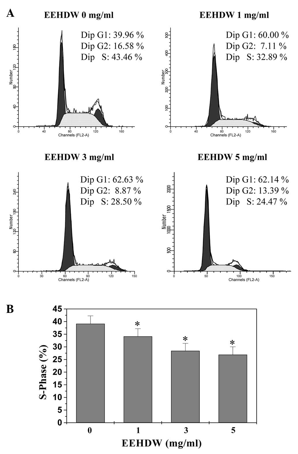
proliferation. We, therefore, investigated the effect of EEHDW on
the G1 to S progression in HT-29 cells via PI staining followed by
FACS analysis. As shown in Fig. 3A and
B, the percentage of S-phase cells following treatment with 0,
1, 3 and 5 mg/ml of EEHDW was 39.13, 34.14, 28.42 and 26.91%,
respectively (P<0.05), indicating that EEHDW inhibits HT-29 cell
proliferation by blocking the cell cycle at the G1 to S
progression.
EEHDW regulates mRNA expression of PCNA,
Cyclin D1, CDK4 and p21
We examined the effect of EEHDW on the mRNA
expression of the pro-proliferative PCNA, Cyclin D1, CDK4, and the
anti-proliferative p21 using RT-PCR. As shown in Fig. 4, EEHDW treatment markedly reduced
the mRNA expression of PCNA, Cyclin D1 and CDK4, but increased that
of p21.
Discussion
Hedyotis diffusa Willd (HDW) is a Chinese
medicinal herb with numerous reported pharmacological applications.
HDW is clinically effective in the treatment of various types of
cancer, including CRC (18–20).
Recently, we reported that the direct cytotoxic effect of HDW on
colon cancer cells is partially due to the induction of
mitochondrion-dependent apoptosis (21). However, the effect of HDW on cancer
cell proliferation is still unclear.
Using MTT and colony formation assays, in the
present study, we demonstrated that the ethanol extract of
Hedyotis diffusa Willd (EEHDW) inhibited the proliferation
of human colon carcinoma HT-29 cells. Cell proliferation is
regulated by the cell cycle, which consists of four periods; S
phase (DNA synthesis), M phase (mitosis), G1 and G2 phase. The G1/S
transition is one of the two main checkpoints of the cell cycle
(2), and is responsible for the
initiation and completion of DNA replication. By using FACS
analysis and PI staining we observed that the inhibitory effect of
EEHDW on HT-29 cell proliferation was associated with the
prevention of G1 to S transition. The G1/S progression is strongly
regulated by Cyclin D1 which forms an active complex with its CDK
major catalytic partners (CDK4/6) (3,4). An
unchecked or hyperactivated Cyclin D1/CDK4 complex may be
responsible for enhanced cellular proliferation. PCNA, a 36-kDa DNA
polymerase delta auxiliary protein that is involved in cell
proliferation and is specifically expressed in proliferating cell
nuclei, has been recognized as a histological marker for the G1/S
phase in the cell cycle (5). As a
proliferation inhibitor, p21 protein plays a role in G1 arrest by
binding to and inhibiting the activity of Cyclin-CDK complexes and
PCNA (6). Therefore, the
expression of PCNA, CDK4, Cyclin D1 and p21 reflects the
proliferation state of HT-29 cells to some extent. As predicted,
following EEHDW treatment the mRNA expression of Cyclin D1, CDK4
and PCNA in HT-29 cells was downregulated but that of p21 was
upregulated.
In conclusion, we report for the first time that
EEHDW inhibits the proliferation of HT-29 cells via cell cycle
arrest. Together with our previous study, these results suggest
that Hedyotis diffusa Willd inhibits cancer progression via
multiple mechanisms, including the induction of cancer cell
apoptosis, inhibition of cell proliferation and tumor
angiogenesis.
Abbreviations:
|
EEHDW
|
ethanol extract of Hedyotis
diffusa Willd
|
|
CRC
|
colorectal cancer
|
|
DMSO
|
dimethyl sulfoxide
|
|
MTT
|
3-(4,5-dimethyl-thiazol-2-yl)-2,5-diphenyltetrazolium bromide
|
Acknowledgements
This work was sponsored by the Natural
Science Foundation of Fujian Province of China (2010J01195), the
Research Foundation of the Education Bureau of Fujian Province of
China (JA10162) and the Developmental Fund of Chen Keji Integrative
Medicine (CKJ 2010030).
References
|
1.
|
Evan GI and Vousden KH: Proliferation,
cell cycle and apoptosis in cancer. Nature. 411:342–348. 2001.
View Article : Google Scholar : PubMed/NCBI
|
|
2.
|
Nurse P: Ordering S phase and M phase in
the cell cycle. Cell. 79:547–550. 1994. View Article : Google Scholar : PubMed/NCBI
|
|
3.
|
Chen Y, Robles AI, Martinez LA, Liu F,
Gimenez-Conti IB and Conti CJ: Expression of G1 cyclins,
cyclin-dependent kinases, and cyclin-dependent kinase inhibitors in
androgen-induced prostate proliferation in castrated rats. Cell
Growth Differ. 7:1571–1578. 1996.PubMed/NCBI
|
|
4.
|
Graña X and Reddy EP: Cell cycle control
in mammalian cells: role of cyclins, cyclin dependent kinases
(CDKs), growth suppressor genes and cyclin-dependent kinase
inhibitors (CKIs). Oncogene. 11:211–219. 1995.PubMed/NCBI
|
|
5.
|
Zhong W, Peng J, He H, Wu D, Han Z, Bi X
and Dai Q: Ki-67 and PCNA expression in prostate cancer and benign
prostatic hyperplasia. Clin Invest Med. 31:E8–E15. 2008.PubMed/NCBI
|
|
6.
|
Harper JW, Adami GR, Wei N, Keyomarsi K
and Elledge SJ: The p21 Cdk-interacting protein Cip1 is a potent
inhibitor of G1 cyclin-dependent kinases. Cell. 75:805–816. 1993.
View Article : Google Scholar : PubMed/NCBI
|
|
7.
|
Gustin DM and Brenner DE: Chemoprevention
of colon cancer: current status and future prospects. Cancer Metast
Rev. 21:323–348. 2002. View Article : Google Scholar : PubMed/NCBI
|
|
8.
|
Gorlick R and Bertino JR: Drug resistance
in colon cancer. Semin Oncol. 26:606–611. 1999.
|
|
9.
|
Longley DB, Allen WL and Johnston PG: Drug
resistance, predictive markers and pharmacogenomics in colorectal
cancer. Biochim Biophys Acta. 1766:184–196. 2006.PubMed/NCBI
|
|
10.
|
Boose G and Stopper H: Genotoxicity of
several clinically used topoisomerase II inhibitors. Toxicol Lett.
116:7–16. 2000. View Article : Google Scholar : PubMed/NCBI
|
|
11.
|
Jemal A, Bray F, Center MM, Ferlay J, Ward
E and Forman D: Global cancer statistics. CA Cancer J Clin.
61:69–90. 2011. View Article : Google Scholar
|
|
12.
|
Kelloff GJ: Perspectives on cancer
chemoprevention research and drug development. Adv Cancer Res.
78:199–334. 2000. View Article : Google Scholar : PubMed/NCBI
|
|
13.
|
Tyagi AK, Singh RP and Agarwal C, Chan DC
and Agarwal C: Silibinin strongly synergizes human prostate
carcinoma DU145 cells to doxorubicin-induced growth inhibition,
G2-M arrest, and apoptosis. Clin Cancer Res. 8:3512–3519. 2002.
|
|
14.
|
Newman DJ, Cragg GM and Snader KM: The
influence of natural products upon drug discovery. Nat Prod Rep.
17:215–234. 2000. View
Article : Google Scholar : PubMed/NCBI
|
|
15.
|
Jeune MA, Kumi-Diaka J and Brown J:
Anticancer activities of pomegranate extracts and genistein in
human breast cancer cells. J Med Food. 8:469–475. 2005. View Article : Google Scholar : PubMed/NCBI
|
|
16.
|
Sausville EA: Versipelostatin: unfolding
an unsweetened death. J Natl Cancer Inst. 96:1266–1267. 2004.
View Article : Google Scholar : PubMed/NCBI
|
|
17.
|
Won HJ, Han CH, Kim YH, Kwon HJ, Kim BW,
Choi JS and Kim KH: Induction of apoptosis in human acute leukemia
Jurkat T cells by Albizzia julibrissin extract is mediated
via mitochondria-dependent caspase-3 activation. J Ethnopharmacol.
106:383–389. 2006. View Article : Google Scholar : PubMed/NCBI
|
|
18.
|
Song LR: Zhonghuabencao. 61. Shanghai
Science and Technology Press; Shanghai: pp. 4331999
|
|
19.
|
Yang JJ, Hsu HY, Ho YH and Lin CC:
Comparative study on the immunocompetent activity of three
different kinds of Peh-Hue-Juwa-Chi-Cao, Hedyotis diffusa,
H. corymbosa and Mollugo pentaphylla after sublethal
whole body X-irradiation. Phytother Res. 11:428–432. 1997.
View Article : Google Scholar
|
|
20.
|
Li R, Zhao HR and Lin YM: Anti-tumor
effect and protective effect on chemotherapeutic damage of water
soluble extracts from Hedyotis diffusa. J Chin Pharm Sci.
11:54–58. 2002.
|
|
21.
|
Lin JM, Chen YQ, Wei LH, Chen XZ, Xu W,
Hong ZF, Sferra TJ and Peng J: Hedyotis Diffusa Willd
extract induces apoptosis via activation of the
mitochondrion-dependent pathway in human colon carcinoma cells. Int
J Oncol. 37:1331–1338. 2010.
|
|
22.
|
Lin JM, Wei LH, Xu W, Hong ZF, Liu XX and
Peng J: Effect of Hedyotis Diffusa Willd extract on tumor
angiogenesis. Mol Med Rep. 4:1283–1288. 2011.
|


