Introduction
Mixed-histology primary lung cancer is a rare type
of lung cancer that shows two or more types of mixed pathology
within the same case or tumor. At present, there are no uniform
standards for its diagnosis. Particularly, in previous studies on
lung cancer, with the application of electron microscopy and
immunohistochemical analysis, scientists have identified that the
percentage of all the types of mixed-histology lung cancer was
higher compared to the proportion of lung cancer, in contrast with
clinical studies (1–4). Data regarding epidemiology, clinical
features and prognosis of survival is limited. Consequently,
mixed-histology lung cancer patients diagnosed between January,
1999 and Decemberm, 2008 at the Xiangya Hospital of Central South
University (Changsha, China) were retrospectively analyzed to
investigate the clinical features, diagnosis and prognosis of
mixed-histology lung cancer.
Patients and methods
Patients
Patients diagnosed with primary lung cancer using
pathologic or cytologic methods between January, 1999 and
September, 2008 at Xiangya Hospital of Central South University
(Changsha, China) were selected. The patients with mixed primary
lung cancer were included in the present study.
This study was approved by the Institutional Review
Board of Xiangya Hospital, The Central South University and written
informed consent was obtained from all the included patients.
The mixed lung cancer patients included in this
study were diagnosed by pathologic examination of surgical
specimens, bronchoscopic biopsy, CT/B ultrasound-guided
percutaneous lung biopsy, superficial lymph node biopsy as well as
sputum smear and serous effusion biopsy (pleural effusion,
pericardial effusion). According to the World Health Organization
(WHO) classification (5), any
tissue composition of malignant mixed tumor must be >20% of
tumor tissues prior to diagnosis, and tissue with a composition of
60–80% was the dominant component.
Variables
For each patient, age, gender, smoking history,
clinical symptoms, histological type [as a subtype of the
adenocarcinoma, bronchioloalveolar (BA) carcinoma has different
biological characteristics from adenocarcinoma, therefore, BA
carcinoma was treated as a heterogeneous tumor and excluded when
referred to lung adenocarcinoma in our study], anatomical location
of tumor, clinical tumor-node-metastasis (TNM) staging [based on
the International Association for the Study of Lung Cancer (IASLC)
classification (6)], metastasis
[includes M1a (malignant pleural effusion, pericardial effusion or
pleural nodules and contralateral lung nodules) and M1b (distant
metastasis, beyond lung/pleura)] (6), treatment and prognosis.
Follow-up
Mixed lung cancer patients with surgical treatment
and randomly selected non-small cell lung cancer (NSCLC) patients,
with resection in the same period were followed up. Follow-up was
performed by telephone, and survival was considered from the day of
lung cancer diagnosis, until the day of death or the endpoint of
follow-up (September 31, 2011).
Statistical analysis
A clinical database was established using Excel
2007, and analyzed using SPSS Statistics 17.0 edition software
(Statsoft, Tulsa, OK, USA). Differences between the frequencies
were examined using the χ2 test. The median survival
time and the 1st, 2nd and 3rd year rate of survival were used to
evaluate prognosis. Survival rates were computed using the
Kaplan-Meier method (7), while the
log-rank test was used to compare the survival curves. P<0.05
was considered to indicate a statistically significant
difference.
Results
Clinicopathological findings
In general, 92 lung tumors represented the
association of two histological types, accounting for 4.99% of the
total population of diagnosed lung tumors (92/1,842) (Table I). The patients comprised 75 men
and 17 women (4.41:1), with a mean age of 56 years (range, 29–74
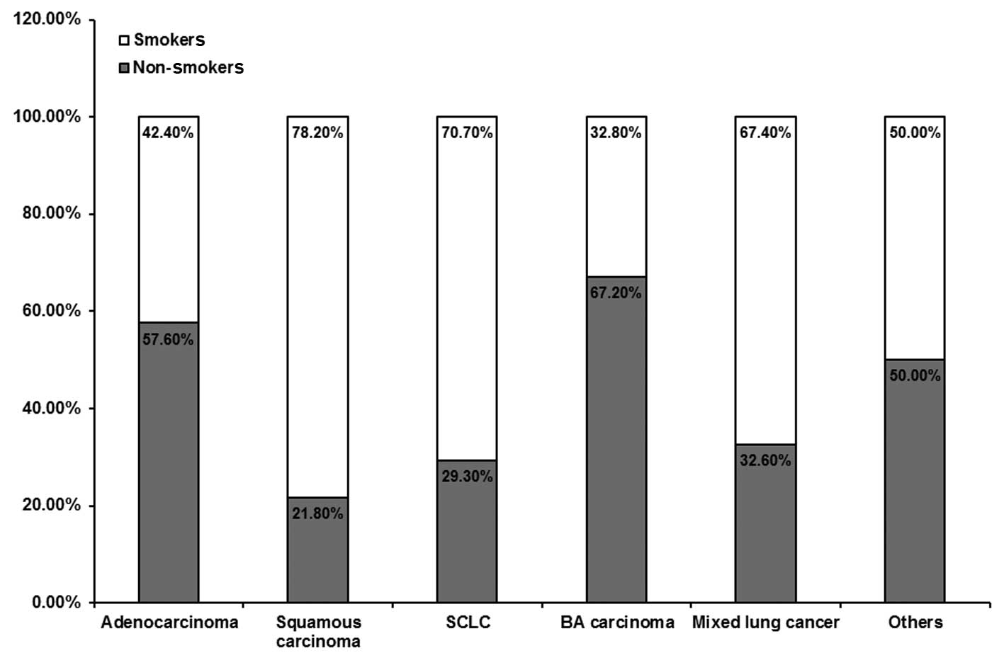
years). Sixty-two patients had a smoking history with a smoking
index of ≥400 (Fig. 1), and 61 of
the 62 cases were male. Regarding mixed pathologic patterns,
adenosquamous carcinoma was the most common (69/92), followed by
adenocarcinoma with BA pattern (6/92); additional pathologic
patterns were rare (Table
II).
 | Table IHistology of 1,842 cancer
patients. |
Table I
Histology of 1,842 cancer
patients.
| Histology | Total (n) | Percentage (%) |
|---|
| Squamous
carcinoma | 701 | 38.05 |
| Adenocarcinoma | 516 | 28.01 |
| SCLC | 273 | 14.82 |
| BA carcinoma | 64 | 3.47 |
| Mixed lung
cancer | 92 | 4.99 |
| Others | 196 | 10.64 |
| Total | 1842 | 100.00 |
| Table IIDifferent patterns of 92 mixed lung
cancer patients. |
Table II
Different patterns of 92 mixed lung
cancer patients.
| Histology | Cases (n) | Percentage (%) |
|---|
| Adenosquamous
carcinoma | 69 | 75.0 |
| Adenocarcinoma
combined with BA pattern | 6 | 6.3 |
| Squamous cell
carcinoma combined with SCLC | 2 | 2.2 |
| Squamous cell
carcinoma combined with BA pattern | 2 | 2.2 |
| Adenosquamous
carcinoma combined with sarcoma | 2 | 2.2 |
| Adenocarcinoma
combined with sarcoma | 2 | 2.2 |
| Adenosquamous
carcinoma combined with clear cell carcinoma | 2 | 2.2 |
| Adenosquamous
carcinoma combined with SCLC | 1 | 1.1 |
| Adenosquamous
carcinoma combined with BA carcinoma | 1 | 1.1 |
| BA carcinoma combined
with large cell carcinoma | 1 | 1.1 |
| BA carcinoma combined
with SCLC | 1 | 1.1 |
| Squamous cell
carcinoma combined with large cell carcinoma | 1 | 1.1 |
| BA carcinoma combined
with neuroendocrine differentiation | 1 | 1.1 |
| Squamous cell
carcinoma with neuroendocrine differentiation | 1 | 1.1 |
The apicoposterior segment of the upper lobe (45/92)
was more commonly involved in tumor location compared to the basal
segment (including posterior, medial and lateral) of the lower lobe
(21/92); epimere of lower lobes (14/92); anterior segment of upper
lobes (11/92); middle lobe, including lingual lobe (6/92). Tumors
were rarely located in the segmental bronchus and above (2/92), the
two sides (2/92) and mediastinum (1/92).
Cough and sputum (59/92) as well as hemoptysis
(28/92) were the most common symptoms of mixed lung cancer
patients. Additional symptoms included chest pain (20/92), chest
tightness and shortness of breath (7/92), dizziness and headache
(2/92), hoarseness (2/92), fever (1/92), neck mass (1/92), fatigue
(1/92), bone pain (1/92) and numbness (1/92).
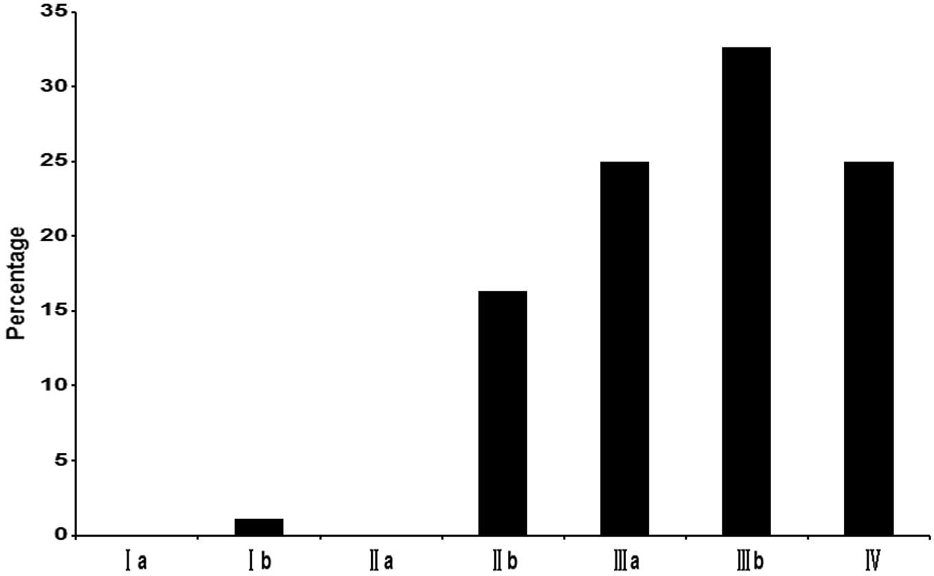
Sixteen patients were diagnosed as having stage
I–IIb tumors, 23 as stage IIIa tumors and 53 as stage IIIb–IV
tumors (Fig. 2). Metastasis was
found in 64 patients following diagnosis, only second to
adenocarcinoma patients. Bone metastasis was observed in 13
patients; pleural or pericardial metastasis in 11 patients;
supraclavicular lymph node metastasis in 10 patients; and brain,
liver and other organ (spleen and pancreas) metastasis in 5
patients each. Single metastasis was observed in 40 cases, and
multiple metastases in 24 cases.
Regarding diagnostic methods utilized, 56/92
patients were diagnosed by pathologic examination of surgical
specimens, 36/92 patients were diagnosed depending on the specimen
obtained from bronchial biopsy (21/92), CT/B ultrasound-guided
percutaneous lung biopsy (8/92), superficial lymph node biopsy
(6/92) and pleural biopsy (1/92).
Of the 92 patients, 56 underwent surgical treatment.
There were 2 N0, 15 N1 and 39 N2 patients. Thirty patients
underwent resection alone, and postoperatively, radiotherapy was
administered to 3 patients, chemotherapy (third-generation
platinum-based chemotherapy) to 18 patients and combined
chemo-radiotherapy to 5 patients. Of the 92 patients, 8 were
administered chemotherapy, and 5 chemoradiotherapy without
resection. No treatment was used in the remaining 23 patients.
Survival and prognosis
By December 31, 2011, there were 52 effective
follow-up cases of mixed lung cancer. No data were available for 4
cases, due to refusal of follow-up, change of contact details, or
mortality unrelated to this study; thus, the dropout rate was 7.1%.
Of the 59 NSCLC cases randomly selected during the same time
period, 54 patients were effectively followed up, while no data
were available for 5 patients (dropout rate, 8.5%). The l-, 2- and
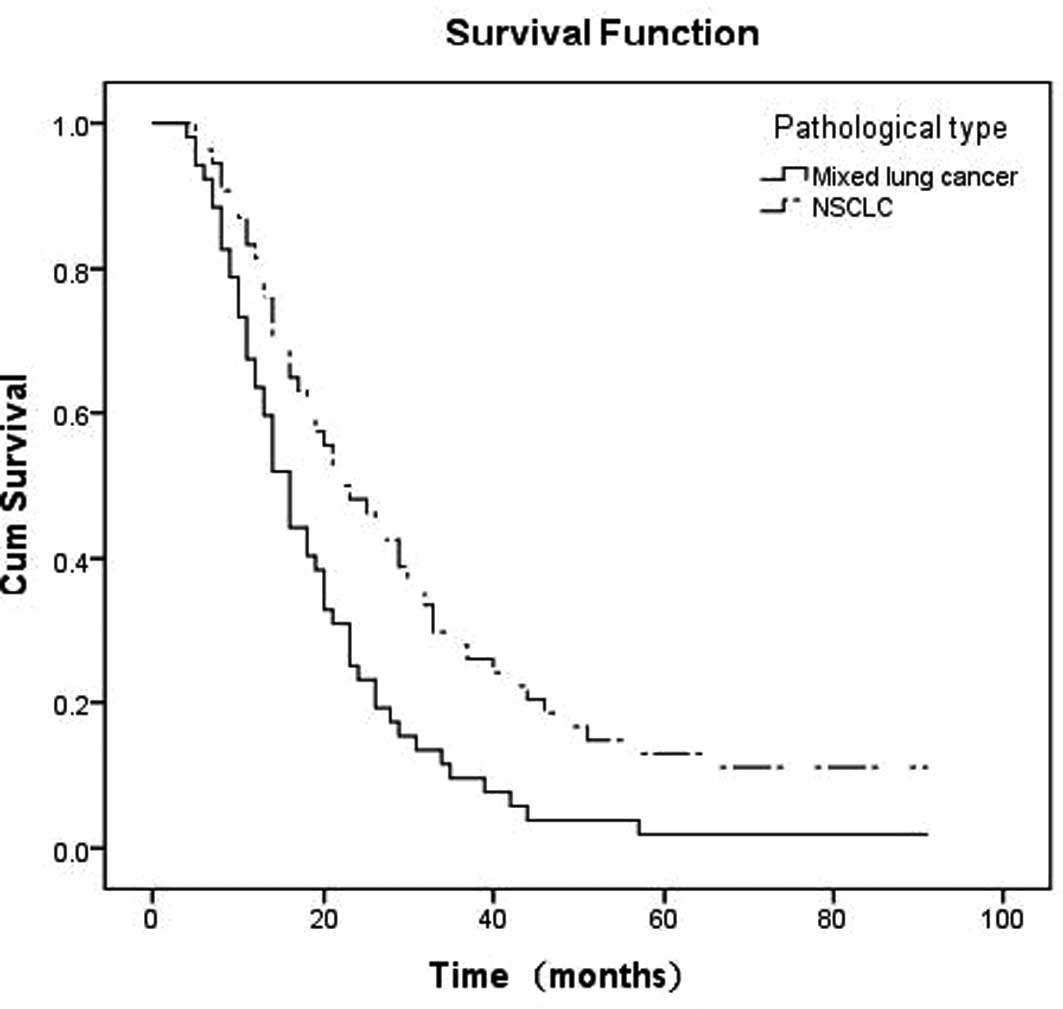
3-year survival rates of the mixed group were 63.5 (33/52), 23.1
(12/52) and 9.6% (5/52), respectively. Survival time was 3–91
months (alive). The median survival time was 15 months
(χ2 value = 7.516; P-value = 0.006) (Fig. 3). The l-, 2- and 3-year survival
rates of the NSCLC group were 81.5 (44/54), 48.1 (26/54) and 27.7%
(15/54), respectively, and the median survival time was 22 months.
The difference among the two groups was significant (P=0.006).
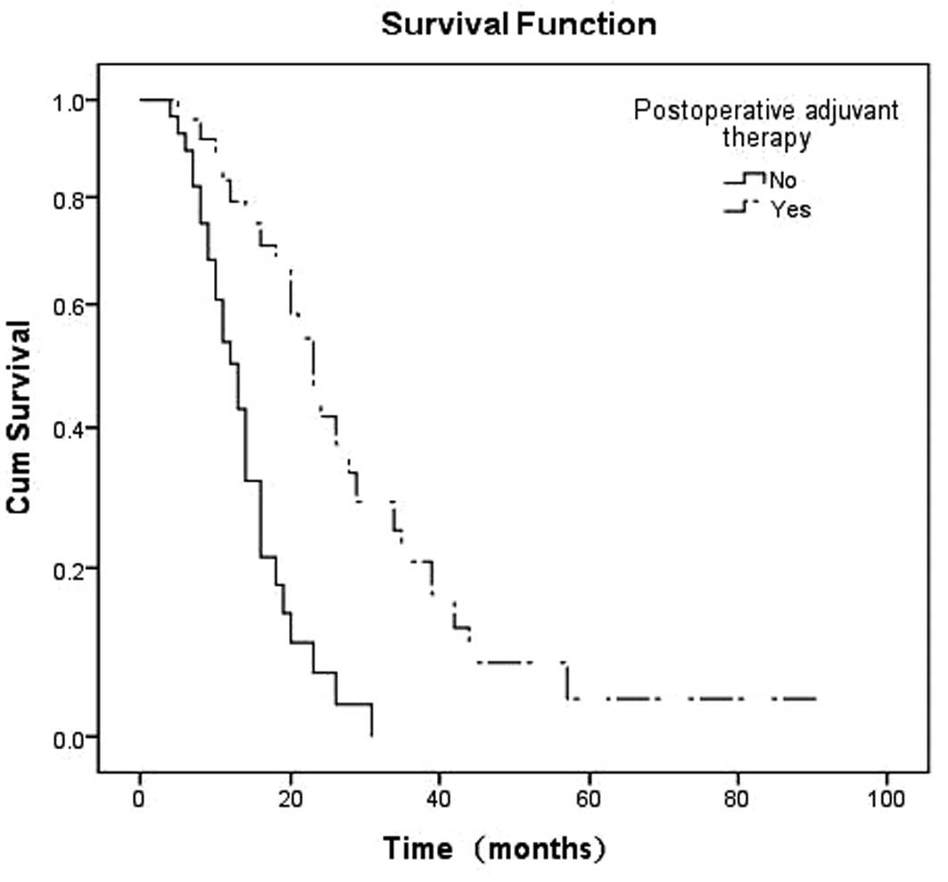
To discuss the advantages of postoperative adjuvant
therapy on survival time by survival analysis, it was found that
the median survival time of the postoperative adjuvant therapy and
the surgery alone groups were 22 and 12 months, respectively, which
was prolonged after postoperative adjuvant therapy (χ2
value = 9.640; P-value = 0.000) (Fig.
4).
Discussion
In previous studies, scientists have found
additional manifestations of the polymorphism of lung cancer in
morphology, karyotype, immunophenotype, genetic markers, growth
rate, potential of metastatic, drug sensitivity and biological
behavior (8–10), which is of great significance in
explaining basic and clinical research of lung cancer, and are
thought to deserve more attention.
In the present study, we selected 92 patients with
mixed lung cancer from 1,842 cases of primary lung cancer diagnosed
using histopathologic methods, which accounts for the 4.99% of the
total number of primary lung cancer in the same period, equal to
the data of a similar study conducted by Ruffini et al
(5–10%) (11) and a study
conducted in China (1.96–5.23%). Adenosquamous carcinoma (69/92) is
the most common pattern of mixed-histology lung cancer, followed by
adenocarcinoma combined with BA pattern (6/92). The tissue biopsies
above were NSCLC, while additional types were rare, suggesting that
most types of lung cancer with a mixed histological pattern may
behave similar to NSCLC regarding clinical characteristics and
therapeutic responses.
Patients with mixed pattern are found mainly in
middle-to-old age individuals, with an age range of 50–59 (28/92)
and 60–69 years (34/92). The incidence of mixed lung cancer was
significantly decreased after the age of 70 years. The percentage
of middle-to-old age patients with all the types of mixed-histology
lung cancer had only less squamous cell carcinoma. The gender ratio
(male:female) was 4.41:1, which was lower compared to squamous cell
carcinoma patients (7.65:1), but higher compared to adenocarcinoma
(1.72:1) and BA carcinoma patients (0.83:1), and similar to small
cell lung cancer (SCLC) patients (3.96:1) during the same time
period. In this study, the ratio of smokers to non-smokers was
2.07:1, and patients who were heavy smokers numbered 62/92, of whom
61/62 were male. As a result, mixed lung cancer may occur more
frequently in middle-to-old age, as well as in heavy and long-term
smoking men.
The apicoposterior segment of upper lobes (45/92),
basal segment of lower lobes (21/92) and epimere of lower lobes
(14/92) were involved more commonly compared to the other segments,
and all were predilection sites of lung cancer with single
histology, a fact which was similar to the study conducted by
Takamori et al(12). All 92
patients had clinical symptoms when they presented to hospital.
Cough and sputum (59/92) were the most common clinical
manifestations, followed by hemoptysis, chest pain, chest tightness
and shortness of breath. The accepted view was that key types of
lung cancer were more common in squamous cell carcinoma and small
cell carcinoma, which were mostly located in the segmental bronchi
near the hilum of the lobes, which leads to an earlier onset of
cough, sputum and hemoptysis. Adenocarcinoma were more frequently
located in the periphery of lung, which involved bronchial to a
lesser degree, and rarely manifested as early cough, sputum, in
contrast to early metastasis. Mixed lung cancer patients included
in this study exhibited no significant tendency of its location in
the central or peripheral lungs, and the majority of them exhibited
a mixed pathological type of squamous cell carcinoma and
adenocarcinoma, resulting in the presence of the biological
characteristics of both squamous cell carcinoma and adenocarcinoma.
Moreover, most of these patients were smokers and at an advanced
stage, involving small bronchi, leading to the manifestation of
symptoms including cough, expectoration and hemoptysis
symptoms.
Since the types of mixed lung cancer are
characterized by the unique mixture of pathologic features, they
have combined malignant biological characteristics of various types
of cancer, including a tendency for local invasion, lymph node
metastasis and hematogenous spread. In 1987, Naunheim et
al(13) concluded that
adenosquamous carcinoma is a subtype of non-small cell lung
carcinoma with an aggressive behavior and poor survival. Moreover,
75% of their patients were at stage III and only 9 patients
underwent surgical treatment (13). In a study conducted by Sridhar
et al(14), only 38/127
patients with adenosquamous carcinoma underwent curative resection
due to the advanced stage and locoregional or distant spread (stage
IIIa–IV). Findings of the present study are in agreement with those
of studies mentioned previously (13,14).
The mean duration of the period from the manifestation of the first
symptom until the patient’s visit to the doctor was 84.8 days. The
clinical TNM stage for diagnosis was common in the advanced stage
of IIIb–IV. Of the 92 patients, 64 patients had metastasis, which
according to severity were bone, pleura or pericardium, as well as
brain, liver and other organs. The metastatic ratio of mixed lung
cancer was equal to adenocarcinoma, but higher compared to that of
other types. As a result, mixed tumors of lung may have a high
malignancy rate.
Since no significant difference was observed in the
symptoms, signs and imaging features between lung tumors with
single- and mixed-histological pattern, it is difficult to make a
diagnosis in the clinical setting. In the present study, 56
patients were diagnosed by pathologic examination of the surgical
specimens, while 47 patients had a history of preoperative
bronchial biopsy and sputum cytology or CT/B ultrasound-guided lung
biopsy. Of the 47 patients, 32 were not diagnosed with cancer and
15 were diagnosed with single-histology lung cancer. Although the
specimens obtained by non-surgical methods had mixed patterns in
several cases in this study, the ratio of positive results was
lower compared to the surgical specimens, which provided relatively
higher positive results. Doctors in China have examined the sputum
of 2,005 cases of lung cancer patients and have only identified two
cases of lung cancer with a mixed histological pattern (15). Peking Union Medical College
Hospital have used specimens obtained from bronchoscopy of 1,315
cases of lung cancer for diagnosis, and only 1.1% had a mixed
pattern (16). Thus, different
methods of obtaining the specimen affect the detection rate of
mixed lung cancer, and the number of mixed lung cancer patients
that have been successfully diagnosed in the clinical setting
constitute a small percentage. Additionally, the heterogeneity of
lung cancer can be analyzed more prominently by using electron
microscopy and immunohistochemical analysis. Previous studies have
shown that the heterogeneity of lung cancer may reach 66% (2). Roggli et al(1) used electron microscopy to observe
specimens of 100 lung cancer cases collected by surgical resection
or bronchoscopy biopsy, which were consecutively cut into 10
slices, and found that 45% showed ≥1 mixed tissue types in addition
to major tissue types of lung cancer. Bombi et al(3) used electron microscopy to examine 110
cases of resected lung cancer, and found that 65% of cases were
single ultrastructural, 27% were adenosquamous carcinoma or
adenocarcinoma with neuroendocrine ultrastructural, and ~3% of all
the cases had three differentiation. Su et al(4) examined a large number of surgical
specimens obtained rom 98 patients with lung cancer under a light
microscope subsequent to hematoxylin and eosin (H&E) staining,
with 32 cases (32/98) revealing tissue heterogeneity. When
specimens were observed by electron microscopy and
immunohistochemical staining, the heterogeneity ratio reached
64/98. In the present study, the mixed pattern of lung cancer
accounted for only 4.99% (92/1,842) of all the primary lung cancer
cases during the same time period, significantly lower compared to
the data reported above. One reason for this finding is that 47.6%
(876/1,842) of patients in this study were diagnosed based on the
specimens collected by bronchial biopsy. However, the specimens
were small and therefore other components were easy to miss.
Additionally, all the specimens in this study were examined under
light microscopy, although few of them were investigated using
immunohistochemistry, leading to the difficulty in finding tissue
heterogeneity. The different proportions of the ingredients, the
difference of the quantity of the specimens, the different methods
employed to collect the specimens, the different locations from
which the specimens were collected and the methods used during
pathologic diagnosis caused difficulty in diagnosing mixed lung
cancer, leading to the low detection rate of this study and the
insufficient attention to mixed lung cancer in clinical study.
The prognosis of mixed lung cancer is also not
optimistic. During 1994, Hofmann et al(17) reported a 28% 3-year survival rate
with no patients with a 5-year survival in a series of 13 patients
with adenosquamous carcinoma. During 1999, Hsia et
al(18) reported a 22% 5-year
survival in 39 patients and, in 2002, Ruffini et al(11) reported a 28% 3-year survival rate
(11). The Beijing Breast Cancer
Research Center (BBCRC) has reported a resected mixed lung cancer
2-, 3- and 5-year survival rate of 25.6, 9.8 and 8.6%,
respectively, which was lower compared to that for squamous cell
carcinoma and adenocarcinoma lung cancer in single-histology and
similar to that of SCLC (19). In
the present study, 52 patients were followed-up successfully, with
l-, 2- and 3-year survival rates of 63.5, 23.1 and 9.6%,
respectively, and a median survival of 15 months, lower compared to
that reported by Hofmann et al(17), Ruffini et al(11), Hsia et al(18) and Peng (19), which was potentially due to the
differences in the number of samples, clinical stage and treatment
after resection of patients. The patients in the studies by Hofmann
et al(17), Ruffini et
al(11) and Hsia et
al(18) were mostly
administered radiotherapy or chemotherapy, or combined chemo- and
radiotherapy, while 30 patients in our study underwent surgery
alone (due to the prognosis of the disease, lack of financial
resources and patient concern regarding the side-effects of
radiotherapy or chemotherapy). The median survival of mixed lung
cancer patients was also lower compared to the survival rate and
the median survival of NSCLC patients in the present study (1-, 2-
and 3-year survival rates were 81.5, 48.1 and 27.7%, respectively,
and the median survival time was 22 months), indicating a poorer
prognosis of mixed- compared to single-histology NSCLC, which was
similar to the studies mentioned above.
For the NSCLC patients in stages I and II, active
surgical treatment was recommended. Early diagnosis and treatment
are essential to obtain satisfactory results. Patients in stage III
require a combination of surgical treatment, adjuvant radiotherapy,
chemotherapy and comprehensive treatment to prolong survival
(20,21). To understand the impact of adjuvant
therapy on prognosis in resected mixed lung cancer, we compared
postoperative adjuvant therapy group to the surgery-alone group by
survival analysis. The findings showed that the median survival
time of the two groups was 22 and 12 months, which was prolonged
subsequent to postoperative adjuvant therapy (χ2 value,
9.640; P=0.002). Similar to NSCLC, mixed lung cancer patients who
underwent surgery plus postoperative adjuvant therapy may
experience improved survival.
A comparison among the present study and other
studies indicated that mixed primary lung cancer has a unique
biological behavior, strong potential of invasion and early
metastasis, leading to rapid progression and poor prognosis.
However, insufficient attention has been given to these types of
cancer due to the limitation of the current methods applied in the
clinical setting, rendering the level of pathological diagnosis of
mixed lung cancer significantly lower in value. Therefore,
pathological examination of large sample size and use of electron
microscopy and immunohistochemical methods should be promoted to
improve the diagnosis of mixed lung cancer. Furthermore, the
survival and prognosis of mixed lung cancer were lower compared to
NSCLC. Consequently, early diagnosis together with treatment by
surgery plus adjuvant therapy should be investigated for the
satisfactory prognosis of mixed lung cancer patients.
References
|
1
|
Roggli VL, Vollmer RT, Greenberg SD,
McGavran MH, Spjut HJ and Yesner R: Lung cancer heterogeneity: a
blinded and randomized study of 100 consecutive cases. Hum Pathol.
16:569–579. 1985. View Article : Google Scholar : PubMed/NCBI
|
|
2
|
Anami Y, Takeuchi T, Mase K, Yasuda J,
Hirohashi S, Perucho M and Noguchi M: Amplotyping of
microdissected, methanol-fixed lung carcinoma by arbitrarily primed
polymerase chain reaction. Int J Cancer. 89:19–25. 2000. View Article : Google Scholar : PubMed/NCBI
|
|
3
|
Bombi JA, Martinez A, Ramirez J, Grau JJ,
Nadal A, Fernandez PL, et al: Ultrastructural and molecular
heterogeneity in non-small cell lung carcinomas: study of 110 cases
and review of the literature. Ultrastruct Pathol. 26:211–218. 2002.
View Article : Google Scholar : PubMed/NCBI
|
|
4
|
Su XD, Wu QL, Zhang X, Zhu ZH, Zhang DK,
Cao Y, et al: The histological heterogeneity of non-small cell lung
cancer. Zhong Liu Xue Za Zhi. 14:877–880. 2008.(In Chinese).
|
|
5
|
World Health Organization. The World
Health Organization histological typing of lung tumours. Second
edition. Am J Clin Pathol. 77:123–136. 1982.PubMed/NCBI
|
|
6
|
Goldstraw P: IASLC Staging Manual in
Thoracic Oncology. Editorial Rx Press; Orange Park, FL: 2009
|
|
7
|
Kaplan EL and Meier P: Nonparametric
estimation from incomplete observations. J Am Stat Assoc.
53:457–481. 1958. View Article : Google Scholar
|
|
8
|
Fidler IJ: Tumor heterogeneity and the
biology of cancer invasion and metastasis. Cancer Res.
38:2651–2660. 1978.PubMed/NCBI
|
|
9
|
Heppner GH: Tumor heterogeneity. Cancer
Res. 44:2259–2265. 1984.PubMed/NCBI
|
|
10
|
Bryne M: Prognostic value of various
molecular and cellular features in oral squamous cell carcinomas: a
review. J Oral Pathol Med. 20:413–420. 1991. View Article : Google Scholar : PubMed/NCBI
|
|
11
|
Ruffini E, Rena O, Oliaro A, Filosso PL,
Bongiovanni M, Arslanian A, et al: Lung tumors with mixed
histologic pattern. Clinico-pathologic characteristics and
prognostic significance. Eur J Cardiothorac Surg. 22:701–707. 2002.
View Article : Google Scholar : PubMed/NCBI
|
|
12
|
Takamori S, Noguchi M, Morinaga S, Goya T,
Tsugane S, Kakegawa T and Shimosato Y: Clinicopathologic
characteristics of adenosquamous carcinoma of the lung. Cancer.
67:649–654. 1991. View Article : Google Scholar : PubMed/NCBI
|
|
13
|
Naunheim KS, Taylor JR, Skosey C, Hoffman
PC, Ferguson MK, Golomb HM and Little AG: Adenosquamous lung
carcinoma: clinical characteristics, treatment, and prognosis. Ann
Thorac Surg. 44:462–466. 1987. View Article : Google Scholar : PubMed/NCBI
|
|
14
|
Sridhar KS, Bounassi MJ, Raub W Jr and
Richman SP: Clinical features of adenosquamous lung carcinoma in
127 patients. Am Rev Respir Dis. 142:19–23. 1990. View Article : Google Scholar : PubMed/NCBI
|
|
15
|
Liu LN and Yang YH: Cytology analysis of
sputum in 2005 cases. Shi Yong Zhong Liu Xue Za Zhi. 8:15–16.
1994.(In Chinese).
|
|
16
|
Zhong W, Wang M, Chen Y, Zhang L and Li L:
Application of bronchoscopy in the diagnosis of lung cancer. Oncol
Prog. 7:308–313. 2009.
|
|
17
|
Hofmann HS, Knolle J and Neef H: The
adenosquamous lung carcinoma: clinical and pathological
characteristics. J Cardiovasc Surg (Torino). 35:543–547.
1994.PubMed/NCBI
|
|
18
|
Hsia JY, Chen CY, Hsu CP, Shai SE and Wang
PY: Adenosquamous carcinoma of the lung. Surgical results compared
with squamous cell and adenocarcinoma. Scand Cardiovasc J.
33:29–32. 1999. View Article : Google Scholar : PubMed/NCBI
|
|
19
|
Peng YS: Surgical treatment of
adenosquamous carcinoma of the lung - analysis of 223 cases.
Zhonghua Jie He He Hu Xi Za Zhi. 14:165–167. 1991.(In Chinese).
|
|
20
|
Gawrychowski J, Brulinski K, Malinowski E
and Papla B: Prognosis and survival after radical resection of
primary adenosquamous lung carcinoma. Eur J Cardiothorac Surg.
27:686–692. 2005. View Article : Google Scholar : PubMed/NCBI
|
|
21
|
Birim O, Kappetein AP, van Klaveren RJ and
Bogers AJ: Prognostic factors in non-small cell lung cancer
surgery. Eur J Surg Oncol. 32:12–23. 2006. View Article : Google Scholar : PubMed/NCBI
|