Introduction
Excessive estrogen exposure is a critical risk
factor for breast cancer (1).
While the ovary is the major site for estrogen biosynthesis in
premenopausal females, adipose stromal cells (ASCs) in the breast
are a significant source for local estrogen production. Estrogens
produced in distal adipose tissues and within the breast tissues
affect the growth of breast epithelial cells (2). Notably, excessive local estrogen
production in the breast promotes estrogen-dependent breast cancer.
At the molecular level, tumor cell-derived soluble factors,
including cytokines and prostaglandin E2, stimulate the
stromal expression of aromatase, a key enzyme in estrogen
biosynthesis (3). Breast quadrants
bearing malignant tumors consistently exhibit high levels of
aromatase activity (4), and breast
adipose tissue adjacent to the tumor has a marked increase in
aromatase expression and activity (5–7). The
clinically proven efficacy of aromatase inhibitors (AIs) in
treating estrogen receptor-positive post-menopausal breast cancer
indicates the important role of excessive local estrogen production
in breast cancer development.
The transcription of the aromatase gene is
controlled by a number of tissue and cell type-specific promoters
that are located upstream of the aromatase coding region. In
cancer-free breast adipose tissue, aromatase mRNA contributions are
mainly from the relatively weak I.4 promoter, with a small amount
of aromatase mRNA arising from the ovary-specific promoters, I.3
and PII. However, in ASCs adjacent to breast tumors, aromatase
expression is activated by the proximally-located promoters, I.3
and PI (2). The switch in promoter
utilization from weak I.4 to strong I.3 and PII promoters results
in elevated aromatase expression and excessive production of local
estrogen (8–9).
It has been shown that mechanical changes, including
elevated extracellular matrix (ECM) stiffness and increased
interstitial pressures, are associated with epithelial carcinomas.
Furthermore, mechanical force due to altered architecture in the
tissue microenvironment can affect the gene expression pattern
(10–14). In the case of breast cancer,
mechanical force significantly affects the invasive behaviors of
breast tumor cells, as well as breast cancer incidence and
mortality (15–18). The current study, aimed to assess
whether ASCs from varying individuals have differential induction
levels of aromatase expression responding to mechanical force.
Materials and methods
Cell culture
Primary human ASCs were isolated from individuals
undergoing elective surgical procedures at the University of
Virginia (Charlottesville, VA, USA), using methods previously
published and approved by the University of Virginia’s Human
Investigation Committee (21). The
cells were cultured in Dulbecco’s modified Eagle’s medium/F12
medium (Gibco, Big Cabin, OK, USA) with 10% fetal bovine serum
(HyClone, Lawrenceville, GA, USA) and 1% antibiotic-antimycotic
solution (Gibco), using these previously described methods.
Forskolin was purchased from Sigma-Aldrich (St. Louis, MO, USA) and
used at a final concentration of 25 μM. 5-Aza-2′-deoxycytidine
(AZA; Sigma-Aldrich) was used at a final concentration of 10
μM.
3D cultures were conducted in 24-well plates using
collagen (collagen bovine type I; BD Bioscience, Franklin Lakes,
NJ, USA). Briefly, 2×10⁵ cells were suspended in 125 μl
medium and mixed with 125 μl collagen. Following gel-like 3D
structure formation, medium was added to the top.
Aromatase activity assay
Aromatase activity was measured using a tritiated
water-release assay as previously described (22). Aromatase
activity was determined by the rate of conversion of
(1β-3H)-androstenedione to estrone by aromatase. The quantity of 3H
in extracts of medium was determined by liquid scintillation
counting.
DNA methylation assay
Genomic DNA was obtained using the GenElute
Mammalian Genomic DNA Miniprep kit from Sigma-Aldrich. BiSulfite
conversion of genomic DNA was performed using the EpiTect Bisulfite
kit (Qiagen, Hilden, Germany). PCR, TOPO TA cloning and sequencing
(Beckman Coulter Genomics, Danvers, MA, USA) were performed.
Quantitative and semi-quantitative
reverse transcription PCR (RT-PCR)
Total RNA was isolated using TRIzol reagent
(Invitrogen Life Technologies, Carlsbad, CA, USA) according to the
manufacturer’s instructions. The concentration of RNA was measured
and the RNA was reverse-transcribed using the ImPrompII kit
(Promega Corporation, Madison, WI, USA). Real-time PCR was
performed using the SYBR-Green fluorescent dye and an ABI7900
Real-Time PCR System (Applied Biosystems, Foster City, CA, USA).
The forward primer was 5′-TGGAATTATGAGGGCACATCC-3′ and the reverse
primer was 5′-GTCCAATTCCCATGCAGTAGC-3′. Semi-quantitative RT-PCR
was performed using pairs of primers for aromatase transcripts that
are specific from promoters I.4, I.3 or PII. GAPDH served as
internal control. The PCR conditions were as follows: 94°C for 2
min, 94°C for 30 sec, 56°C for 30 sec, 72°C for 1 min (35 cycles),
72°C for 10 min and then a hold at 4°C. The forward primer sequence
for promoter I.3 was 5′-CCTTGTTTTGACTTGTAACCA-3′, for promoter I.4
was 5′-GTAGAACGTGACCAACTGG-3′ and for promoter II was
5′-GCAACAGGAGCTATAGAT-3′. The reverse primer sequence for all three
promoters was 5′-ATT CCCATGCAGTAGCCAGG-3′. The GAPDH forward primer
sequence was 5′-CCATCAATGACCCCTTCATTG-3′ and the reverse primer
sequence was 5′-GACGGTGCCATGGAATTT-3′.
Statistical and data analysis
A paired t-test was used to analyze pairwise
comparisons of collagen 3D-cultured cells with control cells. Data
from independent measurements were collected for analysis.
P<0.05 was considered to indicate a statistically significant
difference.
Results
Differential expression of mechanical
force-induced aromatase in ASCs of different individuals
The aim of the current study was to determine
whether individuals respond to mechanical force differently in
terms of aromatase induction.
Stromal cells were isolated from the fat tissue
excised from cancer-free individuals. Once growing confluently in
regular growth medium, the ASCs were seeded in a 2D or collagen 3D
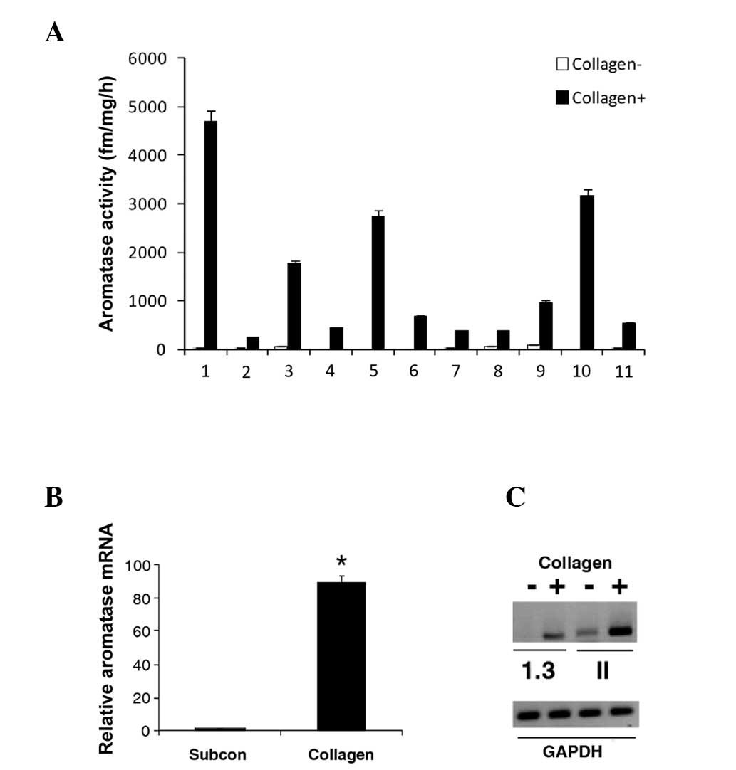
system. The data of the aromatase activity assay showed that there
was differential expression of mechanical force-induced aromatase
in the different individuals (Fig.
1A). Aromatase mRNA was induced in response to mechanical force
(Fig. 1B). In addition, the
induction of aromatase expression was regulated by promoter I.3/PII
(Fig. 1C).
Higher DNA methylation load of certain
CpG sites of the PII promoter corresponds to the lower aromatase
activation
Next, the mechanism of differential expression in
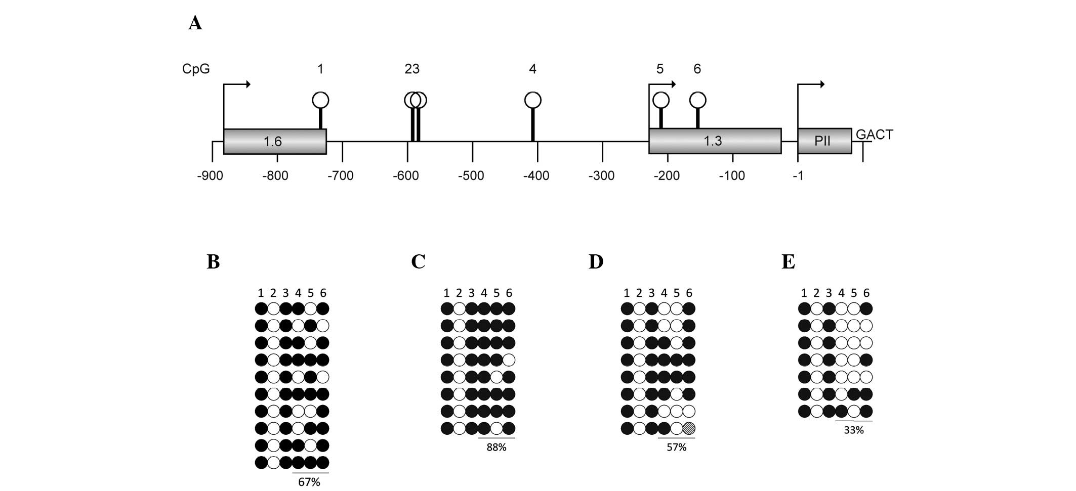
various individuals was determined. A DNA methylation assay was
performed to test the methylation status of aromatase promoter PII
(Fig. 2A). The differential
induction of aromatase expression is associated with the DNA
methylation load of the promoter region. The higher DNA methylation
load of PII promoter is associated with the lower aromatase
activation (Fig. 2B–E).
Reduction of DNA methylation load by AZA
treatment restores aromatase activation
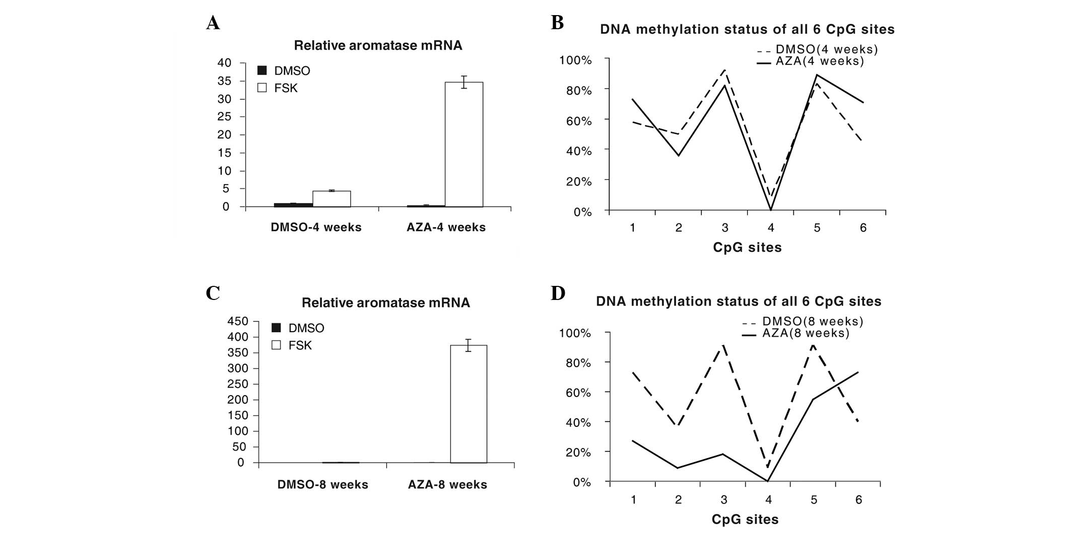
The cells were treated with AZA for 4 (Fig. 3A and B) or 8 weeks (Fig. 3C and D). Aromatase expression and
DNA methylation status were analyzed. Along with reduction of the
DNA methylation load, aromatase activation was restored.
Discussion
The current literature on the mechanical properties
of tumors is almost exclusively focused on tumor cells (10,12).
As altered mechanical homeostasis in breast tumors affects the
epithelial and stromal compartments of the same tumor
microenvironment, it is necessary to look beyond the ‘box’ of tumor
cells by examining the impact of mechanical forces on the
surrounding stroma. Furthermore, ASCs from different individuals
may respond to mechanical forces differently, thus, individuals
respond differently to risk factors of breast cancer. The current
study data showed that there was differential expression of
mechanical force-induced aromatase in differing individuals. It
also showed that mechanical force activates the PII promoter and
that the DNA methylation status of the PII promoter plays an
important role in aromatase induction when ASCs respond to
mechanical forces.
The efficacy of AIs in treating breast cancer has
been clinically proven. However, AIs indiscriminately reduce
estrogen synthesis throughout the body, causing major side-effects,
including bone loss, increased fracture rates and abnormal lipid
metabolism (21). Thus, it is
worthwhile to develop inhibitors that selectively block aromatase
and estrogen production in breast cancer. By contrast, aromatase
I.3/PII promoters have been reported to be activated in tumors, but
not in normal tissues (2).
Logically, the specific inhibition of signals that lead to
activation of promoter I.3/II is likely to inhibit aromatase
expression specifically in tumor tissues. The current study data
enhance our understanding of the regulation of aromatase expression
in an epigenetic manner. These findings may lead to the
identification of novel AIs.
Acknowledgements
This study was supported by the Science and
Technology Project Foundation, Science and Technology Commission,
Guangdong Province, P.R. China (grant no. 2010B031600147).
References
|
1
|
Nass SJ and Davidson NE: The biology of
breast cancer. Hematol Oncol Clin North Am. 13:311–332. 1999.
View Article : Google Scholar
|
|
2
|
Bulun SE, Lin Z, Imir G, et al: Regulation
of aromatase expression in estrogen-responsive breast and uterine
disease: from bench to treatment. Pharmacol Rev. 57:359–383. 2005.
View Article : Google Scholar : PubMed/NCBI
|
|
3
|
Simpson ER and Davis SR: Minireview:
aromatase and the regulation of estrogen biosynthesis - some new
perspectives. Endocrinology. 142:4589–4594. 2001.PubMed/NCBI
|
|
4
|
O’Neill JS, Elton RA and Miller WR:
Aromatase activity in adipose tissue from breast quadrants: a link
with tumour site. Br Med J (Clin Res Ed). 296:741–743.
1988.PubMed/NCBI
|
|
5
|
Bulun SE, Price TM, Aitken J, Mahendroo MS
and Simpson ER: A link between breast cancer and local estrogen
biosynthesis suggested by quantification of breast adipose tissue
aromatase cytochrome P450 transcripts using competitive polymerase
chain reaction after reverse transcription. J Clin Endocrinol
Metab. 77:1622–1628. 1993. View Article : Google Scholar
|
|
6
|
Harada N: Aberrant expression of aromatase
in breast cancer tissues. J Steroid Biochem Mol Biol. 61:175–184.
1997. View Article : Google Scholar : PubMed/NCBI
|
|
7
|
Sasano H and Harada N: Intratumoral
aromatase in human breast, endometrial, and ovarian malignancies.
Endocr Rev. 19:593–607. 1998.PubMed/NCBI
|
|
8
|
Meng L, Zhou J, Sasano H, Suzuki T,
Zeitoun KM and Bulun SE: Tumor necrosis factor alpha and
interleukin 11 secreted by malignant breast epithelial cells
inhibit adipocyte differentiation by selectively down-regulating
CCAAT/enhancer binding protein alpha and peroxisome
proliferator-activated receptor gamma: mechanism of desmoplastic
reaction. Cancer Research. 61:2250–2255. 2001.
|
|
9
|
Zhou J, Gurates B, Yang S, Sebastian S and
Bulun SE: Malignant breast epithelial cells stimulate aromatase
expression via promoter II in human adipose fibroblasts: an
epithelial-stromal interaction in breast tumors mediated by
CCAAT/enhancer binding protein beta. Cancer Research. 61:2328–2334.
2001.
|
|
10
|
Avvisato CL, Yang X, Shah S, Hoxter B, Li
W, Gaynor R, Pestell R, Tozeren A and Byers SW: Mechanical force
modulates global gene expression and beta-catenin signaling in
colon cancer cells. J Cell Sci. 120:2672–2682. 2007. View Article : Google Scholar : PubMed/NCBI
|
|
11
|
Shyy JY and Chien S: Role of integrins in
endothelial mechanosensing of shear stress. Circ Res. 91:769–775.
2002. View Article : Google Scholar : PubMed/NCBI
|
|
12
|
Alenghat FJ and Ingber DE:
Mechanotransduction: all signals point to cytoskeleton, matrix, and
integrins. Sci STKE. 2002:pe62002.PubMed/NCBI
|
|
13
|
Lopez JI, Mouw JK and Weaver VM:
Biomechanical regulation of cell orientation and fate. Oncogene.
27:6981–6993. 2008. View Article : Google Scholar : PubMed/NCBI
|
|
14
|
Nelson CM and Bissell MJ: Of extracellular
matrix, scaffolds, and signaling: tissue architecture regulates
development, homeostasis, and cancer. Annu Rev Cell Dev Biol.
22:287–309. 2006. View Article : Google Scholar : PubMed/NCBI
|
|
15
|
Paszek MJ and Weaver VM: The tension
mounts: mechanics meets morphogenesis and malignancy. J Mammary
Gland Biol Neoplasia. 9:325–342. 2004. View Article : Google Scholar : PubMed/NCBI
|
|
16
|
Ingber DE: Can cancer be reversed by
engineering the tumor microenvironment? Semin Cancer Biol.
18:356–364. 2008. View Article : Google Scholar : PubMed/NCBI
|
|
17
|
Le Beyec J, Xu R, Lee SY, Nelson CM, Rizki
A, Alcaraz J and Bissell MJ: Cell shape regulates global histone
acetylation in human mammary epithelial cells. Exp Cell Res.
313:3066–3075. 2007.PubMed/NCBI
|
|
18
|
Wiseman BS and Werb Z: Stromal effects on
mammary gland development and breast cancer. Science.
296:1046–1049. 2002. View Article : Google Scholar : PubMed/NCBI
|
|
19
|
Katz AJ, Tholpady A, Tholpady SS, Shang H
and Ogle RC: Cell surface and transcriptional characterization of
human adipose-derived adherent stromal (hADAS) cells. Stem Cells.
23:412–423. 2005. View Article : Google Scholar : PubMed/NCBI
|
|
20
|
Yue W and Brodie AM: Mechanisms of the
actions of aromatase inhibitors 4-hydroxyandrostenedione,
fadrozole, and aminoglutethimide on aromatase in JEG-3 cell
culture. J Steroid Biochem Mol Biol. 63:317–328. 1997. View Article : Google Scholar : PubMed/NCBI
|
|
21
|
Morales L, Neven P and Paridaens R:
Choosing between an aromatase inhibitor and tamoxifen in the
adjuvant setting. Curr Opin Oncol. 17:559–565. 2005. View Article : Google Scholar : PubMed/NCBI
|


