Introduction
The adult heart is no longer considered a terminally
differentiated organ, due to the presence of cardiac stem cells
(CSCs) (1). CSCs have been
considered a prospective cell source for myocardial repair
following acute myocardial infarction (AMI) due to their
self-renewing properties, and their multipotent ability to
differentiate into cardiomyocytes and endothelial cells (2,3).
CSCs are positive for several stem cell antigens, including stem
cell antigen (Sca)-1 and c-Kit (4). CSC-based therapy may be considered a
novel strategy to repair infarcted myocardium post-AMI; however,
this method is limited by low regenerative efficiency, due to
insufficient cell viability and a high rate of apoptosis following
transplantation (5).
AMI is associated with inflammation and may result
in the release of high concentrations of cytokines and inflammatory
factors. These molecules may act as chemotactic factors that induce
the homing of several progenitor cells into the infarcted areas to
participate in myocardial repair (6,7). The
paracrine ability of transplanted cells is considered an important
factor in the improvement of cardiac function (8,9).
Previous studies have indicated that the predominant effect of
mesenchymal stem cells in myocardial repair was associated with the
release of several cytokines and chemotactic factors involved in
neovascularization or chemotaxis, including vascular endothelial
growth factor (VEGF) and stromal derived factor (SDF)-1α (10,11).
However, whether transplanted CSCs may also act in the same way and
increase the release of VEGF and SDF-1α, resulting in the induction
of microvascular sprouting, the formation of new blood vessels and
an increase in progenitor cell homing remains unknown.
Resveratrol, which is a compound extracted from red
wine, is considered a strong antioxidative and protective molecule
in the cardiovascular system (12). A previous in vitro study
demonstrated that resveratrol was able to reduce oxidative stress
in several cell types, including endothelial cells, smooth muscle
cells, cardiomyocytes and macrophages (13). Besides its antioxidative effects,
resveratrol has been reported to exert cardioprotective effects
against ischemia-reperfusion injury in animal models. Pretreatment
of AMI rats with resveratrol resulted in an increase in microvessel
density, and preservation of left ventricle (LV) function and blood
flow (14,15). In addition, resveratrol has been
shown to increase the protein levels of VEGF, angiotensin II and
their receptors, in order to fulfill its angiogenic effect
(16). The angiogenic effect of
resveratrol was achieved through increasing the bioviability and
production of nitrogen monoxide, in order to modulate the VEGF
signaling pathway (17).
Furthermore, upregulation of VEGF may directly influence the
expression of SDF-1α and CXC chemokine receptor type 4 (CXCR-4),
which are expressed on the surface of several types of stem cells,
and are required for signal transduction in stem cell-associated
chemotaxis, migration and homing (18,19).
Therefore, the present study hypothesized that
pretreatment with resveratrol may influence the angiogenic and
homing ability of CSCs via the VEGF/SDF-1α pathway, and may improve
their transplantation efficiency following AMI. An AMI mouse model
was generated and the effects of CSC regenerative therapy with
resveratrol pretreatment were determined.
Materials and methods
Ethics statement
The present study was approved by the Soochow
University Scientific and Animal Ethics Committee (Suzhou, China)
and it was conducted in compliance with the Chinese national
regulations on the use of experimental animals. Procedures for the
animal studies were performed in accordance with the Guide for the
Care and Use of Laboratory Animals published by the US National
Institutes of Health (revised in 1996). All animals were purchased
from the Laboratory Animal Center of Soochow University. They were
maintained on a standard diet with access to water, under a 12-h
light/dark cycle at the Animal Center of the First Affiliated
Hospital of Soochow University (Suzhou, China).
CSCs culture and resveratrol
administration
CSCs were isolated from hearts harvested from male
C57/BL6 mice (age, 5 weeks; weight, 10–15 g; two groups n=11 per
group) with 0.1% collagenase B (Sigma-Aldrich; Merck Millipore,
Darmstadt, Germany) and 0.2% trypsin (Invitrogen; Thermo Fisher
Scientific, Inc., Waltham, MA, USA) followed euthanasia with carbon
dioxide. Cells were labeled with antigen-presenting cell-conjugated
anti-Sca-1 (cat. no. 130092529; Miltenyi Biotec, Inc., Auburn, CA,
USA) and were separated using a magnetic selection system employing
anti-antigen-presenting microbeads (Miltenyi Biotec, Inc.).
Selected cells were cultured in Dulbecco's modified Eagle's
medium/F12 supplemented with 10% fetal bovine serum (Gibco; Thermo
Fisher Scientific, Inc.), 10 ng/ml basic fibroblast growth factor,
10 ng/ml leukemia inhibitory factor, 10 ng/ml cardiotroponin, 10
ng/ml epidermal growth factor (PeproTech, Rocky Hill, NJ, USA) and
a 1% antibiotic solution of penicillin and streptomycin (Hyclone;
GE Healthcare, Logan, UT, USA). The C57/BL6 mice (age, 5 weeks, two
groups; n=8 per group) were treated with 2.5 mg/kg.d resveratrol
(Baile Company, China) by intragastric administration with a
stomach needle. The drug was dissolved in 0.5 ml PBS. The control
group received the same volume of PBS. All mice (age, 5 weeks)
received intragastric administration of resveratrol or PBS. Some
(n=3 per group) were sacrificed in order to quantify CSCs after 7
days. Other mice (n=8 per group) underwent AMI construction and
cell transplantation on day 7 and continued to be treated with
administration of resveratrol or PBS for 4 weeks.
Measurement of CSCs after resveratrol
pretreatment
A total of 7 days after resveratrol administration,
mice were sacrificed with CO2 frozen cardiac tissue
specimens (n=3/group) were obtained and sliced into 2 µm sections
to quantify the number of Sca-1+ CSCs in the myocardium using an
anti-Sca-1 immunofluorescent antibody (1:50; eBioscience, Inc., San
Diego, CA, USA; cat. no. 11-5981-81). Positive cells were stained
red and were counted in 10 random 100x fields using a fluorescence
microscope. The average number was obtained for statistical
analysis.
AMI construction and cell
transplantation
An AMI model was constructed by surgically ligating
the left anterior descending coronary artery (LAD) with a prolene
suture. Successful ligation was verified by observation of a color
change from red to white in the infarct area. C57/BL6 mice (age, 6
weeks; weight, 15–20 g) were randomly assigned into two groups
(n=8/group), which received either PBS or 2.5 mg/kg.d resveratrol
via intragastric administration for 7 days prior to and 4 weeks
after LAD ligation. All mice received an injection with 1×10⁶ CSCs
into the peri-ischemic area and were sacrificed with CO2
4 weeks after cell transplantation for histological analysis.
Assessment of cardiac function with
echocardiography
A total of 4 weeks after LAD ligation, transthoracic
echocardiography (SONOS 5500, Philips Medical Systems International
BV, Eindhoven, The Netherlands) was used to evaluate the cardiac
function of experimental mice by an observer blinded to the
experiment. LV end-diastolic diameter (LVEDD), LV end-systolic
diameter (LVESD), interventricular septal thickness in diastole
(IVST), LV posterior wall thickness (LVPWT) and the percentage of
LV fractional shortening (FS) were detected. All measurements were
repeated for at least three consecutive pulsation cycles and the
data were averaged for statistical analysis.
Capillary density assessment
Cluster of differentiation (CD)31 immunofluorescent
staining was conducted to determine capillary density. Frozen
tissue specimens of the infarcted area were sliced into 2 µm
sections and were labeled with rat anti-mouse CD31 (1:800; Abcam,
Cambridge, MA, USA; cat. no. ab8365) and Alexa Fluor 594 anti-rat
immunoglobulin G (Invitrogen; Thermo Fisher Scientific, Inc.; cat.
no. SA000064). CD31-positive cells were stained red and the number
of capillaries was counted using Image Pro Plus Software version
6.0 (Media Cybernetics, Inc., Rockville, MD, USA) in 10 random 100x
fields using a fluorescence microscope and averaged for statistical
analysis.
Terminal deoxynucleotidyl
transferase-mediated dUTP nick end labeling (TUNEL) assay
The present study detected cardiomyocyte apoptosis
in the peri-ischemic myocardium using the TUNEL assay (Promega
Corporation, Madison, WI, USA), according to the manufacturer's
protocol. Nuclei of apoptotic cardiomyocytes were stained dark
brown. The number of TUNEL-positive cardiomyocyte nuclei per 200x
field was counted and used for statistical analysis.
Western blot analysis
Cardiac tissues were lysed using lysis buffer with
phosphatase inhibitor (Roche Diagnostics, Basel, Switzerland). A
bicinchoninic acid protein assay kit (Pierce; Thermo Fisher
Scientific, Inc.) was used to determine protein concentrations.
Proteins (30 µg samples) were separated by 10% SDS-PAGE and were
transferred to a polyvinylidene fluoride membrane (EMD Millipore,
Billerica, MA, USA). The membrane was blocked with 5% nonfat dried
milk dissolved in Tris-buffered saline containing 0.1% Tween-20 at
room temperature for 2 h. Subsequently, the membrane was incubated
with primary antibodies against VEGF (1:2,000; Abcam; cat. no.
ab2350) and SDF-1α (1:1,500; Abcam; cat. no. ab9797) overnight at
4°C, followed by an incubation with peroxidase-conjugated secondary
antibodies for 2 h at room temperature. Bands were detected using a
chemiluminescent western blot detection system (Pierce; Thermo
Fisher Scientific, Inc.). Protein expression was normalized to
GAPDH. Assays were repeated three times and band intensities were
quantified using the Photo-Image system (Siemens AG, Munich,
Germany).
Statistical analysis
Data are presented as the mean ± standard deviation
and were analyzed using SPSS 17.0 statistical software (SPSS, Inc.,
Chicago, IL, USA). Comparisons between two groups were performed
using an unpaired Student's t-test. P<0.05 was considered to
indicate a statistically significant difference.
Results
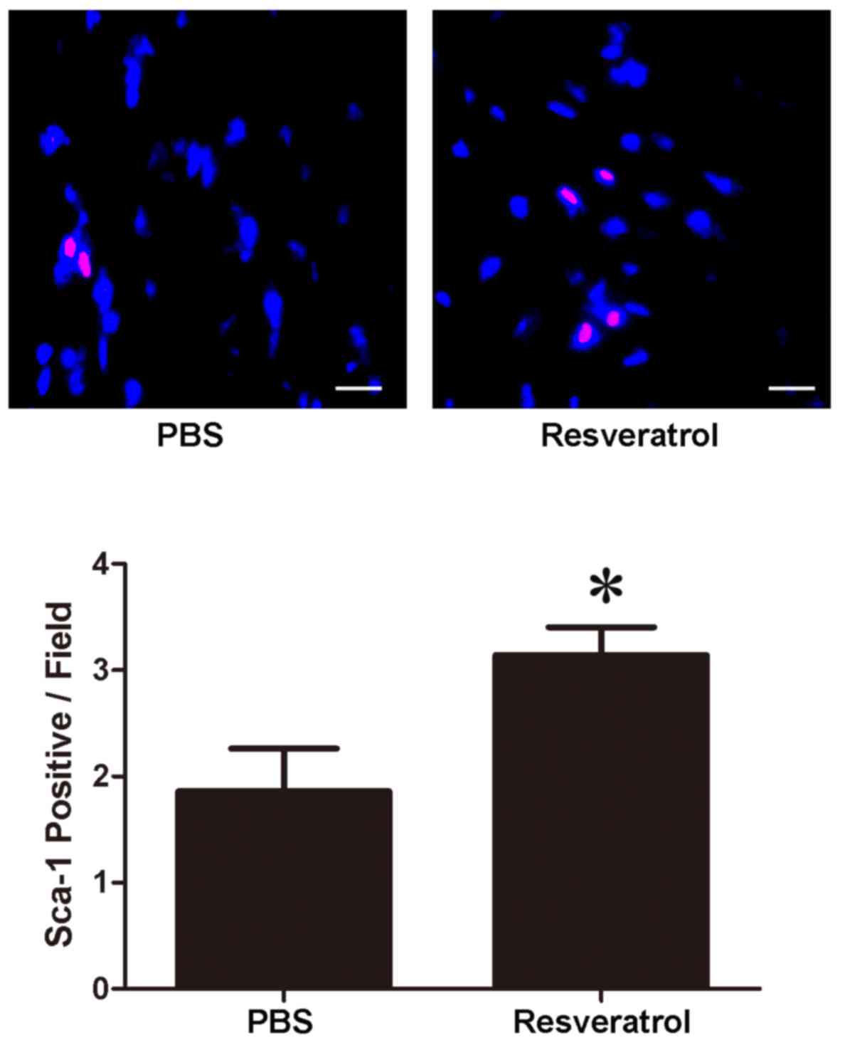
Pretreatment with resveratrol
increases the number of Sca-1+ CSCs in myocardium
The present study examined the number of Sca-1+ CSCs
following resveratrol pretreatment. Sca-1+ CSCs were stained red
and the nuclei were stained blue. Pretreatment with resveratrol for
7 consecutive days resulted in an increase in the number of Sca-1+
CSCs in the myocardium (PBS vs. resveratrol, 1.85±0.41/field vs.
3.14±0.26/field, P<0.05, Fig.
1).
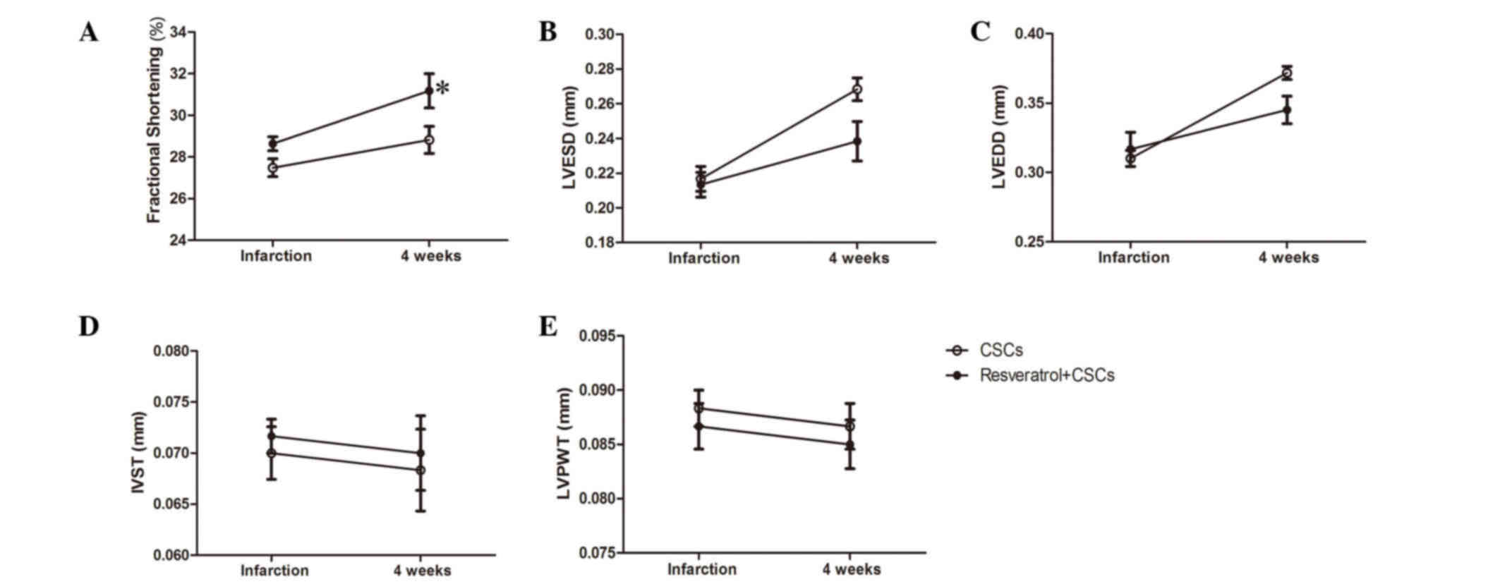
CSCs combined with resveratrol protect
cardiac function post-AMI
A total of 4 weeks after LAD ligation, cardiac
function of the mice was evaluated by echocardiography; the results
are presented in Fig. 2. CSCs
transplantation combined with resveratrol administration exerted a
protective effect on mice, as compared with CSCs transplantation
only. Resveratrol and CSCs transplantation preserved LV FS, and
reduced LVEDD and LVESD (CSCs vs. resveratrol + CSCs, P<0.05,
Fig. 2). The detailed data are
presented in Table I.
 | Table I.Echocardiography parameters following
acute myocardial infarction. |
Table I.
Echocardiography parameters following
acute myocardial infarction.
| Group | %FS | LVESD (mm) | LVEDD (mm) | IVST (mm) | LVPWT (mm) |
|---|
| CSCs
(baseline) | 27.48±1.05 | 0.21±0.02 | 0.31±0.01 | 0.07±0.006 | 0.09±0.004 |
| Res + CSCs
(baseline) | 28.64±0.83 | 0.21±0.02 | 0.32±0.03 | 0.07±0.004 | 0.09±0.005 |
| CSCs (4w) | 28.82±1.58 | 0.26±0.01 | 0.37±0.01 | 0.07±0.009 | 0.09±0.005 |
| Res + CSCs
(4w) |
31.18±2.02a |
0.23±0.02a |
0.35±0.02a | 0.07±0.008 | 0.09±0.005 |
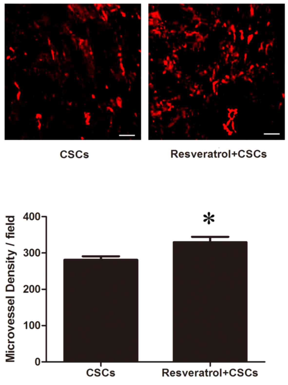
CSCs combined with resveratrol
increase capillary density in infarcted myocardium
CD31 immunofluorescent staining was used to measure
capillary density in the infarcted myocardium. As presented in
Fig. 3, capillaries were stained
red. A total of 4 weeks after LAD ligation, an increased number of
CD31-positive cells was detected in the resveratrol-treated
myocardium (CSCs vs. resveratrol + CSCs, 281.02±24.08/field vs.
329.75±36.69/field, P<0.05).
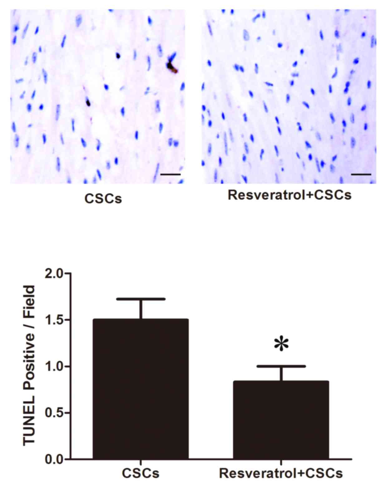
CSCs combined with resveratrol reduce
cardiomyocyte apoptosis
Cardiomyocyte apoptosis was measured by TUNEL assay.
TUNEL-positive cells were stained brown. As presented in Fig. 4, the number of TUNEL-positive cells
in the peri-ischemic area was reduced in the resveratrol + CSCs
group compared with in the CSCs group (CSCs vs. resveratrol + CSCs,
1.5±0.54/field vs. 0.83±0.40/field, P<0.05).
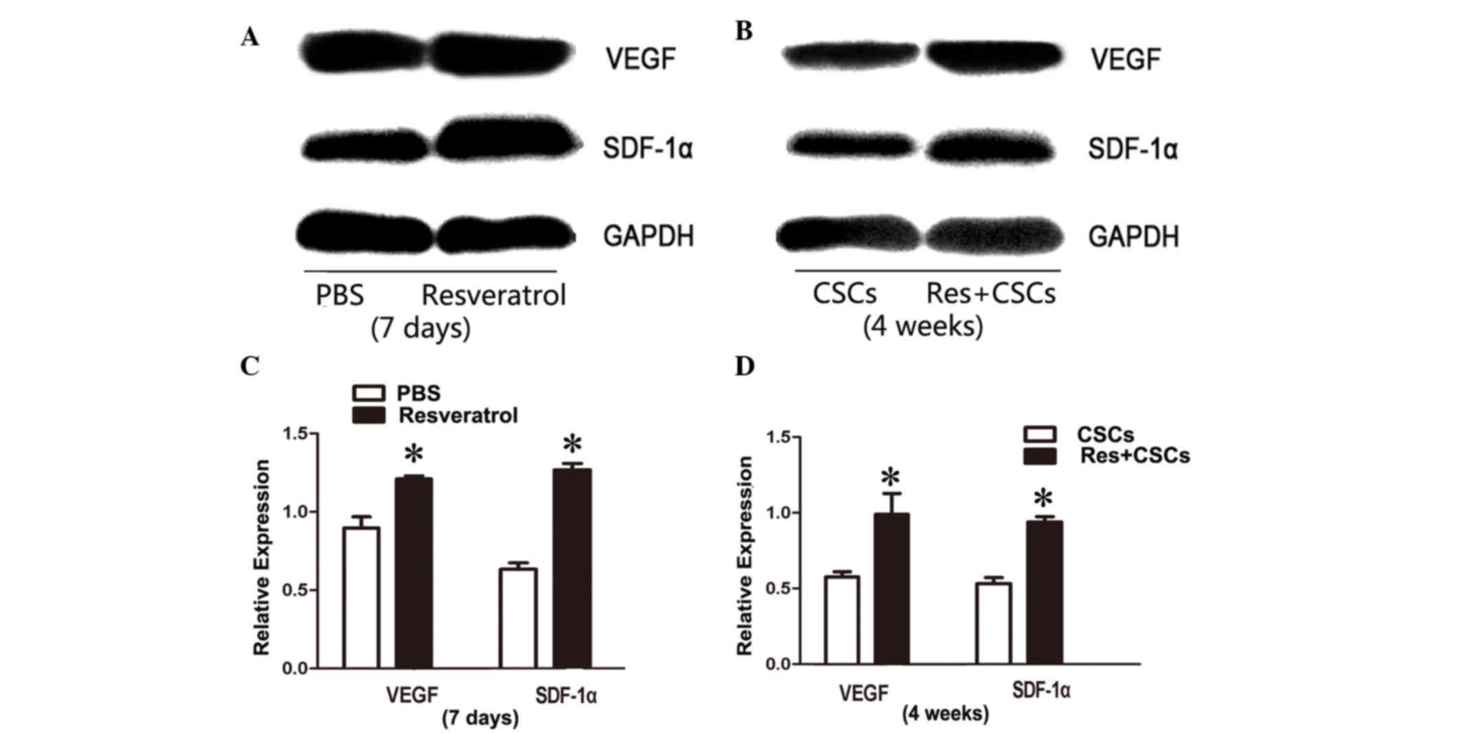
VEGF and SDF-1α expression is
upregulated in resveratrol-treated myocardium
Western blotting was used to evaluate the protein
expression levels of VEGF and SDF-1α in myocardium 7 days after
resveratrol administration, and 4 weeks after AMI. Western blot
analysis indicated that VEGF and SDF-1α were upregulated in
resveratrol-treated myocardium after 7 days of treatment, and 4
weeks after AMI (7 days VEGF PBS vs. resveratrol, 0.89±0.07 vs.
1.21±0.02, P<0.05; SDF-1α PBS vs. resveratrol, 0.66±0.04 vs.
1.33±0.04, P<0.05; 4 weeks VEGF CSCs vs. resveratrol + CSCs,
0.54±0.03 vs. 0.93±0.13, P<0.05; SDF-1α CSCs vs. resveratrol +
CSCs, 0.53±0.03 vs. 0.93±0.03, P<0.05, Fig. 5).
Discussion
Since the adult heart was recognized as containing
its own stem cells (20), CSCs
therapy for AMI has been considered as a novel treatment strategy.
CSCs are able to regenerate several types of cell, including
cardiomyocytes, endothelial cells and smooth muscle cells, and
directly participate in myocardial and endothelial repair (21,22).
Cell therapy of AMI with CSCs has been reported to ameliorate
cardiac function and tissue remodeling (23). However, just like other stem
cell-based therapies for AMI, CSCs-based therapy is hindered by
limited transplantation efficiency, and researchers have aimed to
solve this problem with appropriate cell modification (24). Resveratrol is a compound that
exerts numerous effects, including anti-inflammatory, antitumor and
immunomodulatory activities. Resveratrol has also been reported to
exert strong cardioprotective effects in the cardiovascular system
(25,26). The present study combined this
cardioprotective agent with CSCs-based therapy. A mouse model of
AMI treated with CSCs transplantation plus resveratrol exhibited
preserved cardiac function, increased capillary density and
decreased cardiomyocyte apoptosis in the peri-ischemic myocardium.
In addition, VEGF and SDF-1α expression was upregulated. These
results suggested that this combination therapy may augment the
effects of CSCs-mediated myocardial regeneration, and may fulfill
the potential of CSCs cell therapy for AMI.
VEGF is widely known as an angiogenic molecule that
serves an important role in neovascularization and angiogenesis in
several physiological and pathological conditions (27). AMI is a process associated with
severe ischemia and hypoxia, which induces upregulation of the
transcription factor hypoxia-inducible factor-1 (HIF) (28). HIF is an upstream signaling
molecule of VEGF, which can modulate the synthesis and release of
VEGF and downstream signaling pathways, such as endothelial nitric
oxide synthase (29). Upregulation
of VEGF directly leads to increased angiogenesis, as well as
capillary density, in the ischemic myocardium. Resveratrol has been
revealed to elevate the protein expression levels of VEGF, thus
modulating the interaction between VEGF and VEGF receptors in an
AMI model, which may lead to improved vessel collateralization and
angiogenesis in infarcted myocardium (30,31).
The present study also demonstrated that CSCs transplantation plus
resveratrol pretreatment upregulated the synthesis and release of
VEGF. This increase in VEGF may subsequently lead to increased
capillary density after cell transplantation, which is evidence of
further angiogenesis.
Besides VEGF, SDF-1α has been reported to modulate
the homing of stem cells to the site of injury by chemokinesis and
chemotaxis, through uniquely binding to the specific cell surface
receptor CXCR-4 (32,33). The SDF-1α/CXCR-4 targeting system
is vital in cardiac development, as confirmed by the fetal
lethality of SDF-1α or CXCR-4 knockout in mice (34). SDF-1α is able to direct several
types of progenitor or stem cell to the targeted area with accurate
chemotaxis, and maintain these stem cells in the regional ischemic
area (35). It has previously been
reported that gene expression of SDF-1α is significantly increased
in infarcted myocardium following AMI (36). Overexpression of the SDF-1α gene in
damaged heart tissue may induce hematopoietic stem cell homing to
local ischemic myocardium. Similar to VEGF, SDF-1α gene expression
is also regulated by HIF (37).
Regional overexpression of HIF in the ischemic and hypoxic
myocardium has been shown to result in selective expression of
SDF-1α in the ischemic myocardium, and the level of SDF-1α was in
direct proportion to local reduced oxygen tension (38). This overexpression of SDF-1α
increases the targeting, migration and homing of circulating and
resident progenitor cells to gather at the local ischemic
myocardium and participate in tissue recovery. In addition,
insufficient release of SDF-1α has been reported to decrease the
mobilization of progenitor cells (39). SDF-1α has been confirmed to induce
migration of Sca-1+ CSCs in vitro. Upregulation
of VEGF may also elevate the expression of CXCR-4 and SDF-1α to
further enhance their effects on stem cell homing, migration and
chemotaxis (40).
In conclusion, the present study demonstrated that
CSCs therapy combined with resveratrol may augment the effects of
CSCs transplantation through increasing the synthesis of VEGF and
SDF-1α. CSCs transplantation combined with resveratrol pretreatment
improved cardiac performance in a mouse model of AMI, as compared
with CSCs therapy alone. The results suggested that
resveratrol-modified CSCs therapy may further augment the
efficiency of CSCs therapy for AMI, and may provide an improved
strategy for cell-based treatment of AMI.
Acknowledgements
The present study was supported by the Youth Science
and Technology of Suzhou Science and Education Project (grant no.
KJXW2013004), the Youth Science Foundation of Jiangsu Province,
China (grant no. BK20140296) and the Science Foundation for Youth
Teacher of Soochow University (grant no. SDY2013A29).
Glossary
Abbreviations
Abbreviations:
|
Sca-1+ CSCs
|
stem cell antigen-1-positive cardiac
stem cells
|
|
AMI
|
acute myocardial infarction
|
|
LVEDD
|
left ventricular end-diastolic
diameter
|
|
LVESD
|
left ventricular end-systolic
diameter
|
|
IVST
|
interventricular septal thickness in
diastole
|
|
LVPWT
|
left-ventricular posterior wall
thickness
|
|
FS
|
percent LV fractional shortening
|
References
|
1
|
Waring CD, Vicinanza C, Papalamprou A,
Smith AJ, Purushothaman S, Goldspink DF, Nadal-Ginard B, Torella D
and Ellison GM: The adult heart responds to increased workload with
physiologic hypertrophy, cardiac stem cell activation, and new
myocyte formation. Eur Heart J. 35:2722–2731. 2014. View Article : Google Scholar : PubMed/NCBI
|
|
2
|
Noseda M, Abreu-Paiva M and Schneider MD:
The quest for the adult cardiac stem cell. Circ J. 79:1422–1430.
2015. View Article : Google Scholar : PubMed/NCBI
|
|
3
|
Nadal-Ginard B, Ellison GM and Torella D:
The cardiac stem cell compartment is indispensable for myocardial
cell homeostasis, repair and regeneration in the adult. Stem Cell
Res. 13:615–630. 2014. View Article : Google Scholar : PubMed/NCBI
|
|
4
|
Hattori F: CD117, adult cardiac stem cell
marker, is transiently expressed in methothelial epicardial cells.
J Mol Cell Cardiol. 49:711–712. 2010. View Article : Google Scholar : PubMed/NCBI
|
|
5
|
Müller-Ehmsen J, Krausgrill B, Burst V,
Schenk K, Neisen UC, Fries JW, Fleischmann BK, Hescheler J and
Schwinger RH: Effective engraftment but poor mid-term persistence
of mononuclear and mesenchymal bone marrow cells in acute and
chronic rat myocardial infarction. J Mol Cell Cardiol. 41:876–884.
2006. View Article : Google Scholar : PubMed/NCBI
|
|
6
|
Duran JM, Makarewich CA, Sharp TE,
Starosta T, Zhu F, Hoffman NE, Chiba Y, Madesh M, Berretta RM, Kubo
H and Houser SR: Bone-derived stem cells repair the heart after
myocardial infarction through transdifferentiation and paracrine
signaling mechanisms. Circ Res. 113:539–552. 2013. View Article : Google Scholar : PubMed/NCBI
|
|
7
|
Doyle B, Sorajja P, Hynes B, Kumar AH,
Araoz PA, Stalboerger PG, Miller D, Reed C, Schmeckpeper J, Wang S,
et al: Progenitor cell therapy in a porcine acute myocardial
infarction model induces cardiac hypertrophy, mediated by paracrine
secretion of cardiotrophic factors including TGFbeta1. Stem Cells
Dev. 17:941–951. 2008. View Article : Google Scholar : PubMed/NCBI
|
|
8
|
Gnecchi M, Zhang Z, Ni A and Dzau VJ:
Paracrine mechanisms in adult stem cell signaling and therapy. Circ
Res. 103:1204–1219. 2008. View Article : Google Scholar : PubMed/NCBI
|
|
9
|
Mazo M, Cemborain A, Gavira JJ, Abizanda
G, Araña M, Casado M, Soriano M, Hernández S, Moreno C, Ecay M, et
al: Adipose stromal vascular fraction improves cardiac function in
chronic myocardial infarction through differentiation and paracrine
activity. Cell Transplant. 21:1023–1037. 2012. View Article : Google Scholar : PubMed/NCBI
|
|
10
|
Boyle AJ, Yeghiazarians Y, Shih H, Hwang
J, Ye J, Sievers R, Zheng D, Palasubramaniam J, Palasubramaniam D,
Karschimkus C, et al: Myocardial production and release of MCP-1
and SDF-1 following myocardial infarction: Differences between mice
and man. J Transl Med. 9:1502011. View Article : Google Scholar : PubMed/NCBI
|
|
11
|
Takahashi M: Role of the SDF-1/CXCR4
system in myocardial infarction. Circ J. 74:418–423. 2010.
View Article : Google Scholar : PubMed/NCBI
|
|
12
|
Escoté X, Miranda M, Menoyo S,
Rodríguez-Porrata B, Carmona-Gutiérrez D, Jungwirth H, Madeo F,
Cordero RR, Mas A, Tinahones F, et al: Resveratrol induces
antioxidant defence via transcription factor Yap1p. Yeast.
29:251–263. 2012. View
Article : Google Scholar : PubMed/NCBI
|
|
13
|
Wang Z, Chen Y, Labinskyy N, Hsieh TC,
Ungvari Z and Wu JM: Regulation of proliferation and gene
expression in cultured human aortic smooth muscle cells by
resveratrol and standardized grape extracts. Biochem Biophys Res
Commun. 346:367–376. 2006. View Article : Google Scholar : PubMed/NCBI
|
|
14
|
Bradamante S, Barenghi L and Villa A:
Cardiovascular protective effects of resveratrol. Cardiovasc Drug
Rev. 22:169–188. 2004. View Article : Google Scholar : PubMed/NCBI
|
|
15
|
Das S and Das DK: Resveratrol: A
therapeutic promise for cardiovascular diseases. Recent Pat
Cardiovasc Drug Discov. 2:133–138. 2007. View Article : Google Scholar : PubMed/NCBI
|
|
16
|
Fukuda S, Kaga S, Zhan L, Bagchi D, Das
DK, Bertelli A and Maulik N: Resveratrol ameliorates myocardial
damage by inducing vascular endothelial growth factor-angiogenesis
and tyrosine kinase receptor Flk-1. Cell Biochem Biophys. 44:43–49.
2006. View Article : Google Scholar : PubMed/NCBI
|
|
17
|
Das S, Alagappan VK, Bagchi D, Sharma HS,
Maulik N and Das DK: Coordinated induction of iNOS-VEGF-KDR-eNOS
after resveratrol consumption: A potential mechanism for
resveratrol preconditioning of the heart. Vascul Pharmacol.
42:281–289. 2005. View Article : Google Scholar : PubMed/NCBI
|
|
18
|
Brunner S, Winogradow J, Huber BC, Zaruba
MM, Fischer R, David R, Assmann G, Herbach N, Wanke R,
Mueller-Hoecker J and Franz WM: Erythropoietin administration after
myocardial infarction in mice attenuates ischemic cardiomyopathy
associated with enhanced homing of bone marrow-derived progenitor
cells via the CXCR-4/SDF-1 axis. FASEB J. 23:351–361. 2009.
View Article : Google Scholar : PubMed/NCBI
|
|
19
|
Shen L, Gao Y, Qian J, Sun A and Ge J: A
novel mechanism for endothelial progenitor cells homing: The
SDF-1/CXCR4-Rac pathway may regulate endothelial progenitor cells
homing through cellular polarization. Med Hypotheses. 76:256–258.
2011. View Article : Google Scholar : PubMed/NCBI
|
|
20
|
Lyngbaek S, Schneider M, Hansen JL and
Sheikh SP: Cardiac regeneration by resident stem and progenitor
cells in the adult heart. Basic Res Cardiol. 102:101–114. 2007.
View Article : Google Scholar : PubMed/NCBI
|
|
21
|
Torella D, Ellison GM, Karakikes I and
Nadal-Ginard B: Resident cardiac stem cells. Cell Mol Life Sci.
64:661–673. 2007. View Article : Google Scholar : PubMed/NCBI
|
|
22
|
Torella D, Ellison GM, Méndez-Ferrer S,
Ibanez B and Nadal-Ginard B: Resident human cardiac stem cells:
Role in cardiac cellular homeostasis and potential for myocardial
regeneration. Nat Clin Pract Cardiovasc Med. 3:(Suppl 1). S8–S13.
2006. View Article : Google Scholar : PubMed/NCBI
|
|
23
|
Chamuleau SA, Vrijsen KR, Rokosh DG, Tang
XL, Piek JJ and Bolli R: Cell therapy for ischaemic heart disease:
Focus on the role of resident cardiac stem cells. Neth Heart J.
17:199–207. 2009. View Article : Google Scholar : PubMed/NCBI
|
|
24
|
Tang JM, Wang JN, Zhang L, Zheng F, Yang
JY, Kong X, Guo LY, Chen L, Huang YZ, Wan Y and Chen SY: VEGF/SDF-1
promotes cardiac stem cell mobilization and myocardial repair in
the infarcted heart. Cardiovasc Res. 91:402–411. 2011. View Article : Google Scholar : PubMed/NCBI
|
|
25
|
Mokni M, Limam F, Elkahoui S, Amri M and
Aouani E: Strong cardioprotective effect of resveratrol, a red wine
polyphenol, on isolated rat hearts after ischemia/reperfusion
injury. Arch Biochem Biophys. 457:1–6. 2007. View Article : Google Scholar : PubMed/NCBI
|
|
26
|
Wu JM and Hsieh TC: Resveratrol: A
cardioprotective substance. Ann N Y Acad Sci. 1215:16–21. 2011.
View Article : Google Scholar : PubMed/NCBI
|
|
27
|
Zhao T, Zhao W, Chen Y, Ahokas RA and Sun
Y: Vascular endothelial growth factor (VEGF)-A: Role on cardiac
angiogenesis following myocardial infarction. Microvasc Res.
80:188–194. 2010. View Article : Google Scholar : PubMed/NCBI
|
|
28
|
Lakkisto P, Kytö V, Forsten H, Siren JM,
Segersvärd H, Voipio-Pulkki LM, Laine M, Pulkki K and Tikkanen I:
Heme oxygenase-1 and carbon monoxide promote neovascularization
after myocardial infarction by modulating the expression of
HIF-1alpha, SDF-1alpha and VEGF-B. Eur J Pharmacol. 635:156–164.
2010. View Article : Google Scholar : PubMed/NCBI
|
|
29
|
Knudsen AR, Kannerup AS, Grønbaek H,
Andersen KJ, Funch-Jensen P, Frystyk J, Flyvbjerg A and Mortensen
FV: Effects of ischemic pre- and postconditioning on HIF-1α, VEGF
and TGF-β expression after warm ischemia and reperfusion in the rat
liver. Comp Hepatol. 10:32011. View Article : Google Scholar : PubMed/NCBI
|
|
30
|
Kaga S, Zhan L, Matsumoto M and Maulik N:
Resveratrol enhances neovascularization in the infarcted rat
myocardium through the induction of thioredoxin-1, heme oxygenase-1
and vascular endothelial growth factor. J Mol Cell Cardiol.
39:813–822. 2005. View Article : Google Scholar : PubMed/NCBI
|
|
31
|
Penumathsa SV, Thirunavukkarasu M, Koneru
S, Juhasz B, Zhan L, Pant R, Menon VP, Otani H and Maulik N: Statin
and resveratrol in combination induces cardioprotection against
myocardial infarction in hypercholesterolemic rat. J Mol Cell
Cardiol. 42:508–516. 2007. View Article : Google Scholar : PubMed/NCBI
|
|
32
|
Cottler-Fox MH, Lapidot T, Petit I, Kollet
O, DiPersio JF, Link D and Devine S: Stem cell mobilization.
Hematology Am Soc Hematol Educ Program. 419–437. 2003.PubMed/NCBI
|
|
33
|
Zou YR, Kottmann AH, Kuroda M, Taniuchi I
and Littman DR: Function of the chemokine receptor CXCR4 in
haematopoiesis and in cerebellar development. Nature. 393:595–599.
1998. View Article : Google Scholar : PubMed/NCBI
|
|
34
|
Lapidot T: Mechanism of human stem cell
migration and repopulation of NOD/SCID and B2mnull NOD/SCID mice.
The role of SDF-1/CXCR4 interactions. Ann N Y Acad Sci. 938:83–95.
2001. View Article : Google Scholar : PubMed/NCBI
|
|
35
|
Askari AT, Unzek S, Popovic ZB, Goldman
CK, Forudi F, Kiedrowski M, Rovner A, Ellis SG, Thomas JD,
DiCorleto PE, et al: Effect of stromal-cell-derived factor 1 on
stem-cell homing and tissue regeneration in ischaemic
cardiomyopathy. Lancet. 362:697–703. 2003. View Article : Google Scholar : PubMed/NCBI
|
|
36
|
Pillarisetti K and Gupta SK: Cloning and
relative expression analysis of rat stromal cell derived factor-1
(SDF-1)1: SDF-1 alpha mRNA is selectively induced in rat model of
myocardial infarction. Inflammation. 25:293–300. 2001. View Article : Google Scholar : PubMed/NCBI
|
|
37
|
Loh SA, Chang EI, Galvez MG, Thangarajah
H, El-ftesi S, Vial IN, Lin DA and Gurtner GC: SDF-1 alpha
expression during wound healing in the aged is HIF dependent. Plast
Reconstr Surg. 123:(Suppl 2). S65–S75. 2009. View Article : Google Scholar
|
|
38
|
Ceradini DJ, Kulkarni AR, Callaghan MJ,
Tepper OM, Bastidas N, Kleinman ME, Capla JM, Galiano RD, Levine JP
and Gurtner GC: Progenitor cell trafficking is regulated by hypoxic
gradients through HIF-1 induction of SDF-1. Nat Med. 10:858–864.
2004. View
Article : Google Scholar : PubMed/NCBI
|
|
39
|
Hattori K, Heissig B, Tashiro K, Honjo T,
Tateno M, Shieh JH, Hackett NR, Quitoriano MS, Crystal RG, Rafii S
and Moore MA: Plasma elevation of stromal cell-derived factor-1
induces mobilization of mature and immature hematopoietic
progenitor and stem cells. Blood. 97:3354–3360. 2001. View Article : Google Scholar : PubMed/NCBI
|
|
40
|
Tang J, Wang J, Kong X, Yang J, Guo L,
Zheng F, Zhang L, Huang Y and Wan Y: Vascular endothelial growth
factor promotes cardiac stem cell migration via the PI3K/Akt
pathway. Exp Cell Res. 315:3521–3531. 2009. View Article : Google Scholar : PubMed/NCBI
|