Introduction
Sepsis is a complex clinical syndrome generated by
the interaction between the host and infectious pathogens and their
toxic products, such as endotoxin and the host immune system, and
inflammation and the coagulation response (1,2).
Severe sepsis may lead to multiple organ failure and is a common
cause of mortality in ICU patients (3,4).
Endotoxin is a component of the outer membrane of the cell wall of
Gram-negative bacteria. The chemical essence of endotoxin is
lipopolysaccharide (LPS) (5). LPS
is the main triggering factor of sepsis and systemic inflammatory
response syndrome (6). Monocytes
are one of the important immune cells involved in inflammatory
responses in the body (7,8). Therefore, monocytes often serve as
effector cells for the study of the inflammatory response.
Propofol, an intravenous anesthetic with a rapid
onset of action and no accumulation during continuous infusion, is
widely used in anesthesia induction, maintenance and sedative
treatment of ICU patients, such as patients with sepsis (9). In recent years, the anti-inflammatory
effects of propofol have gradually attracted more attention.
Propofol can inhibit the release of the inflammatory cytokines,
IL-1β, IL-6 and TNF-α (10,11);
it is capable of removing oxygen free radicals and inhibiting
oxidative damage generated by respiratory burst after neutrophil
activation (12); it also inhibits
the release of PF4 in the blood of rats with endotoxemia and partly
alleviates hypercoagulability in endotoxemia (13). Although we have reached a consensus
on the anti-inflammatory effects of propofol, its effects on
monocyte protein expression remain unclear. In this study,
comparative proteomic techniques were used to explore the impact of
propofol on monocyte protein expression in rats with endotoxemia,
in order to further clarify the molecular mechanisms of the
anti-inflammatory action of propofol.
Materials and methods
Model building
The experiments were approved by the local ethics
committee and performed in accordance with the Guidelines for the
Care and Use of Animals of Guangdong General Hospital. In
accordance with the guidelines of the International Association for
the Study of Pain (14), all
surgery was performed after the animals were anesthetized with
urethane, so they did not feel pain or discomfort during the
experiments, and the minimum possible pain or stress was imposed on
the animals. In total, 18 male Sprague-Dawley rats weighing between
180 and 220 g anesthetized with urethane (1.0 g/kg)
intraperitoneally (i.p.) were divided into a control group, a LPS
(Escherichia coli; Sigma, St. Louis, MO, USA) + intralipid
(Sino-Swed Pharmaceutical Co., Ltd., Beijing, China) group and an
LPS + propofol (FJ685; commercial name, Diprivan; AstraZeneca,
London, UK) group. In the LPS + intralipid group, rats were infused
with intralipid (10 mg·kg−1h−1) immediately
following 10 mg·kg−1 LPS intravenously (i.v.), and in
the LPS + propofol group rats were injected with LPS followed by
propofol at 10 mg·kg−1h−1. In the control
group, rats were injected the same amount of balanced saline. After
6 h, 3–4 ml of blood from the carotid artery of each rat were
collected into Eppendorf tubes and the rats were sacrificed
thereafter.
Isolation of leukocytes from rat
peripheral blood
In total, 3.0 ml Histopaque 10831 (Sigma) were added
to a 15-ml centrifuge tube, then 3.0 ml whole blood were carefully
layered onto the Histopaque 10831 surface and centrifuged at 400 ×
g for 30 min at room temperature. After centrifugation, the upper
layer to within 2–3 mm of the opaque interface contained the
mononuclear cells. The upper layer was carefully aspirated with a
Pasteur pipet and discarded. The opaque interface, containing the
mononuclear cell band, was carefully transferred with a Pasteur
pipet into a clean 15 ml conical centrifuge tube. Isotonic PBS (10
ml) was then added to the mononuclear cells, the tube was mixed by
gentle inversion several times and centrifuged at 250 × g for 10
min. The supernatant was aspirated and discarded. After
resuspending the cell pellet with 0.5 ml of isotonic PBS, an
additional 4.5 ml of isotonic PBS was added and the mixture was
centrifuged at 250 × g for 10 min. The supernatant was aspirated
and discarded. This step was repeated 2–3 times to remove any
remaining Histopaque 10831 from the mononuclear cells. After the
final wash, the cells were added to 300 μl of lysis buffer
consisting of 7 M urea, 2 M thiourea, 4% CHAPS, 65 mM DTT and 2%
Pharmalyte (pH 3–10; GE Healthcare, Piscataway, NJ, USA) by
sonication on ice. The lysates were cleared by centrifugation at
12,000 rpm for 30 min at 4°C, twice. Subsequently, the protein
concentration of the supernatants was determined by the modified
Bradford method and the protein samples were stored at −80°C.
Two-dimensional (2D) electrophoresis
An Immobiline DryStrip (pH 3.0–10.0, length 24 cm,
GE Healthcare) was rehydrated with 1,500 μg protein in 450 ml
rehydration buffer containing 7 M urea, 2 M thiourea, 4% CHAPS, 65
mM DTT, 20 mM Trizma base, 1% IPG buffer and 0.002% bromophenol
blue for 14 h at room temperature. Isoelectric focusing (IEF) was
performed using the Ettan IPGphor 3 IEF System (GE Healthcare) for
a total of 70 kVh. The strip was then subjected to two-step
equilibration in a buffer containing 6 M urea, 20% glycerol, 2% SDS
and 50 mM Tris-HCl (pH 8.8) with 2% w/v DTT for the first step, and
2.5% w/v iodoacetamide for the second step. The second-dimension
SDS-PAGE (12% T, 260×200×1.5 mm3) was carried out using
a Ettan DALTsix Large Vertical system (Amersham Pharmacia Biotech,
Piscataway, NJ, USA) according to the following procedures: 45 min
at a constant power of 5 watt followed by 20 watt per gel until the
bromophenol blue front reached the bottom of the gel. Subsequently,
the gels were stained with 0.12% w/v Coomassie Brilliant Blue G250.
Each group was run in triplicate to minimize run-to-run variation.
The Coomassie Blue-stained protein 2D gels were scanned using an
Amersham Biosciences ImageScanner and analyzed using a DeCyder
software package (GE Healthcare).
In-gel digestion
Protein spots were excised from the gel with an
operating knife blade, destained twice with 30 mM potassium
ferricyanide and 100 mM sodium thiosulfate (1:1 v/v) and then
equilibrated in 100 mM NH4HCO3 to pH 8.0.
After dehydrating with acetonitrile (ACN) and drying in nitrogen at
37°C for 20 min, the gel pieces were rehydrated in 10 μl trypsin
solution (12.5 ng/μl in 50 mM NH4HCO3) at 4°C
for 30 min and incubated at 37°C overnight. Peptides were extracted
twice using 0.1% trifluoroacetic acid (TFA) in 60% CAN and dried
with the RCT60 (Jouan S.A., Jouan, France).
Matrix-assisted laser desorption
ionization time-of-flight mass spectrometry (MALDI-TOF-MS)
identification
The peptide mixtures were solubilized with 0.1% TFA
and desalted with C18 ZipTip (Millipore, USA). The peptide was then
eluted by saturated a-cyano-4-hydroxy-trans-cinnamic (CHCA)
solution in 0.1% TFA/60% acetonitrile as the matrix and analyzed
using a 4800 MALDI TOF/TOF analyzer (Applied Biosystems, USA). Mass
spectra were internally calibrated with angiotensin I (Mr:
1296.6853).
Protein identification and database
searching
Protein identification using peptide mass
fingerprinting (PMF) and peptide sequence tag (PST) was performed
using the Mascot search engine (http://www.matrixscience.com, MatrixSicence Ltd.,
London, UK) against the SwissProt protein database. The errors in
peptide masses were in the range of 50 ppm. One missed tryptic
cleavage site per peptide was allowed during the search. Proteins
matching more than 4 peptides and with a MASCOT score higher than
64 were considered significant (P<0.05). Carboamidomethylation
of cysteine was selected as the fixed modification, and oxidation
of methionine as the variable modification. Protein identification
results were filtered with GPS software.
Western blot analysis
In order to verify the 2D electrophoresis protein
expression data, another 36 Sprague-Dawley rats were divided into 6
groups: the control, intralipid, propofol, LPS, propofol + LPS and
intralipid + LPS groups. In the intralipid and propofol group, rats
were infused with intralipid or propofol at 10
mg·kg−1h−1. The LPS group was injected with
LPS followed by the same amount of balanced saline and the other 3
groups were treated the same as above. S100A9 in the mononuclear
cells of the rats was detected by western blot analysis. The
mononuclear cells of the rats were lysed on ice in 300 μl cell
lysis buffer [1X PBS, 1% NP40, 0.1% sodium dodecyl sulfate (SDS), 5
mM EDTA, 0.5% sodium deoxycholate and 1 mM sodium orthovanadate]
with protease inhibitors. Protein concentration was determined by
the modified Bradford method. Equal amounts of protein were
separated electrophoretically on 12% SDS/polyacrylamide gels and
transferred onto polyvinylidene difluoride membranes (PVDF)
(Amersham Pharmacia Biotech). For S100A9 detection, the membrane
was probed with anti-S100A9 rabbit polyclonal antibody (1:1000;
Abcam) and horseradish peroxidase-conjugated anti-rabbit
immunoglobulin G (1:2000; Cell Signaling Technology, Danvers, MA,
USA). The immunoreactive bands were visualized on a Kodak 2000M
camera system (Eastman Kodak, Rochester, NY, USA) according to the
manufacturer’s instructions.
Statistical analysis
All the data were examined for normal distribution
prior to statistical analysis and the statistical analysis was
carried out using SPSS 13.0. All data were expressed as the means ±
SD. Multiple groups were compared using analysis of variance
(ANOVA) followed by the Student-Newman-Keuls post-hoc procedure.
P<0.05 was considered to indicate statistically significant
differences.
Results
Quantitative comparison and
identification of protein spots on 2D gels
To determine the change in the monocyte protein
profile in response to LPS and propofol, gel-based comparative
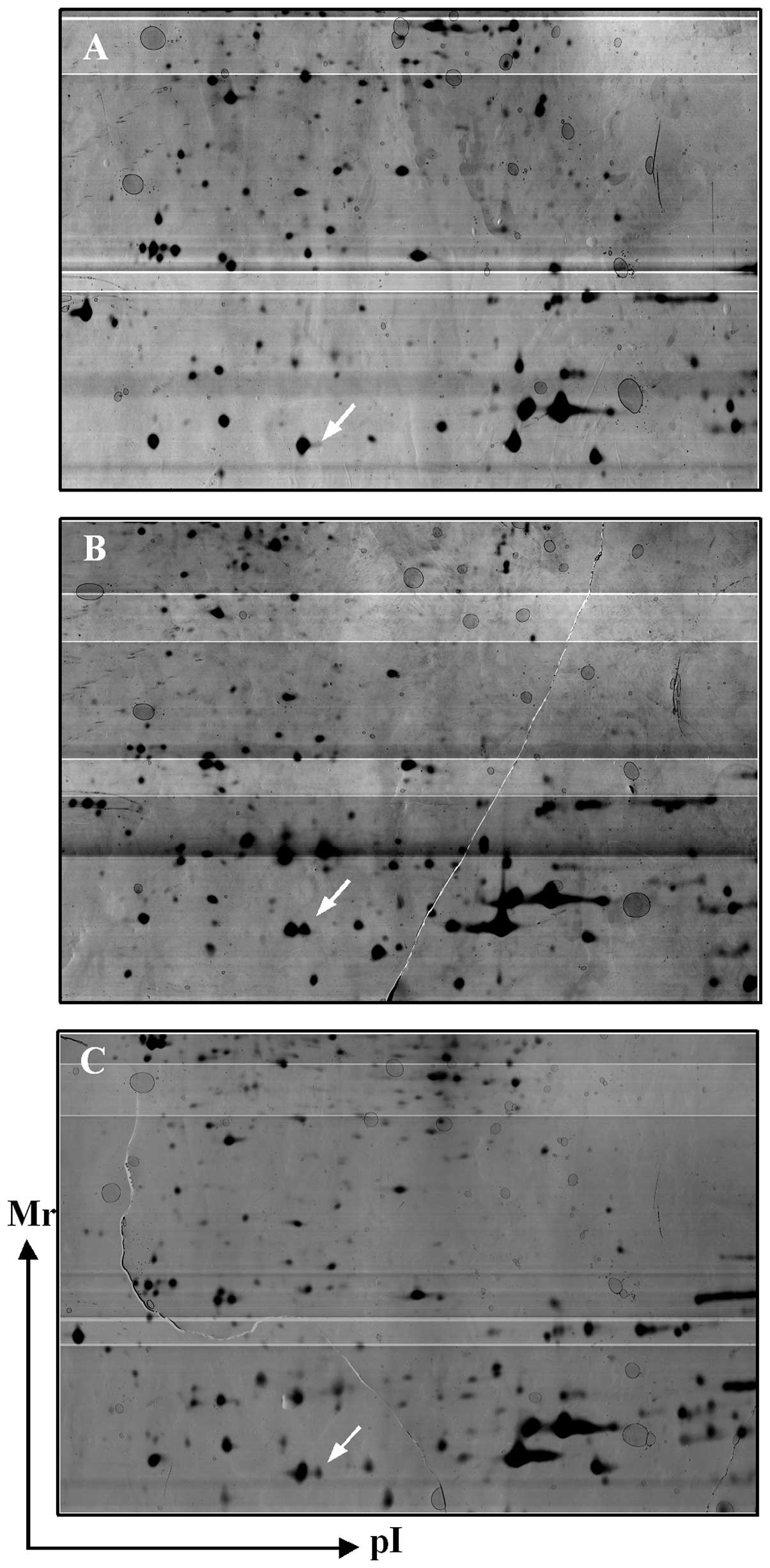
proteomic analysis was performed. Thirteen protein spots were found
to be significantly altered (data not shown). One of them was
S100A9 identified by MALDI-TOF MS and by a subsequent comparative
sequence search in the Mascot database (Fig. 1).
Verification of downregulation of S100A9
in the LPS + propofol group by western blot analysis in endotoxemic
rats
As gel-based proteomic analysis showed that the
expression of S100A9 decreased markedly in the LPS + propofol
group, S100A9 was elected for further investigation. The
corresponding differential expression patterns identified by 2D
electrophoresis and the MALDI-TOF mass spectra are shown in
Fig. 2. S100A9 is a 13,200
molecular weight pro-inflammatory protein expressed abundantly in
the cytosol of monocytes and neutrophils (1). To more rigorously study the effect of
propofol on the expression of S100A9 in the monocytes, 36 rats were
divided into 6 groups: control group, intralipid group, propofol
group, LPS group, LPS + propofol group and LPS + intralipid group.
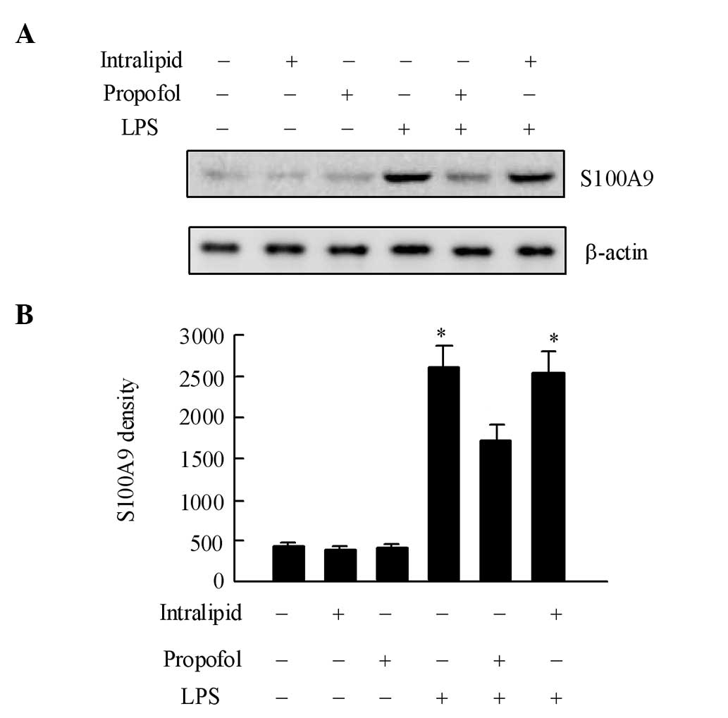
S100A9 expression in the monocytes of each group was determined by
western blot analysis. As shown in Fig. 3A, the amount of S100A9 in monocytes
showed no statistical difference in the control, intralipid,
propofol and LPS + propofol groups (P>0.05). However, in the
monocytes of the LPS and LPS + intralipid group, the expression of
S100A9 increased markedly in accordance with our 2D gels results.
These results indicate that propofol is capable of inhibiting the
expression of S100A9 in the monocytes of endotoxemic rats. Total
β-actin loaded on SDS-PAGE was used as the internal control
(Fig. 3B).
Discussion
The anti-inflammatory effect of the intravenous
anesthetic, propofol, has drawn much attention (10–12).
It has previously been reported that propofol is capable of
inhibiting the release of inflammatory cytokines, such as IL-1β,
IL-6 and TNF-α (12); however, the
mechanisms by which propofol inhibits the secretion of inflammatory
cytokines and the specific signal transduction mechanisms remain
unclear. This also limits the clinical application of the
anti-inflammatory effect of propofol. Monocytes are the most
important effector cells in the systemic inflammatory response
(7). Therefore, a rat model of
endotoxemia by LPS was established and 2D electrophoresis and
proteomic techniques were used to detect monocyte protein
expression in serum to find propofol-related inflammatory proteins.
By mass spectrometry, it was found that the inflammation-associated
protein, S100A9, was significantly reduced in the LPS + propofol
treatment group.
S100A9 is a key member of the S100 calcium-binding
protein family. Its relative molecular weight is 13.2 kDa (16). S100A9 is mainly expressed in human
and rat monocyte/macrophage cell lines, neutrophils and
keratinocytes under certain pathological conditions (16,17).
Previous studies have indicated that S100A9 levels are
significantly increased in inflammatory states: after inflammation
is induced by carrageenan in rats, S100A9 concentration is
increased in exudates, and it plays a major role in chronic
inflammation by stimulating the proliferation of fibroblasts
(18). In the late stage of
endotoxin-induced uveitis, S100A9 may clear inflammation in cells
(17). It is also closely related
to systemic lupus erythematosus, giant cell arteritis, multiple
sclerosis and many other immune inflammations (19). Studies have suggested that S100A9
is a specifically overexpressed marker in asthma (20). In the initial stage of
inflammation, S100A9-positive cells express higher levels of CD11b.
Studies have shown that the lack of S100A9 in cells in vitro
reduces the response of white blood cells to chemical stimuli
(21). A S100A9 gene knockout
mouse model of pancreatitis showed that leukocyte infiltration was
reduced in lung tissue and pancreatic tissue in mice, and serum
amylase levels were also decreased (22). These findings have prompted the
theory that S100A9 is closely related to the inflammatory
response.
Therefore, in the present study, we chose the S100A9
protein for subsequent validation and research. In addition, we
selected 36 Sprague-Dawley rats to give endotoxin, propofol or
intralipid for stimulation and used western blot analysis to detect
S100A9 protein expression in monocytes. The results showed that
after LPS stimulation, S100A9 expression in monocytes was
significantly increased, which to a certain extent, indicated that
S100A9 is a marker of inflammatory response; following propofol
treatment, S100A9 expression was significantly reduced, indicating
that propofol can significantly inhibit the expression of the
inflammatory protein, S100A9. Intralipid is a solvent of propofol.
Certain studies have suggested that intralipid has
anti-inflammatory effects and can inhibit the release of
inflammatory cytokines (23);
however, our results showed that the administration of intralipid
after LPS stimulation did not inhibit the expression of S100A9
protein.
Mitogen-activated protein kinase (MAPK) pathways are
one of the important signal transduction systems in organisms. They
are capable of mediating the signal responses of cells to a variety
of external stresses. p38MAPK, ERK and JNK are 3 relatively
important signaling pathways (24). These MAPK pathways may be activated
by a variety of inflammatory stimuli, such as LPS and play an
important role in regulating the occurrence and development of
inflammation. Previous studies have suggested that the effects of
S100A9 and MAPK signal transduction pathways are very similar:
S100A9, as a reliable marker of inflammation in macrophages, is
capable of specifically activating JNK and ERK pathways through
TLR4, and at the gene and protein levels, can induce nitric oxide
synthase expression (25). Under
pathological conditions, S100A9 can be specifically phosphorylated
by p38MAPK kinase, decreasing the aggregating ability of cell
membranes, thus promoting the travelling of inflammatory cells.
S100A9 can also have a reverse effect on p38 kinase, inhibiting
cell migration (26). The study of
Tang et al (27) showed
that propofol plays an anti-inflammatory role by inhibiting p38
phosphorylation. S100A9 not only affects inflammatory responses,
but can also regulate tumor cell proliferation, metabolism and
migration. It plays a promoting role in the physiological and
pathological process of tumors. El-Rifai et al (28) found that the S100A9 gene was
overexpressed in gastric cancer epithelial cells using serial
analysis of gene expression; the immunohistochemical results showed
that S100A9 was overexpressed in the macrophages and
leukocyte-infiltrated tissue edge in colorectal cancer (29). Propofol has also been shown to have
anti-tumor properties (30). It is
speculated from the literature that the anti-inflammatory and
anti-tumor properties of propofol correlate with the S100A9 protein
and the MAPK signaling pathway.
In conclusion, the results from this study suggest
that propofol inhibits the expression of the S100A9 protein;
however, the upstream and downstream acting elements in the
inhibition of S100A9 protein expression are not yet clear. In a
subsequent experiment, we will investigate the correlation between
S100A9 and the MAPK pathway and observe whether there is an
interaction between them and further improve the molecular basis of
the anti-inflammatory effects of propofol.
Acknowledgements
This study was supported by the Guangdong Science
and Technology Plan (2006B36007015).
References
|
1
|
Brunkhorst FM and Reinhart K: Diagnosis
and causal treatment of sepsis. Internist (Berl). 50:810–816. 2009.
View Article : Google Scholar
|
|
2
|
Sharma S and Kumar A: Septic shock,
multiple organ failure, and acute respiratory distress syndrome.
Curr Opin Pulm Med. 9:199–209. 2003. View Article : Google Scholar : PubMed/NCBI
|
|
3
|
Schneider J and Muleta M: Septic shock.
Ethiop Med J. 41:89–104. 2003.
|
|
4
|
Tabbutt S: Heart failure in pediatric
septic shock: utilizing inotropic support. Crit Care Med. 29(Suppl
10): S231–S236. 2001. View Article : Google Scholar : PubMed/NCBI
|
|
5
|
Heine H, Rietschel ET and Ulmer AJ: The
biology of endotoxin. Mol Biotechnol. 19:279–296. 2001. View Article : Google Scholar : PubMed/NCBI
|
|
6
|
Leslie DB, Vietzen PS, Lazaron V, et al:
Comparison of endotoxin antagonism of linear and cyclized peptides
derived from limulus anti-lipopolysaccharide factor. Surg Infect
(Larchmt). 7:45–52. 2006. View Article : Google Scholar : PubMed/NCBI
|
|
7
|
Wu JF, Ma J, Chen J, et al: Changes of
monocyte human leukocyte antigen-DR expression as a reliable
predictor of mortality in severe sepsis. Crit Care. 15:R2202011.
View Article : Google Scholar : PubMed/NCBI
|
|
8
|
Monneret G, Lepape A, Voirin N, et al:
Persisting low monocyte human leukocyte antigen-DR expression
predicts mortality in septic shock. Intensive Care Med.
32:1175–1183. 2006. View Article : Google Scholar : PubMed/NCBI
|
|
9
|
Sztark F and Lagneau F: Agents for
sedation and analgesia in the intensive care unit. Ann Fr Anesth
Reanim. 27:560–566. 2008.(In French).
|
|
10
|
Lee CJ, Subeq YM, Lee RP, et al: Low-dose
propofol ameliorates haemorrhagic shock-induced organ damage in
conscious rats. Clin Exp Pharmacol Physiol. 35:766–774. 2008.
View Article : Google Scholar : PubMed/NCBI
|
|
11
|
Chu CH, David LD, Hsu YH, et al: Propofol
exerts protective effects on the acute lung injury induced by
endotoxin in rats. Pulm Pharmacol Ther. 20:503–512. 2007.
View Article : Google Scholar : PubMed/NCBI
|
|
12
|
Tsao CM, Ho ST, Chen A, et al: Propofol
ameliorates liver dysfunction and inhibits aortic superoxide level
in conscious rats with endotoxic shock. Eur J Pharmacol.
477:183–193. 2003. View Article : Google Scholar : PubMed/NCBI
|
|
13
|
Tang J, Sun Y, Wu WK, et al: Propofol
lowers serum PF4 level and partially corrects hypercoagulopathy in
endotoxemic rats. Biochim Biophys Acta. 1804:1895–1901. 2010.
View Article : Google Scholar : PubMed/NCBI
|
|
14
|
Zimmermann M: Ethical guidelines for
investigations of experimental pain in conscious animals. Pain.
16:109–110. 1983. View Article : Google Scholar : PubMed/NCBI
|
|
15
|
Simard JC, Girard D and Tessier PA:
Induction of neutrophil degranulation by S100A9 via a
MAPK-dependent mechanism. J Leukoc Biol. 87:905–914. 2010.
View Article : Google Scholar : PubMed/NCBI
|
|
16
|
Hessian PA, Edgeworth J and Hogg N: MRP-8
and MRP-14, two abundant Ca(2+)-binding proteins of neutrophils and
monocytes. J Leukoc Biol. 53:197–204. 1993.PubMed/NCBI
|
|
17
|
Chi ZL, Hayasaka Y, Zhang XY, et al:
S100A9-positive granulocytes and monocytes in
lipopolysaccharide-induced anterior ocular inflammation. Exp Eye
Res. 84:254–265. 2007. View Article : Google Scholar : PubMed/NCBI
|
|
18
|
Shibata F, Miyama K, Shinoda F, et al:
Fibroblast growth-stimulating activity of S100A9 (MRP-14). Eur J
Biochem. 271:2137–2143. 2004. View Article : Google Scholar : PubMed/NCBI
|
|
19
|
Gebhardt C, Nemeth J, Angel P, et al:
S100A8 and S100A9 in inflammation and cancer. Biochem Pharmacol.
72:1622–1631. 2006. View Article : Google Scholar : PubMed/NCBI
|
|
20
|
Yin LM, Jiang GH, Wang Y, et al: Use of
serial analysis of gene expression to reveal the specific
regulation of gene expression profile in asthmatic rats treated by
acupuncture. J Biomed Sci. 16:462009. View Article : Google Scholar : PubMed/NCBI
|
|
21
|
Manitz MP, Horst B, Seeliger S, et al:
Loss of S100A9 (MRP14) results in reduced interleukin-8-induced
CD11b surface expression, a polarized microfilament system, and
diminished responsiveness to chemoattractants in vitro. Mol Cell
Biol. 23:1034–1043. 2003. View Article : Google Scholar : PubMed/NCBI
|
|
22
|
Schnekenburger J, Schick V, Kruger B, et
al: The calcium binding protein S100A9 is essential for pancreatic
leukocyte infiltration and induces disruption of cell-cell
contacts. J Cell Physiol. 216:558–567. 2008. View Article : Google Scholar : PubMed/NCBI
|
|
23
|
Yang YH, Toh ML, Clyne CD, et al: Annexin
1 negatively regulates IL-6 expression via effects on p38 MAPK and
MAPK phosphatase-1. J Immunol. 177:8148–8153. 2006. View Article : Google Scholar : PubMed/NCBI
|
|
24
|
Kaminska B: MAPK signalling pathways as
molecular targets for anti-inflammatory therapy--from molecular
mechanisms to therapeutic benefits. Biochim Biophys Acta.
1754:253–262. 2005. View Article : Google Scholar : PubMed/NCBI
|
|
25
|
Pouliot P, Plante I, Raquil MA, et al:
Myeloid-related proteins rapidly modulate macrophage nitric oxide
production during innate immune response. J Immunol. 181:3595–3601.
2008. View Article : Google Scholar : PubMed/NCBI
|
|
26
|
Hiratsuka S, Watanabe A, Aburatani H, et
al: Tumour-mediated upregulation of chemoattractants and
recruitment of myeloid cells predetermines lung metastasis. Nat
Cell Biol. 8:1369–1375. 2006. View
Article : Google Scholar : PubMed/NCBI
|
|
27
|
Tang J, Chen X, Tu W, et al: Propofol
inhibits the activation of p38 through up-regulating the expression
of annexin A1 to exert its anti-inflammation effect. PLoS One.
6:e278902011. View Article : Google Scholar : PubMed/NCBI
|
|
28
|
El-Rifai W, Moskaluk CA, Abdrabbo MK, et
al: Gastric cancers overexpress S100A calcium-binding proteins.
Cancer Res. 62:6823–6826. 2002.PubMed/NCBI
|
|
29
|
Stulik J, Osterreicher J, Koupilova K, et
al: The analysis of S100A9 and S100A8 expression in matched sets of
macroscopically normal colon mucosa and colorectal carcinoma: the
S100A9 and S100A8 positive cells underlie and invade tumor mass.
Electrophoresis. 20:1047–1054. 1999. View Article : Google Scholar : PubMed/NCBI
|
|
30
|
Tsuchiya M, Asada A, Arita K, et al:
Induction and mechanism of apoptotic cell death by propofol in
HL-60 cells. Acta Anaesthesiol Scand. 46:1068–1074. 2002.
View Article : Google Scholar : PubMed/NCBI
|


