Introduction
Cervical cancer ranks third among the most
frequently detected cancer in women around the globe. Every year
more than 500,000 women are diagnosed for this disease which
approximately accounts for about 9% of the total newly diagnosed
cancer cases (1). Nevertheless,
existing treatment options including radical hysterectomy and
radiotherapy have decent clinical outcomes, still around 300,000
deaths are accredited to cervical cancer annually. Moreover,
surgery is lone and appropriate option for early stage cervical
cancer and radiotherapy exhibits severe side effects which badly
influence the quality of life (2).
Natural products have gained tremendous importance as anticancer
agents due to their lower side effects. Among these, anticancer
marine plants form one of the important sources for isolation of
anti-cancerous molecules (3).
Recently, a compound fucosterol (Fig.
1A) has been shown toexhibit tremendous pharmacological
potential activities have been attributed to this molecule which
include, but are not limited to anticancer, antidepressant,
anticonvulsant, anti-inflammatory, and antimicrobial (3,4). Here in
the present study a natural product, fucosterol was evaluated
against human cervical cells. Moreover, the probable underlying
mechanism was assessed with particular emphasis on the effect of
this natural product on PI3K/Akt/mTOR cascade. Except for p53
signaling pathway, the PI3K/Akt/mTOR cascade is probably the most
recurrently changed signaling pathway in cancer (5). Consistent with this, first generation
mTOR inhibitors exhibit significant anti-cancer properties and
several have even been approved for the management of several types
of cancers which include, but are not limited to pancreatic,
cervical, renal and breast cancers. Additionally, PI3K, Akt
together with second generation inhibitors of mTOR are undergoing
clinical trials (5,6). Of note, the results of the present study
indicated that fucosterol exhibits a significant anticancer
activity by inducing apoptosis in human cervical HeLa cancer cell
line by reactive oxygen species (ROS) mediated alterations in
mitochondrial membrane potential (ΔΨm) and cell cycle
arrest. It was also found to downregulate the expression key
proteins of PI3K/Akt/mTOR signalling pathway. Additionally,
fucosterol also caused significant inhibition on cell migration.
Taken together, we propose that fucosterol may prove a potential
candidate towards the management of cervical cancer.
Materials and methods
Chemicals and reagents
Fucosterol, propidium iodide (PI), RNase A triton
X-100 dimethyl and sulfoxide (DMSO), were obtained from
Sigma-Aldrich Co. (St. Louis, MO, USA). All primary and secondary
antibodies were purchased from Santa Cruz Biotechnology Inc. (Santa
Cruz, CA, USA). The fluorescent probes DCFH-DA, DiOC6,
4′-6-diamidino-2-phenylindole (DAPI), Fetal bovine serum (FBS),
RPMI-1640 medium, L-glutamine, antibiotics were obtained from
Invitrogen Life Technologies (Carlsbad, CA, USA).
Cell line and culture conditions
Human cancer cell lines, human lung cancer cell line
(A-549), pancreas (MiaPaca-2), prostate (PC-3, CVCL_0035), breast
(MCF-7), gastric cancer cell line (SNU-5), cervical cancer cell
line (HeLa) and human normal cell line (fR2) were procured from
Cancer Research Institute of Beijing, China, and it was maintained
in DMEM and was supplemented with 10% FBS and antibiotics (100
µg/ml streptomycin and 100 U/ml penicillin G) in a incubator at
37°C (5% CO2 and 95% air).
MTT assay
The anti-proliferation effect of fucosterol was
evaluated against a panel of human cancer cell linesby MTT assay.
Cells were grown at 1×10⁶ cells per well in 96-well
plates for a time period of 12 h and then exposed to different
concentration of fucosterol (0–160 µM) for 48–72 h. To each well,
MTT solution (20 µl) was added. Prior to the addition of 500 µl of
DMSO, the medium was completely removed. To solubilize MTT formazan
crystals, 500 µl DMSO was added. ELISA plate reader was used for
the determination of optical density. Since an lowest IC50 was
observed for HeLa cervical cancer cell line, they were subjected to
further evaluation at the doses of 0, 20, 40 and 80 µM of
fucosterol dose for 24, 48 and 72 h. We selected one dose lower and
one dose higher than the IC50 of fucosterol.
Colony formation assay
For clonogenic assay, cervical cancer HeLa cells at
the exponential growth phase were harvested and counted with a
hemocytometer. Seeding of the cells was done at 200 cells per well,
incubated for a time period of 48 h to allow the cells to attach
and then to the cell culture different doses (0, 20 40 and 80 µM)
of fucosterol was added. After treatment, the cells were again
incubated for 6 days, washing was done with PBS and methanol was
used to fix colonies and then stained with crystal violet for about
30 min before being counted under light microscope.
DAPI staining
HeLa cells at a density of 2×10⁵
cells/well were seeded in 6-well plates were administrated with 0,
20, 40 and 80 µM fucosterol for 48 h. The cells were then subjected
to DAPI staining. Afterwards, the cell sample was studied and
photographs taken under fluorescence microscopy as previously
described (7).
Determination of ROS, and
ΔΨm
HeLa cells were seeded at a density of
2×10⁵ cells/wel in a 6-well plate and kept for 24 h and
treated with (0–100 µM) fucosterol for 48 h at 37°C in 5%
CO2 and 95% air. Thereafter cells from all samples were
collected, washed 2 times by PBS and re-suspended in 500 µl of
DCFH-DA (10 µM) for ROS estimation and DiOC6 (1 µmol/l)
for ΔΨm at 37°C indark room for 30 min. The samples were
then examined instantly using flow cytometer as described
previously in literature (8).
Determination of cell cycle
distribution of HeLa cells
The cells seeded in 6 well plates (2×10⁵
cells/well) and fucosterol was administrated to the cells at the
doses of 0, 20,40 and 80 µM followed by 24 h of incubation. DMSO
was used as a control. For estimation of DNA content, PBS was used
to wash the cells and fixed in ethanol at −20°C. This was followed
by re-suspension in PBS holding 40 µg/ml PI and, RNase A (0.1
mg/ml) and Triton X-100 (0.1%) for 30 min in a dark room at 37°C.
Afterwards, analysis was carried out by flow cytometry as reported
previously (9).
Cell migration assay
Cell migration assay was carried out by Boyden
chamber assay with some modifications. Cells at the density of
5×10⁴ cells/well were suspended in 2% FBS medium and
placed in the upper chamber of 8 µm pore size transwells. After
wards, medium supplemented with 10% FBS was added to lower chamber.
This was followed by an incubation of 48 h. On the upper surface of
the membrane, unmigrated cells were removed while as on the lower
surface of the membrane the migrated cells were fixed in methanol
(100%) and Giemsa stained. The cell migration was estimated by
counting the number of the migrated cells under a microscope.
Western blotting analysis
The fucosterol administrated cells were harvested
and lysed. The protein concentrations of the lysates were
quantified by BCA assay using specific antibodies. β-actin was used
as a control. From each sample equal amounts of protein were loaded
and separated by electrophoresis on a 12% denaturing SDS gel.
Afterwards, the proteins were then electroblotted on polyvinylidene
difluoride membranes (0.45 m pore size).
Statistical analysis
All experiments were carried out in triplicates and
expressed as mean ± standard deviation (SD). Statistical analysis
was carried by Students t-test and one way ANOVA (in case of
comparisons between more than two groups) using Tukey's HSD test.
GraphPad prism 7 software (GraphPad Software, Inc, USA). The values
were considered significant at *P<0.01, ** P<0.001,
***P<0.0001.
Results
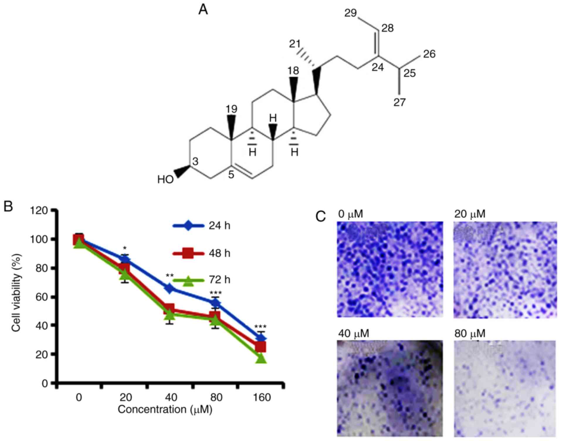
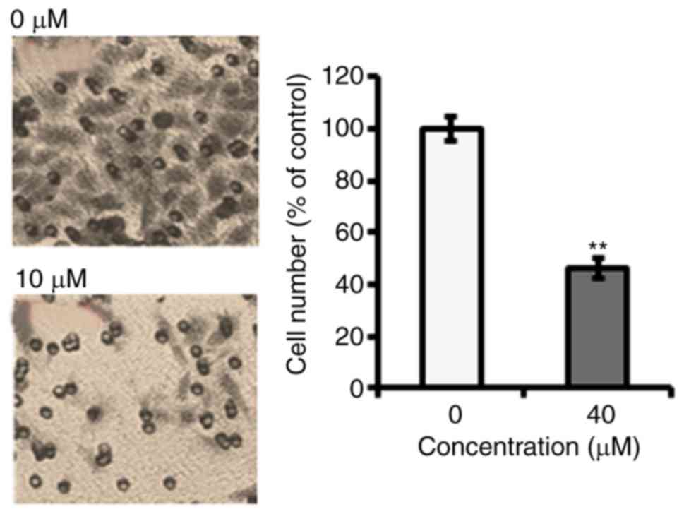
Anti-proliferative potential of
fucosterol on Cervical HeLa cancer cell line
To identify the anti-proliferative role of
fucosterol was evaluated against a panel of human cancer cell lines
(Table I). However, fucosterol
exhibited selective anticancer activity against cervical cancer
HeLa cells in a dose dependent manner and exhibited an
IC50 40 µM (Table I and
Fig. 1B). In the colony formation
assay, we observed that fucosterol administration reduced the
number of colonies in a dose-dependent manner (Fig. 1C).
 | Table I.IC50 of fucosterol against
different cancer cell lines as determined by MTT assay. |
Table I.
IC50 of fucosterol against
different cancer cell lines as determined by MTT assay.
| Cell line | IC50
(µM) |
|---|
| Gastric cancer
SNU-5 | 125 |
| Lung cancer
A-549 | 125 |
| Cervical cancer
HeLa | 40 |
| Prostate PC-3 | 125 |
| Breast MCF-7 | 125 |
| Pancreas
MiaPaca-2 | 250 |
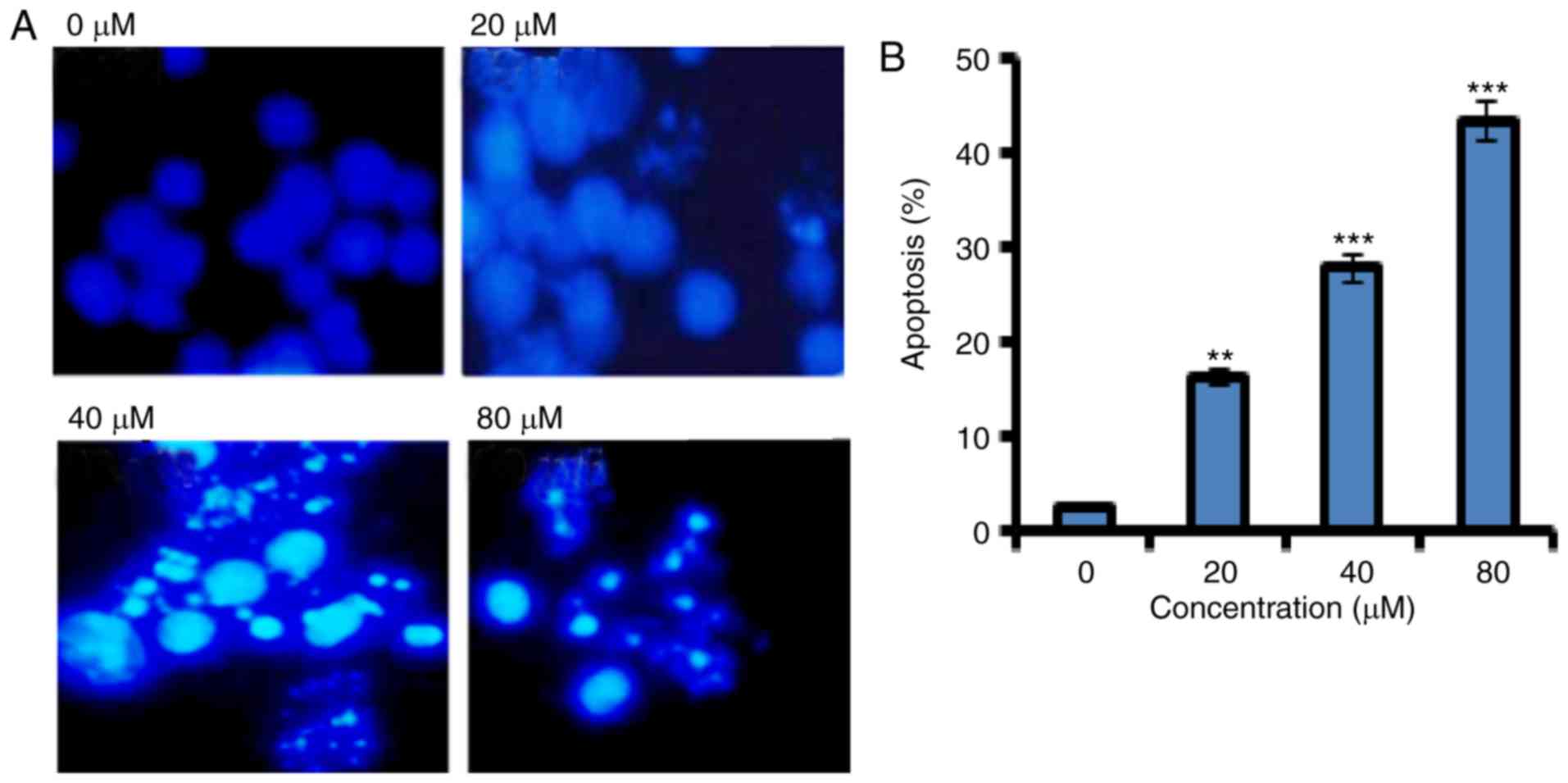
Fucosterol induced apoptosis in human
HeLa cervical cancer cells
In order to confirm apoptotic cell death induced by
fucosterol Annexin V/PI staining was performed. Flow cytometric
results showed that the percentage of apoptotic cell population
increased to 12.2, 37 and 62 % in HeLa cancer cells after 48 h at
the concentrations of 20, 40 and 80 µM, respectively as compared to
untreated control (Fig. 2). Thus the
results indicate that the extract caused apoptotic cell death in a
concentration dependent manner.
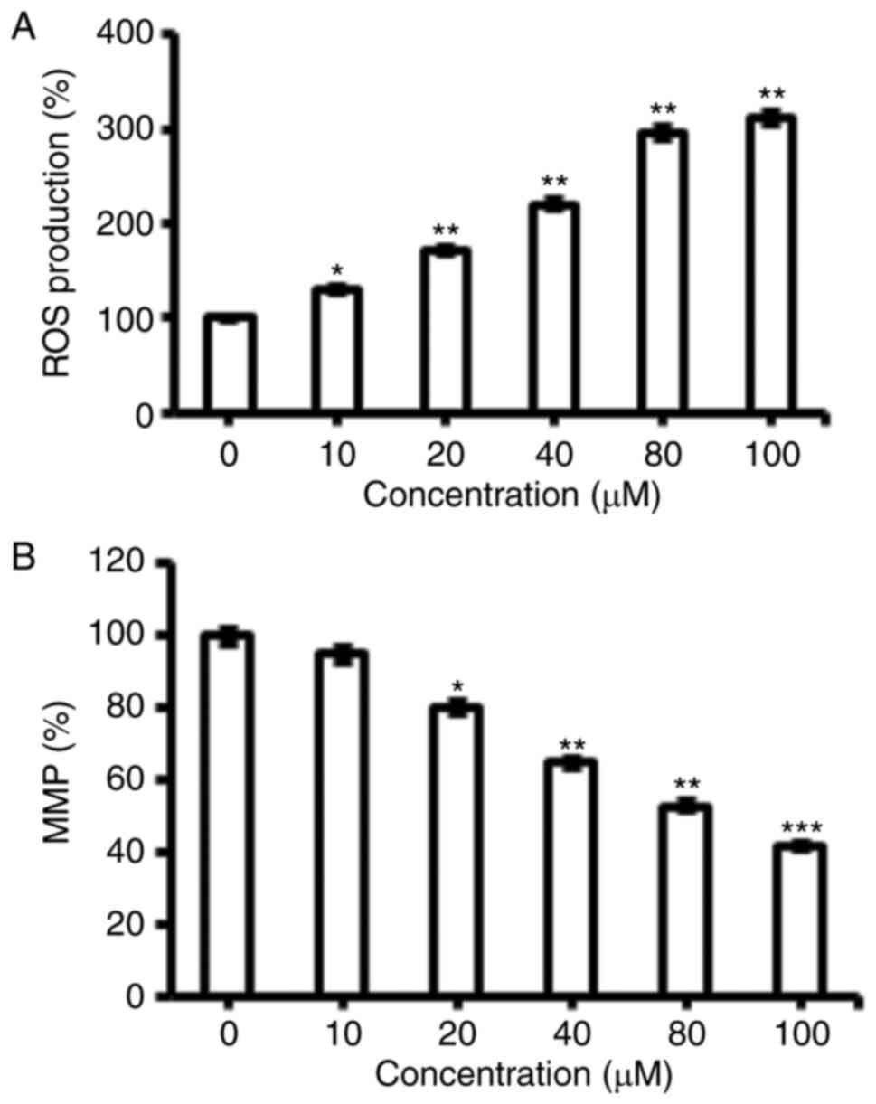
Fucosterol triggered the ROS
activation in human HeLa cervical cancer cells
The pro-apoptotic potential of fucosterol observed
through DAPI staining study suggested that fucosterol might induce
generation of intracellular ROS. Therefore, we calculated the ROS
level at varied concentrations of fucosterol for 48 h. The results
showed that the intracellular ROS levels of treated cells increased
110 to 305% as compared to untreated cells (Fig. 3A). Our result suggested that
fucosterol is a potent molecule for activating ROS in HeLa cells to
trigger the apoptosis.
Fucosterol reduces the mitochondrial
membrane potential (ΔΨm)
ROS generationis related to mitochondrial
dysfunction. It disrupts the outer mitochondrial potential to
release the death-promoting proteins (10). Therefore, we examined whether
fucosterol reduces the ΔΨm in HeLa cells treated with
fucosterol at varied concentrations. Fucosterol treated HeLa cells
showed a significant reduction in ΔΨm in a
dose-dependent manner (Fig. 3B).
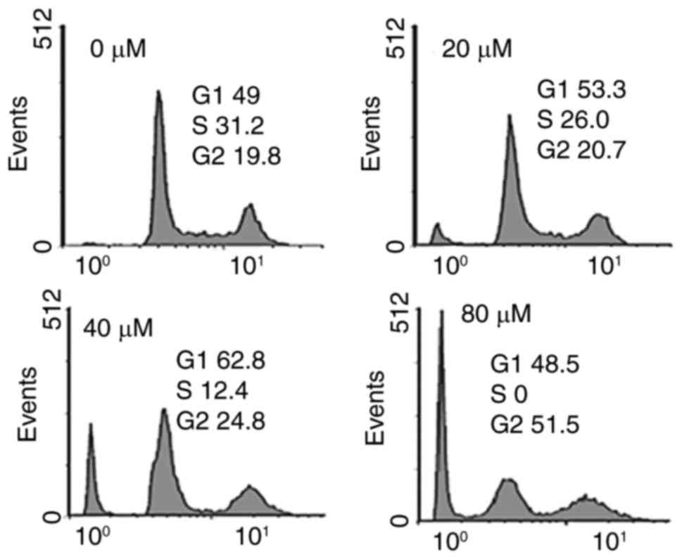
Fucosterol caused alterations in cell
cycle distribution of HeLa cancer cells
It was observed that the percentage of cells was
considerably increased in G2 at the concentrations of 0 to 80 µM
concentrations of fucosterol causing G2 arrest (Fig. 4). Additionally the populations of HeLa
cells G2 phase were marginally increased at a dose of 20 µM,
reasonably increased at 20 µM, and dramatically increased at 40 µM.
This fucosterol-induced G2 increase of HeLa cancer cells was
observed to exhibit a dose-dependent pattern.
Fucosterol inhibits cell
migration
Further, we investigated fucosterol can inhibit the
migration of cervical cancer cells at the IC50
concentration (40 µM). The results of transwell assays showed that
fucosterol reduced the migratory capability of cervical cancer HeLa
cells (Fig. 5).
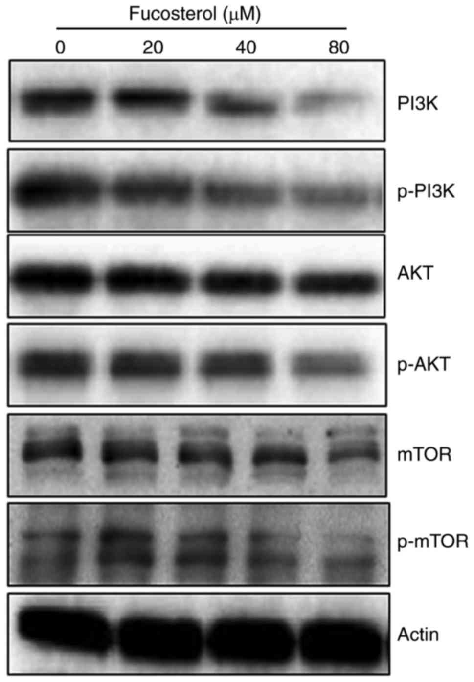
Fucosterol targets m-TOR/PI3K/Akt
signalling pathway
The fact that fucosterol could modulate the protein
expressions of m-TOR/PI3K/Akt signalling pathway, we carried out
the western blot analysis. The findings are shown in (Fig. 6) and indicate an interesting outcome.
Compared to the untreated control cells, fucosterol treated cells
showed a concentration-dependent downregulation of m-TOR and pm-TOR
proteins. It also caused marked downregulation of PI3K/Akt protein
expressions. Thus it may be concluded that fucosterol induced
anticancer and apoptotic effects partly via m-TOR/PI3K/Akt
signalling pathway.
Discussion
Cervical cancer is one of the major cancers detected
in women around the globe. Around 500,000 women are diagnosed for
this disease annually (1).
Nevertheless, the treatment options for cervical cancer are
limited. Moreover, surgery is the only appropriate choiceif the
cancer is detected at an early stage. Other options such as
radiotherapy have severe side effects which badly influence the
quality of life (2). Against this
backdrop, molecules from natural sources with limited side effects
may prove handy. In the current study, fucosterol showed potential
and selective growth inhibiting activity against HeLa cervical
cancer cells as evident from the proliferation assay. The selective
anticancer activity of fucosterol on HeLa cells is interesting. It
may be explained by the fact that several anticancer agents tend
exhibit selective anticancer effects against particular cell line
due to the involvement specific signalling pathways in different
cancer types (10). However, it would
be too early to delimit any particular reason for the selective
anticancer effects of fucosterol and thus will require further
investigation in future. As reported previously, many drugs exhibit
antiproliferative effects via induction of apoptosis. For instance,
several chemotherapeutic drugs, such as cisplatin, taxol and
5-fluorouracil (11–17) have been reported to alter explicit
apoptotic pathways. Additionally, resistance to drug is partially
explained by the ability of cancer cells to flee apoptosis
(18). To asses weather fucosterol
induces apoptosis in HeLa cells, we carried out the DAPI staining
of the fucosterol treated cells. It was observed that fucosterol
induces apoptosis in a concentration dependent manner. Further it
was observed that fucosterol treated cells displayed ROS mediated
MMP reduction. Our results are in agreement with studies carried
out previously (17). Therefore the
results suggest that fucosterol may induce apoptosis through
increasing intracellular ROS and reduction in MMP. Several
anti-cancer drugs target cancer cells partly by accretion of high
levels of ROS (18). Moreover,
mitochondria play a key role in ROS (19). For example, capsaicin disrupts MMP and
mediates oxidative stress resulting in apoptosis in pancreatic
cancer cells (11–17). Flow cytometry using propidium iodide
as a probe was used to study effects of fucosterolon cell cycle
progression. Fucosterol induced G2/M cell cycle arrest and led to a
significant increase of G2 cells in a dose dependently. Further, it
was shown that fucosterol could inhibit HeLa cancer cell in a
concentration dependent manner. These findings are promising since
it is well established that cervical cancer is one of the most
lethal cancers and fucosterol could inhibit this behavior.
Additionally, fucosterol also inhibited the cell migration of HeLa
cells as evident from the transwell assays. This migration
inhibiting potential indicates that fucosterol may prove beneficial
in inhibiting the metastasis of cancer cells in vivo and
therefore deserves further investigation.
Akt and mTOR are well-known major regulatory
signaling cascade that control cell proliferation, metabolism and
survival of cancer cells. Therefore, several inhibitors, such as
everolimus, have been developed and used for treatment to induce
apoptosis in cancer cells. To inhibit the mTOR signaling pathway,
rapamycin has been used in several studies. However, rapamycin only
inhibits mTOR complex (TORC) 1, and it consequently induces Akt
phosphorylation via feedback activation (20,21)
Thefore we investigated the effects of fucosterol on PI3/Akt/mTOR
pathway. Our results indicated that the expression levels of
various proteins including m-TOR, pm-TOR, PI3K, p-PI3K and Akt were
downregulated as evident from the western blot assay. These results
indicate the potential of fucosterol to inhibit cancer cell growth
via inhibition of PI3K/Akt/mTOR pathway. Though our results showed
promising activity of fucosterol, the feasibility of uses of
fucosterol in human and its bioavailability will require further
in vivo studies However, the low toxicity of fucosterol
towards normal cancer cells indicated that it could be used at even
at the concentrations that 4 times higher than its
IC50.
Taken together, we conclude that fucosterol may
prove a potential candidate for the treatment of cervical cancer by
regulating m-TOR/PI3K/Akt signalling pathway. With limited drug
options available and limited toxicity associated with this
naturally occurring fucosterol, this molecule seems a strongoption
and deserves further research endeavors.
References
|
1
|
Jemal A, Bray F, Center MM, Ferlay J, Ward
E and Forman D: Global cancer statistics. CA Cancer J Clin.
61:69–90. 2011. View Article : Google Scholar : PubMed/NCBI
|
|
2
|
Cadron I, Van Gorp T, Amant F, Leunen K,
Neven P and Vergote I: Chemotherapy for recurrent cervical cancer.
Gynecol Oncol. 107(1 Suppl 1): S113–S118. 2007. View Article : Google Scholar : PubMed/NCBI
|
|
3
|
Blunden G: Biologically active compounds
from marine organisms. Phytother Res. 15:89–94. 2001. View Article : Google Scholar : PubMed/NCBI
|
|
4
|
Zhen XH, Quan YC, Jiang HY, Wen ZS, Qu YL
and Guan LP: Fucosterol, a sterol extracted from sargassum
fusiforme, shows antidepressant and anticonvulsant effects. Eur J
Pharmacol. 768:131–138. 2015. View Article : Google Scholar : PubMed/NCBI
|
|
5
|
Engelman JA: Targeting PI3K signalling in
cancer: Opportunities, challenges and limitations. Nat Rev Cancer.
9:550–562. 2009. View
Article : Google Scholar : PubMed/NCBI
|
|
6
|
Romashkova JA and Makarov SS: NF-kappaB is
a target of AKT in anti-apoptotic PDGF signalling. Nature.
401:86–90. 1999. View
Article : Google Scholar : PubMed/NCBI
|
|
7
|
Chiang LC, Ng LT, Lin IC, Kuo PL and Lin
CC: Anti-proliferative effect of apigenin and its apoptotic
induction in human Hep G2 cells. Cancer Lett. 237:207–214. 2006.
View Article : Google Scholar : PubMed/NCBI
|
|
8
|
Chiang JH, Yang JS, Ma CY, Yang MD, Huang
HY, Hsia TC, Kuo HM, Wu PP, Lee TH and Chung JG: Danthron, an
anthraquinone derivative, induces DNA damage and caspase
cascades-mediated apoptosis in SNU-1 human gastric cancer cells
through mitochondrial permeability transition pores and
bax-triggered pathways. Chem Res Toxicol. 24:20–29. 2011.
View Article : Google Scholar : PubMed/NCBI
|
|
9
|
Sun SY, Hail N Jr and Lotan R: Apoptosis
as a novel target for cancer chemoprevention. J Natl Cancer Inst.
96:662–672. 2004. View Article : Google Scholar : PubMed/NCBI
|
|
10
|
Stierle AA, Stierle DB and Kelly K:
Berkelic acid, a novel spiroketal with selective anticancer
activity from an acid mine waste fungal extremophile. J Org Chem.
71:5357–5360. 2006. View Article : Google Scholar : PubMed/NCBI
|
|
11
|
Maitra R, Porter MA, Huang S and Gilmour
BP: Inhibition of NFkappaB by the natural product Withaferin A in
cellular models of Cystic Fibrosis inflammation. J Inflamm (Lond).
6:152009. View Article : Google Scholar : PubMed/NCBI
|
|
12
|
Hissin PJ and Hilf R: A fluorometric
method for determination of oxidized and reduced glutathione in
tissues. Anal Biochem. 74:214–226. 1976. View Article : Google Scholar : PubMed/NCBI
|
|
13
|
Chipuk JE, Bouchier-Hayes L and Green DR:
Mitochondrial outer membrane permeabilization during apoptosis: The
innocent bystander scenario. Cell Death Diff. 13:1396–1402. 2006.
View Article : Google Scholar
|
|
14
|
Azuma M, Tamatani T, Ashida Y, Takashima
R, Harada K and Sato M: Cisplatin induces apoptosis in oral
squamous carcinoma cells by the mitochondria-mediated but not the
NF-kappaB-suppressed pathway. Oral Oncol. 39:282–289. 2003.
View Article : Google Scholar : PubMed/NCBI
|
|
15
|
Yoneda K, Yamamoto T and Osaki T: p53- and
p21-independent apoptosis of squamous cell carcinoma cells induced
by 5-fluorouracil and radiation. Oral Oncol. 34:529–537. 1998.
View Article : Google Scholar : PubMed/NCBI
|
|
16
|
Abal M, Andreu JM and Barasoain I:
Taxanes: Microtubule and centrosome targets, and cell cycle
dependent mechanisms of action. Curr Cancer Drug Targets.
3:193–203. 2003. View Article : Google Scholar : PubMed/NCBI
|
|
17
|
Ferreira CG, Epping M, Kruyt FA and
Giaccone G: Apoptosis: Target of cancer therapy. Clin Cancer Res.
8:2024–2034. 2002.PubMed/NCBI
|
|
18
|
Malaguarnera L: Implications of apoptosis
regulators in tumorigenesis. Cancer Metastasis Rev. 23:367–387.
2004. View Article : Google Scholar : PubMed/NCBI
|
|
19
|
Ding H, Han C, Guo D, Chin YW, Ding Y,
Kinghorn AD and D'Ambrosio SM: Selective induction of apoptosis of
human oral cancer cell lines by avocado extracts via a ROS-mediated
mechanism. Nutr Cancer. 61:348–356. 2009. View Article : Google Scholar : PubMed/NCBI
|
|
20
|
Radhakrishnan P, Baraneedharan U,
Veluchamy S, Dhandapani M, Pinto DD, Thiyagarajan S, Thayakumar A,
Prasath A, K A, Velu A, et al: Inhibition of rapamycin-induced AKT
activation elicits differential antitumor response in head and neck
cancers. Cancer Res. 73:1118–1127. 2013. View Article : Google Scholar : PubMed/NCBI
|
|
21
|
Khursheed A, Rather MA and Rashid R:
Plant-based natural compounds and herbal extracts as promising
apoptotic agents: Their implications for cancer prevention and
treatment. Bio Med Pharma. 3:245–269. 2016.
|