Introduction
The potential use of bacteria for cancer treatment
has been extensively investigated in previous years. Bacteria,
including Bifidobacterium (1,2),
Clostridium (3) and
Salmonella have been demonstrated to preferentially target
and replicate in the hypoxic and necrotic regions of a tumor,
resulting in tumor repression (4–7). In a
previous study, a synthetic biology approach was used to generate
the novel Salmonella typhimurium strain YB1 (YB1) (8). This bacterium specifically colonizes and
proliferates in the hypoxic/necrotic areas of the tumor, but avoids
normal organs and retards tumor growth (8). Furthermore, a previous study reported
that numerous macrophages accumulate in breast tumors and are
associated with a poor prognosis (9).
Macrophages are heterogeneous cells that respond
differently to various stimulating signals and display numerous
different phenotypes (5). The M1 and
M2 macrophage phenotypes represent the two extremes of a broad
range of macrophage functional states. Fully polarized M1 (or
classically activated) macrophages are stimulated by microbial
agents or pro-inflammatory factors, including lipopolysaccharide
(LPS), whereas M2 (or alternatively activated) macrophages respond
to anti-inflammatory molecules, including interleukin-4 (IL-4)
(10,11). Macrophages located in the stroma of
breast cancer tissues [known as tumor-associated macrophages
(TAMs)] are primarily M2 macrophages activated by IL-4-producing
cluster of differentiation (CD)4+ T cells (12). TAMs are the most notable migratory
hematopoietic cell type in the tumor microenvironment and promote
the invasiveness of breast cancer cells (13).
Clinically, a large amount of macrophage
infiltration in tumor sections from patients with breast cancer has
been observed using CD68 immunohistochemical staining. TAMs are
associated with breast cancer aggressiveness and promote cancer
metastasis, whereas M1 macrophages are prone to killing cancer
cells and devouring bacteria (14).
Furthermore, studies have revealed that TAMs (which are primarily
M2 macrophages activated by IL-4) exhibit a
CD206high/human leukocyte antigen-antigen D related
(HLA-DR)low phenotype that is associated with immune
suppression (15–17). Therefore, CD206 and HLA-DR may be used
as markers for M1 and M2 macrophage phenotype analysis (15). In the present study, the newly
engineered tumor-targeting YB1 strain was used in order to attempt
to redirect M2 macrophages into the M1 phenotype. More than half of
the M2 macrophages devoured the bacteria after 2 h of co-culture.
These M2 macrophages exhibited a decreased CD206 expression and an
increased HLA-DR expression. Therefore, the IL-4-activated M2
macrophages switched from the
CD206high/HLA-DRlow phenotype to the
CD206low/HLA-DRhigh phenotype subsequent to
co-culture with the engineered YB1 strain. The present study
indicates that differentiated M2 macrophages may be redirected into
an M1 phenotype following exposure to different stimuli. This
finding may reflect a potential mechanism by which bacteria retard
tumor growth. Therefore, these engineered bacteria may be used as a
vector to target tumors.
Materials and methods
Patient samples and macrophage
immunohistochemistry staining
All tumor samples from breast-infiltrating ductal
carcinomas were obtained from female patients (mean age, 45 years;
age range, 35–55 years) at the Guangdong Women and Children's
Hospital (Guangdong, China). The samples were used with written
informed consent and ethical approval was obtained from the
Internal Review and the Ethics Boards of Guangdong Women and
Children's Hospital (Guangdong, China).
The samples were fixed in 10% formalin for >2 h
at room temperature, paraffin-embedded (3 min at 56°C) and
sectioned into 5 µM-thick slices. The macrophages were visualized
by immunohistochemistry staining using an anti-CD68 antibody (cat.
no. M0814; dilution, 1:200; Dako; Agilent Technologies, Inc., Santa
Clara, CA, USA), and sections were treated using this antibody
overnight at 4°C. For details, please refer to reference (18).
Bacterial culture
The bacterial YB1 strain was cultured in lysogeny
broth medium overnight (12 to 16 h) (Sigma-Aldrich; Merck KGaA,
Darmstadt, Germany) supplemented with chloramphenicol and
2,3-diaminopropionic acid (Sigma-Aldrich; Merck KGaA) at 37°C
(8).
Isolation and activation of human
monocyte-derived macrophages
Institutional ethical approval was obtained from the
Internal Review and the Ethics Boards of Guangdong Women and
Children's Hospital, Guangdong, China prior to conducting the
study. Human mononuclear cells were isolated from 100 ml peripheral
blood of healthy donors by Ficoll density gradient centrifugation
(20°C at 250 × g for 20 min), as previously described (18). The resulting monocyte-derived
macrophages were activated by the addition of IL-4 (45 ng/ml) to
the culture medium for 3 days (19),
and LPS (20 ng/ml) was added as a control.
Bacteria and macrophage
co-culture
Isolated macrophages were activated by IL-4. The
bacterial YB1 strain, which carried the green fluorescent protein
(GFP)-tagged plasmid, was added to the macrophage culture for a
final bacterial concentration of 5×10⁶/ml. The cultures
were incubated at 37°C under hypoxic conditions as previously
described (8) for 2 h. Then, the
bacteria were washed away and the macrophages were harvested for
further analysis.
Flow cytometry
Following the co-culture, the macrophages were fixed
in paraformaldehyde (4%) at 4°C for 0.5–2 h. The macrophage
phagocytic rate of YB1 (GFP) was detected using flow cytometry.
CD206 or HLA-DR expression levels in the macrophages were also
determined using flow cytometry. Briefly, the macrophages
(105-106/ml) were collected, fixed in 4%
paraformaldehyde at 4°C for 0.5–2 h, blocked with 5% bovine serum
albumin for 20 min at room temperature and stained using a
phycoerythrin-conjugated CD206 (dilution, 1:20; cat no. 321105;
BioLegend, Inc., San Diego, CA, USA) or allophycocyanin-conjugated
HLA-DR (dilution, 1:20; cat no. 307609; BioLegend, Inc.) antibody
for 1 h at 4°C. The corresponding isotype control was included in
each test. Then, CD206 or HLA-DR expression was analyzed using flow
cytometry. FlowJo software (version 7.6.1; FlowJo LLC, Ashland, OR,
USA) was used to analyze the data.
Western blotting
Cells were lysed using radioimmunoprecipitation
assay lysis buffer (50 mM TrisHCl pH 7.4, 150 mM NaCl, 2 mM EDTA,
1% NP-40, 0.1% SDS) and protease inhibitors (Sigma-Aldrich; Merck
KGaA). Cells were lysed with lysis buffer for 15–20 min at 4°C.
HLA-DR expression was determined according to a previously
described protocol (18). The
proteins (20 µl per lane) were separated on 10% SDS-PAGE gels and
transferred onto nitrocellulose membranes. The membranes were
blocked in 5% non-fat milk for 2 h at room temperature and then
incubated with antibodies against HLA-DR (1:5,000; rabbit
monoclonal IgG antibody; cat no. ab92511; Abcam, Cambridge, UK)
overnight at 4°C. Subsequent to washing using 0.1% TBST (50 mM
tris, 150 mM NaCl, 0.1% Tween 20, pH 7.4) three times (10 min
each), the membrane was incubated with a horseradish peroxidase
(HRP)-conjugated secondary antibody (GE Healthcare, Chicago, IL,
USA) for 1 h at room temperature. Then, the membranes were washed
again using 0.1% TBST three times (10 min each). The HRP signal was
visualized using enhanced chemiluminescence (ChemiDoc MP Imaging
System; Bio-Rad Laboratories, Inc., Hercules, CA, USA), and
analyzed using Image Lab (version 5.2.1; Bio-Rad Laboratories,
Inc.).
Results
Macrophage infiltration in breast
tumor tissues
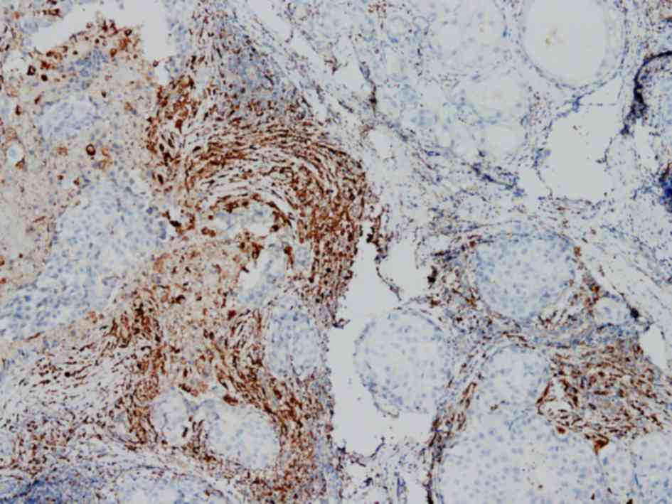
The engineered YB1 strain may specifically target
and survive in a solid tumor. To confirm macrophage infiltration in
the breast tumor, clinical breast cancer samples were collected and
CD68 immunohistochemistry staining was used to demonstrate the
macrophage distribution. As presented in Fig. 1, a large number of macrophages
(CD68-positive) infiltrated the breast tumor, particularly in the
tumor-associated stromal border, a result consistent with previous
reports (12). This result implies
that bacteria may be able to target macrophages localized in the
breast tumor.
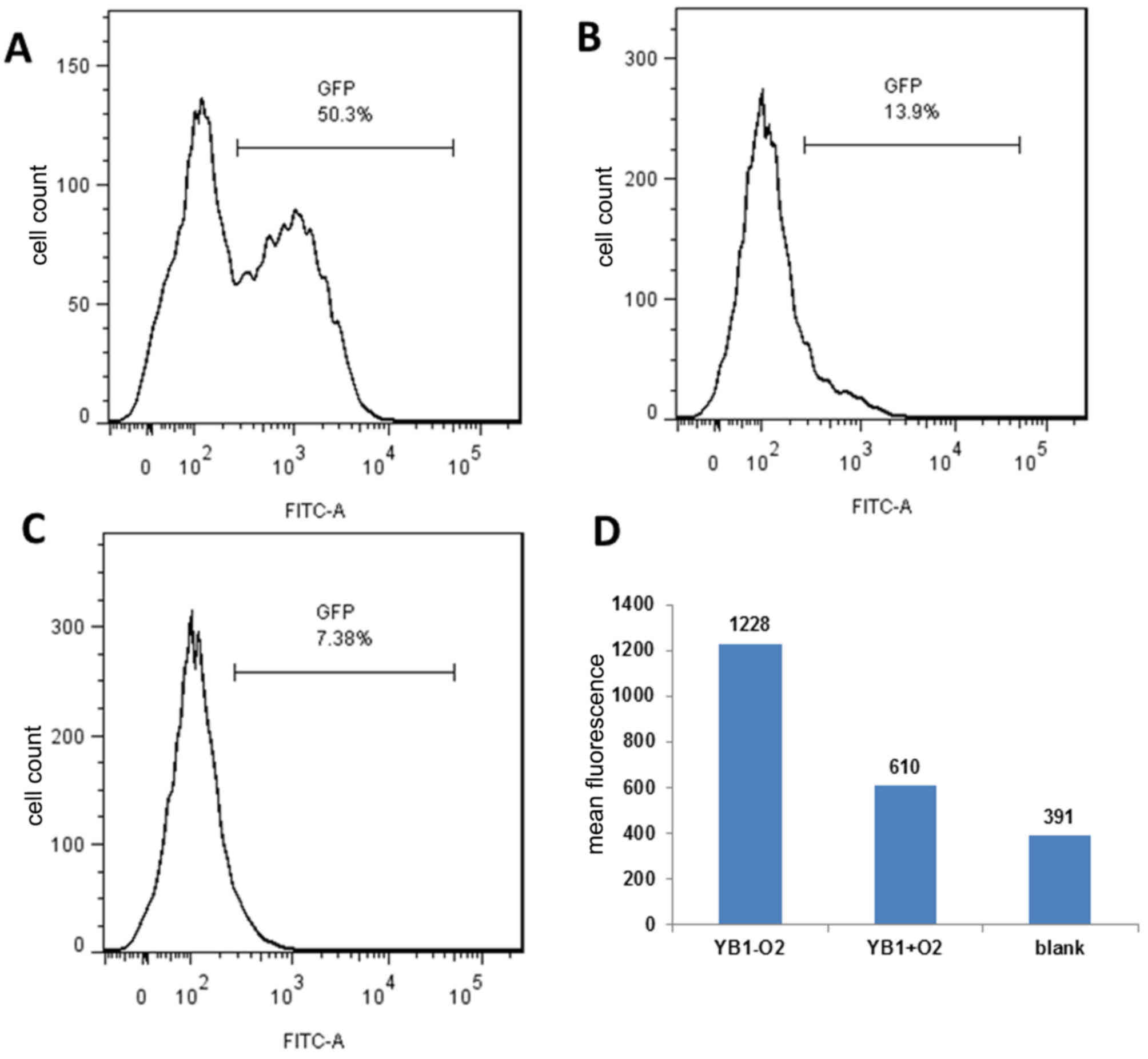
YB1 invaded M2 macrophages under
hypoxic conditions
The engineered YB1 strain only survives under
hypoxic conditions (8). However,
whether YB1 may invade M2 macrophages under hypoxic conditions
remains unknown. Thus, an anaerobic microenvironment was stimulated
in vitro and co-cultured M2 macrophages with the YB1 strain
with the GFP-tagged plasmid for 2 h. Flow cytometry analysis was
used to determine the YB1 invasion rate. As presented in Fig. 2A, YB1 had a high invasion rate under
hypoxic conditions; after 2 h, >50% of the macrophages were
invaded. In contrast, the invasion rate was very low under normal
conditions (21% oxygen) (Fig. 2B) as
YB1 may not survive. Fig. 2C
presented the results of the blank control. It was additionally
identified that the mean fluorescence was substantially higher
under hypoxic conditions than under normal conditions, indicating
that >1 bacterium invaded each macrophage (Fig. 2D).
M2 macrophages exhibited increased
HLA-DR and decreased CD206 expression
Subsequent to co-culturing M2 macrophages with YB1,
the macrophages were collected and flow cytometry was used to
determine the HLA-DR expression levels (Fig. 3A). LPS activated the macrophages to
express higher levels of HLA-DR compared with those in the
IL-4-activated macrophages. Notably, the YB1 alone strain induced
the highest HLA-DR expression in macrophages, suggesting the high
efficiency of YB1 in activating the M1 macrophage phenotype. When
the macrophages were activated in advance with IL-4 and then
co-cultured with YB1, an increase in HLA-DR expression was observed
compared with LPS or IL-4 alone activated macrophages. Western
blotting confirmed these results (Fig.
3B). Next, the M2 macrophage phenotype marker CD206 was
examined (Fig. 3C) and it was
identified that IL-4-activated macrophages expressed higher CD206
levels compared with LPS-activated macrophages. The YB1 strain
alone reduced the CD206 expression levels compared with every other
group. Notably, the YB1 strain reduced CD206 expression in the
IL-4-activated M2 macrophages.
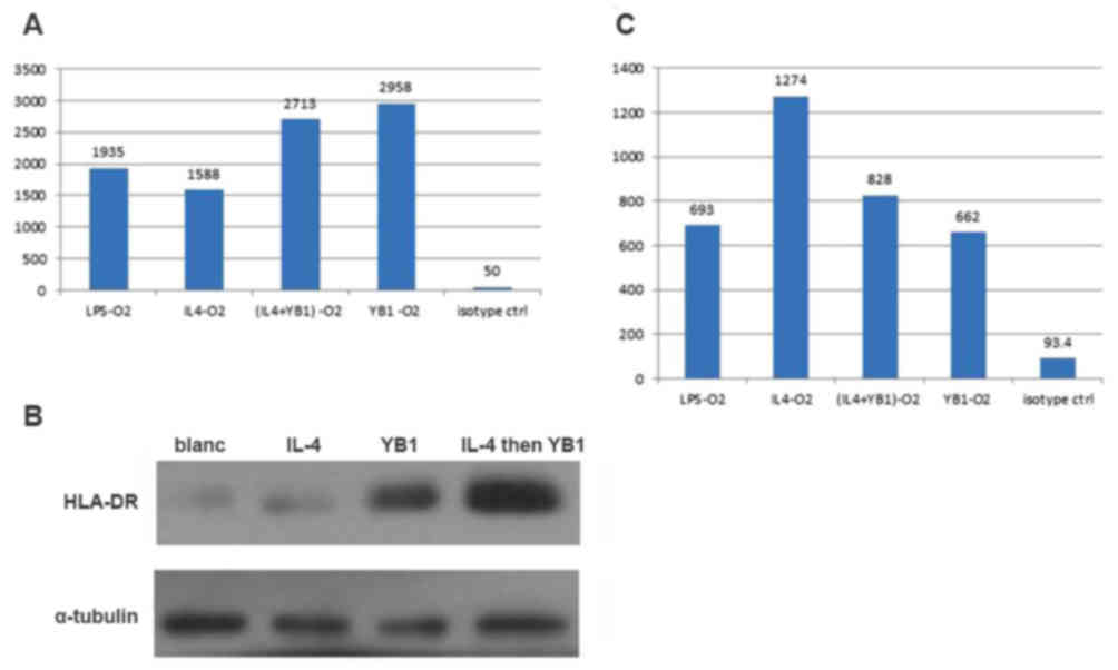 | Figure 3.M2 macrophages exhibited increased
HLA-DR and decreased CD206 expression levels. (A) HLA-DR expression
levels in M2 macrophages activated using LPS, IL-4, YB1, a
combination of YB1 and IL-4 or an isotype. (B) HLA-DR expression
confirmed using western blotting. (C) CD206 expression levels in M2
macrophages activated using LPS, IL-4, YB1, a combination of YB1
and IL-4 or an isotype. HLA-DR, human leukocyte antigen-antigen D
related; CD206, mannose receptor; LPS, lipopolysaccharide; IL,
interleukin; YB1, Salmonella typhimurium strain YB1. |
Discussion
Although a broad range of macrophage subsets have
been identified, the two major macrophage populations are the M1
(classically activated) macrophages and M2 (alternatively
activated) macrophages (20). These
two different types of macrophages have different effects on tumor
progression. As early as 1980, studies have demonstrated that
bacterial LPS activates macrophages (21–23) to
specifically kill tumor cells including breast cancer cells but has
no effect on normal cells (24,25). This
effect on macrophages requires LPS for maintenance (26). Additionally, macrophages themselves
possess a phagocytic ability. When a tumor occurs, macrophages
migrate to the tumor location, as directed by the action of
chemokines, and devour the tumor cells (27). Macrophages may kill tumor cells,
though it remains unknown why this killing effect halts tumor
growth and distant metastasis (28).
Beyond the tumor immune escape mechanism (29), studies have identified that
macrophages are induced by tumor cells in the tumor
microenvironment and develop tumor-promoting properties (M2 type,
otherwise known as TAMs) (30,31).
Statistical data analyses have revealed that the proportion of TAMs
in solid tumor tissues may be as high as 80% (29). Likewise, numerous macrophages have
been detected in clinical breast cancer samples and, furthermore,
macrophage infiltration and breast cancer metastasis are associated
(32–34). However, although M1 and M2 macrophages
serve different functions in tumor progression, there is no
absolute boundary between the two types of macrophages. In the
tumor microenvironment, factors including the MHC expression level
in tumor cells and the oxygen pressure in the microenvironment
affect the macrophage phenotype (29). Therefore, the phenotype of
differentiated macrophages may change. This finding indicates a
novel target of tumor treatment: Macrophages in the tumor
microenvironment. If M2 macrophages may be directed to become the
M1 type, one of the drivers of tumor progression would be
eliminated, and result in the gain of one more helper to kill tumor
cells.
Deng et al (35) has demonstrated that suppression of
heme oxygenase-1 in TAMs in a breast cancer mouse model
alternatively activates the switching of the M2 macrophage type to
the classically activated M1 macrophage type. However, the
‘weapons’ used to specifically target macrophages in breast cancer
are still lacking.
In the present study, a novel engineered
Salmonella strain (YB1) was reported to induce increased
HLA-DR expression and decreased CD206 expression in differentiated
M2 macrophages. These M2 macrophages changed from the
CD206high/HLA-DRlow phenotype to the
CD206low/HLA-DRhigh phenotype, indicating an
M2 to M1-type switch. This result suggests a potential use for the
engineered tumor-targeting bacteria YB1 in redirecting M2 type
macrophages into the M1 type and thus suppressing tumor growth.
Overall, the results suggest that the engineered
bacterial YB1 strain may be a good candidate for targeting and
redirecting M2 macrophages into the M1 type. In addition to its
tumor targeting ability, these bacteria may survive and proliferate
in the tumor microenvironment; therefore, the effects would be
long-lasting, and the activation of the M1 type would be sustained.
Furthermore, for safety, this engineered Salmonella YB1
strain is controllable and may be eliminated by antibiotics.
Finally, the genetic background of YB1 is clear and may be
engineered to carry further ‘weapons’ in order to kill cancer
cells.
Acknowledgements
The present study was supported by the National
Natural Science Foundation of China (grant no. 81202076) and the
Guangzhou Science and Technology Program (grant no. 2014J2200007).
The authors would like to thank Dr Jian-dong Huang (Hong Kong
University) for supplying the bacteria strain.
References
|
1
|
Yazawa K, Fujimori M, Nakamura T, Sasaki
T, Amano J, Kano Y and Taniguchi S: Bifidobacterium longum as a
delivery system for gene therapy of chemically induced rat mammary
tumors. Breast Cancer Res Treat. 66:165–170. 2001. View Article : Google Scholar : PubMed/NCBI
|
|
2
|
Fujimori M: Anaerobic bacteria as a gene
delivery system for breast cancer therapy. Nihon Rinsho.
66:1211–1218. 2008.(In Japanese). PubMed/NCBI
|
|
3
|
Liu SC, Ahn GO, Kioi M, Dorie MJ,
Patterson AV and Brown JM: Optimized clostridium-directed enzyme
prodrug therapy improves the antitumor activity of the novel DNA
cross-linking agent PR-104. Cancer Res. 68:7995–8003. 2008.
View Article : Google Scholar : PubMed/NCBI
|
|
4
|
Zhao M, Yang M, Ma H, Li X, Tan X, Li S,
Yang Z and Hoffman RM: Targeted therapy with a Salmonella
typhimurium leucine-arginine auxotroph cures orthotopic human
breast tumors in nude mice. Cancer Res. 66:7647–7652. 2006.
View Article : Google Scholar : PubMed/NCBI
|
|
5
|
Low KB, Ittensohn M, Le T, Platt J, Sodi
S, Amoss M, Ash O, Carmichael E, Chakraborty A, Fischer J, et al:
Lipid A mutant Salmonella with suppressed virulence and TNFalpha
induction retain tumor-targeting in vivo. Nat Biotechnol. 17:37–41.
1999. View Article : Google Scholar : PubMed/NCBI
|
|
6
|
Felgner S, Kocijancic D, Frahm M, Curtiss
R III, Erhardt M and Weiss S: Optimizing salmonella enterica
serovar typhimurium for bacteria-mediated tumor therapy. Gut
microbes. 7:171–177. 2016. View Article : Google Scholar : PubMed/NCBI
|
|
7
|
Frahm M, Felgner S, Kocijancic D, Rohde M,
Hensel M, Curtiss R III, Erhardt M and Weiss S: Efficiency of
conditionally attenuated Salmonella enterica serovar Typhimurium in
bacterium-mediated tumor therapy. mBio. 6:pii: e00254–15. 2015.
View Article : Google Scholar
|
|
8
|
Yu B, Yang M, Shi L, Yao Y, Jiang Q, Li X,
Tang LH, Zheng BJ, Yuen KY, Smith DK, et al: Explicit hypoxia
targeting with tumor suppression by creating an ‘obligate’
anaerobic Salmonella Typhimurium strain. Sci Rep. 2:4362012.
View Article : Google Scholar : PubMed/NCBI
|
|
9
|
Tang X: Tumor-associated macrophages as
potential diagnostic and prognostic biomarkers in breast cancer.
Cancer Lett. 332:3–10. 2013. View Article : Google Scholar : PubMed/NCBI
|
|
10
|
Luo Y, Zhou H, Krueger J, Kaplan C, Lee
SH, Dolman C, Markowitz D, Wu W, Liu C, Reisfeld RA and Xiang R:
Targeting tumor-associated macrophages as a novel strategy against
breast cancer. J Clin Invest. 116:2132–2141. 2006. View Article : Google Scholar : PubMed/NCBI
|
|
11
|
Leek RD, Lewis CE, Whitehouse R, Greenall
M, Clarke J and Harris AL: Association of macrophage infiltration
with angiogenesis and prognosis in invasive breast carcinoma.
Cancer Res. 56:4625–4629. 1996.PubMed/NCBI
|
|
12
|
DeNardo DG, Barreto JB, Andreu P, Vasquez
L, Tawfik D, Kolhatkar N and Coussens LM: CD4(+) T cells regulate
pulmonary metastasis of mammary carcinomas by enhancing protumor
properties of macrophages. Cancer cell. 16:91–102. 2009. View Article : Google Scholar : PubMed/NCBI
|
|
13
|
Zhang C, Gao L, Cai Y, Liu H, Gao D, Lai
J, Jia B, Wang F and Liu Z: Inhibition of tumor growth and
metastasis by photoimmunotherapy targeting tumor-associated
macrophage in a sorafenib-resistant tumor model. Biomaterials.
84:1–12. 2016. View Article : Google Scholar : PubMed/NCBI
|
|
14
|
Khabbazi S, Goumon Y and Parat MO:
Morphine modulates interleukin-4- or breast cancer cell-induced
pro-metastatic activation of macrophages. Sci Rep. 5:113892015.
View Article : Google Scholar : PubMed/NCBI
|
|
15
|
Su S, Liu Q, Chen J, Chen J, Chen F, He C,
Huang D, Wu W, Lin L, Huang W, et al: A positive feedback loop
between mesenchymal-like cancer cells and macrophages is essential
to breast cancer metastasis. Cancer cell. 25:605–620. 2014.
View Article : Google Scholar : PubMed/NCBI
|
|
16
|
Dangaj D, Abbott KL, Mookerjee A, Zhao A,
Kirby PS, Sandaltzopoulos R, Powell DJ Jr, Lamazière A, Siegel DL,
Wolf C and Scholler N: Mannose receptor (MR) engagement by
mesothelin GPI anchor polarizes tumor-associated macrophages and is
blocked by anti-MR human recombinant antibody. PLoS One.
6:e283862011. View Article : Google Scholar : PubMed/NCBI
|
|
17
|
Kuang DM, Wu Y, Chen N, Cheng J, Zhuang SM
and Zheng L: Tumor-derived hyaluronan induces formation of
immunosuppressive macrophages through transient early activation of
monocytes. Blood. 110:587–595. 2007. View Article : Google Scholar : PubMed/NCBI
|
|
18
|
Yang M, Chen J, Su F, Yu B, Su F, Lin L,
Liu Y, Huang JD and Song E: Microvesicles secreted by macrophages
shuttle invasion-potentiating microRNAs into breast cancer cells.
Mol cancer. 10:1172011. View Article : Google Scholar : PubMed/NCBI
|
|
19
|
Chen J, Yao Y, Gong C, Yu F, Su S, Chen J,
Liu B, Deng H, Wang F, Lin L, et al: CCL18 from tumor-associated
macrophages promotes breast cancer metastasis via PITPNM3. Cancer
Cell. 19:541–555. 2011. View Article : Google Scholar : PubMed/NCBI
|
|
20
|
Castoldi A, Naffah de Souza C, Câmara NO
and Moraes-Vieira PM: The macrophage switch in obesity development.
Front Immunol. 6:6372016. View Article : Google Scholar : PubMed/NCBI
|
|
21
|
Hamaidia M, Staumont B, Duysinx B, Louis R
and Willems L: Improvement of malignant pleural mesothelioma
immunotherapy by epigenetic modulators. Curr Top Med Chem.
16:777–787. 2016. View Article : Google Scholar : PubMed/NCBI
|
|
22
|
Murray PJ and Wynn TA: Protective and
pathogenic functions of macrophage subsets. Nat Rev Immunol.
11:723–737. 2011. View
Article : Google Scholar : PubMed/NCBI
|
|
23
|
Hambleton J, Weinstein SL, Lem L and
DeFranco AL: Activation of c-Jun N-terminal kinase in bacterial
lipopolysaccharide-stimulated macrophages. Proc Natl Acad Sci USA.
93:pp. 2774–2778. 1996; View Article : Google Scholar : PubMed/NCBI
|
|
24
|
Chimal-Ramírez GK, Espinoza-Sánchez NA,
Chávez-Sánchez L, Arriaga-Pizano L and Fuentes-Pananá EM: Monocyte
differentiation towards protumor activity does not correlate with
M1 or M2 phenotypes. J Immunol Res. 2016:60314862016. View Article : Google Scholar : PubMed/NCBI
|
|
25
|
Guo H, Liu Y, Gu J, Wang Y, Liu L, Zhang P
and Li Y: Endostatin inhibits the growth and migration of 4T1 mouse
breast cancer cells by skewing macrophage polarity toward the M1
phenotype. Cancer Immunol Immunother. 65:677–688. 2016. View Article : Google Scholar : PubMed/NCBI
|
|
26
|
Cameron DJ and Churchill WH: Cytotoxicity
of human macrophages for tumor cells: Enhancement by bacterial
lipopolysaccharides (LPS). J Immunol. 124:708–712. 1980.PubMed/NCBI
|
|
27
|
Sinha P, Clements VK, Miller S and
Ostrand-Rosenberg S: Tumor immunity: A balancing act between T cell
activation, macrophage activation and tumor-induced immune
suppression. Cancer Immunol Immunother. 54:1137–1142. 2005.
View Article : Google Scholar : PubMed/NCBI
|
|
28
|
Mills CD, Lenz LL and Harris RA: A
breakthrough: Macrophage-directed cancer immunotherapy. Cancer Res.
76:513–516. 2016. View Article : Google Scholar : PubMed/NCBI
|
|
29
|
Mitra R, Singh S and Khar A: Antitumour
immune responses. Expert Rev Mol Med. 5:1–19. 2003. View Article : Google Scholar : PubMed/NCBI
|
|
30
|
Mantovani A, Sozzani S, Locati M, Allavena
P and Sica A: Macrophage polarization: Tumor-associated macrophages
as a paradigm for polarized M2 mononuclear phagocytes. Trends
Immunol. 23:549–555. 2002. View Article : Google Scholar : PubMed/NCBI
|
|
31
|
Wahl LM and Kleinman HK: Tumor-associated
macrophages as targets for cancer therapy. J Natl Cancer Inst.
90:1583–1584. 1998. View Article : Google Scholar : PubMed/NCBI
|
|
32
|
Ueno T, Toi M, Saji H, Muta M, Bando H,
Kuroi K, Koike M, Inadera H and Matsushima K: Significance of
macrophage chemoattractant protein-1 in macrophage recruitment,
angiogenesis, and survival in human breast cancer. Clin Cancer Res.
6:3282–3289. 2000.PubMed/NCBI
|
|
33
|
Leek RD and Harris AL: Tumor-associated
macrophages in breast cancer. J Mammary Gland Biol Neoplasia.
7:177–189. 2002. View Article : Google Scholar : PubMed/NCBI
|
|
34
|
Valković T, Dobrila F, Melato M, Sasso F,
Rizzardi C and Jonjić N: Correlation between vascular endothelial
growth factor, angiogenesis, and tumor-associated macrophages in
invasive ductal breast carcinoma. Virchows Arch. 440:583–588. 2002.
View Article : Google Scholar : PubMed/NCBI
|
|
35
|
Deng R, Wang SM, Yin T, Ye TH, Shen GB, Li
L, Zhao JY, Sang YX, Duan XG and Wei YQ: Inhibition of tumor growth
and alteration of associated macrophage cell type by an HO-1
inhibitor in breast carcinoma-bearing mice. Oncol Res. 20:473–482.
2013. View Article : Google Scholar : PubMed/NCBI
|