Introduction
Laryngeal squamous cell carcinoma (LSCC) is one of
the most common head and neck malignancies and has been reported to
account for ~2.4% of all new malignancies worldwide each year
(1). The prognosis for LSCC has not
shown any improvement in the last 30 years (2), due to lymphatic metastasis.
Additionally, the supraglottic type frequently undergoes lymphatic
metastasis in LSCC. A better understanding of the molecular
pathways that result in lymphatic metastasis of supraglottic LSCC
is essential in the identification of novel molecular biomarkers
which have clinical utility in predicting prognosis and therapeutic
efficacy, as well as in designing targeted therapy for this
disease.
MicroRNAs (miRNAs) are a class of short non-coding
RNAs that modulate gene expression by targeting mRNAs and
triggering either the repression of translation or RNA degradation
(3). Due to the partial
complementarity between miRNAs and their target transcripts, a
single miRNA is capable of simultaneously regulating hundreds of
genes, and therefore carries a significant modulatory potential
(4). miRNAs are involved in tissue
differentiation during both physiological and pathological
processes (5). Previous studies on
different cancer types have identified the emergence of distinct
miRNA expression profiles between tumor tissues and their
corresponding normal tissues (6,7).
Another study identified miRNA expression profiles capable of
distinguishing the different tumor subtypes or developmental
lineages (8). Increasing data
support the value of miRNA expression profiles as a novel biomarker
in diagnosis, prognosis and as a new target in therapy.
In this study, the miRNA expression of normal
laryngeal epithelia was compared with primary human supraglottic
LSCC at advanced stage to define those miRNA that are most capable
of differentiating disease, thus having the greatest potential as
biomarkers and therapy targets.
Materials and methods
Tissue samples
Five pairs of tumor and adjacent normal epithelial
tissues were obtained from supraglottic LSCC patients undergoing
total laryngectomy in the Shengjing Hospital, China Medical
University in January, 2012. Tissue samples were used for
microarray analysis. Another 48 patients with supraglottic LSCC,
who were treated at the Department of Otorhinolaryngology,
Shengjing Hospital, China Medical University, from July, 2011 to
November, 2012, were included for qRT-PCR. All the patients were
diagnosed pathologically to be supraglottic LSCC and received no
radiation and chemotherapy preoperatively. All the patients
provided informed consent preoperatively. The study was approved by
the ethics committee of the Shengjing Hospital of China Medical
University. Ten patients underwent total laryngectomy and 38
underwent partial laryngectomy (except surgery by laser), 17
patients had while 31 did not have lymphatic metastasis. Samples
were immediately snap-frozen in liquid nitrogen and stored at
−80°C. Mucosas, which were obtained from 10 patients with total
laryngectomy and were >2.0 cm away from the tumor margin, were
used as the control. The mucosas were histologically normal. TNM
classification definitions were according to the UICC (2002).
RNA isolation
Total RNA was isolated from either 100 mg normal
epithelia or 100 mg tumor using TRIzol reagent (Invitrogen,
Paisley, UK) according to the manufacturer’s instructions. Quality
of isolated RNA was assessed on a 1% agarose gel based on the
relative abundance of 18S and 28S subunits of ribosomal RNA.
Isolated RNA was quantified using the Nanodrop ND-1000
spectrophotometer (Nanodrop, Wilmington, DE, USA), and stored at
−80°C briefly until use.
Microarray experiment
Total RNA (8 μg) from 5 patients (tumor and normal
epithelial, respectively) was sent for miRNA profiling analysis.
miRNA expression profiles were determined at Capital-Bio Corp.
(Beijing, China) by using mammalian miRNA arrays (version 3.0),
which were designed based on the miRBase release 10.0 and contained
924 probes from humans, mice and rats. The arrays were scanned
using a LuxScan™ laser confocal scanner and the images obtained
were analyzed using the LuxScan 3.0™ image analysis package
(Capital-Bio Corp., Beijing, China). Raw signal data were
normalized by first log2 transformation of signal
intensity followed by global variance stabilization normalization
(9) of all the arrays within the
project.
Quantitative real-time polymerase chain
reaction (qRT-PCR)
SYBR Premix Ex Taq (Takara Bio, Dalian, China) was
used to quantify mature miRNAs which were differentially expressed
in the microarray experiment. cDNA was synthesized by priming with
a pool of gene-specific looped primers (RiboBio Biotechnology,
Guangzhou, China) including the primers of the miRNA of interest
and U6, as an endogenous control. Total RNA (1 μg) was reverse
transcribed using Takara Reverse Transcription kit (Takara Bio,
Inc.) and specific primers for each reverse transcription (RT)
reaction. qRT-PCRs were performed in triplicate with SYBR Premix Ex
Taq (Takara Bio, Inc.) in 10 μl mixtures. qRT-PCRs were conducted
by the two-step method on a LightCycler (Roche Diagnostics,
Indianapolis, IN, USA). Data for qRT-PCR were analyzed using the
comparative CT method, which was normalized against the
expression of U6.
Statistical analysis
Differential expression of miRNAs by microarray was
determined with the Significance Analysis of Microarray software
(Stanford University Labs). A miRNA was determined as
differentially expressed if its expression change was >2-fold,
and it was identified as significantly changed with q≤5%. A
two-tailed Student’s t-test was used to compare miRNA expression
levels determined by qRT-PCR between supraglottic LSCC tumors with
and without lymphatic metastasis.
Results
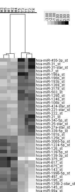
In total, the expression of 38 miRNAs was
significantly altered between normal laryngeal epithelial and
supraglottic LSCC, with 22 being upregulated and 16 being
downregulated in tumors (q≤5%) (Table
I). All 38 miRNAs were human miRNAs. The expression pattern of
the miRNAs listed in Table I with
>2-fold change is presented as a clustered heat map (Fig. 1), showing a distinct cluster between
normal and supraglottic LSCC samples. The most significant
aberration was >9.5-fold upregulation of miR-1290 and
>7.8-fold downregulation of miR-375.
 | Table IMicroarray of supraglottic laryngeal
squamous cell carcinomaa. |
Table I
Microarray of supraglottic laryngeal
squamous cell carcinomaa.
| miRNA | FC | q-value (%) |
|---|
| hsa-miR-1290_st | 9.59 | 0 |
| hsa-miR-196a_st | 3.61 | 0 |
|
hsa-miR-455-3p_st | 2.79 | 2.130 |
| hsa-miR-1269_st | 6.96 | 0 |
| hsa-miR-31*_st | 3.45 | 0 |
| hsa-miR-1246_st | 6.04 | 0 |
| hsa-miR-31_st | 3.11 | 0 |
| hsa-miR-21_st | 2.94 | 0 |
| hsa-miR-424*_st | 3.89 | 0 |
| hsa-miR-193b_st | 4.03 | 0 |
| hsa-miR-503_st | 3.44 | 0 |
| hsa-miR-27a*_st | 2.28 | 4.825 |
|
hsa-miR-34c-5p_st | 2.52 | 3.502 |
| hsa-miR-196b_st | 2.99 | 0 |
|
hsa-miR-339-5p_st | 2.74 | 2.130 |
| hsa-miR-25*_st | 2.41 | 3.502 |
| hsa-miR-222_st | 3.26 | 0 |
| hsa-miR-3178_st | 3.35 | 0 |
| hsa-miR-181b_st | 2.87 | 0 |
| hsa-miR-221-*_st | 2.77 | 2.130 |
| hsa-miR-106b_st | 4.36 | 0 |
| hsa-miR-7_st | 2.27 | 4.825 |
| hsa-miR-125b_st | −3.57 | 0 |
|
hsa-miR-3065-5p_st | −2.67 | 2.96 |
|
hsa-miR-199b-5p_st | −2.42 | 4.93 |
| hsa-miR-126_st | −2.91 | 1.36 |
|
hsa-miR-139-3p_st | −4.23 | 0 |
| hsa-miR-143_st | −2.78 | 2.25 |
| hsa-miR-497_st | −3.26 | 0 |
|
hsa-miR-125b-2*_st | −2.68 | 2.96 |
| hsa-miR-617_st | −2.82 | 2.25 |
| hsa-miR-145_st | −2.92 | 1.36 |
|
hsa-miR-1224-5p_st | −5.65 | 0 |
|
hsa-miR-139-5p_st | −3.67 | 0 |
| hsa-miR-675_st | −3.40 | 0 |
| hsa-miR-195_st | −3.32 | 0 |
| hsa-miR-99a_st | −2.75 | 2.250 |
| hsa-miR-375_st | −7.80 | 0 |
To confirm the microarray results, Taqman qRT-PCR
and normalized miRNA expression levels by snRNA U6 were used in 48
supraglottic LSCC patient samples. The three most up- or
downregulated miRNAs were selected for further analysis using
qRT-PCR. All the miRNAs tested were reliably confirmed, showing
significant differential expression between tumor and mucosa.
miR-375, miR-139-3P, miR-1290 and miR-106b showed significantly
differential expression between patients with and without lymphatic
metastasis (Table II).
| Table IIExpression of some miRNAs detected by
quantitative real-time polymerase chain reactiona. |
Table II
Expression of some miRNAs detected by
quantitative real-time polymerase chain reactiona.
| miRNA | Tumor (48 cases) | P-value | Mucosa (10
cases) |
|---|
|
|---|
| Lymphatic metastasis
(17/48) | No lymphatic
metastasis (31/48) |
|---|
| miR-375 | 3.773±0.104 | 3.201±0.553 | 0.004 | 0.273±0.024 |
| miR-139-3P | 2.986±0.099 | 2.327±0.026 | 0.000 | 0.339±0.014 |
| miR-1224 | 2.915±0.582 | 2.893±0.068 | 0.755 | 0.529±0.017 |
| miR-1290 | 0.223±0.028 | 0.510±0.010 | 0.000 | 3.579±0.023 |
| miR-1269 | 0.825±0.016 | 0.830±0.021 | 0.924 | 3.630±0.028 |
| miR-106b | 0.476±0.038 | 0.716±0.320 | 0.017 | 2.922±0.056 |
Discussion
The global expression of miRNAs in head and neck
squamous cell carcinoma (HNSCC) has recently been reported
(10,11). However, HNSCC includes various
subtypes, including nasopharyngeal, oropharyngeal, hypopharyngeal,
laryngeal and tongue carcinomas. Each subtype is individually
characterized in clinic, particularly with regard to metastasis and
prognosis, indicating a potential difference in their miRNA
expression. Supraglottic LSCC is the most common type of LSCC
identified in northeast China (12), and its relatively early lymphatic
metastasis and poor prognosis distinguish it from other types of
LSCC. To the best of our knowledge, this is the first study
regarding miRNA expression profiles in supraglottic LSCC and
adjacent normal mucosa. Microarray profiling of >900 miRNAs
identified 38 miRNAs that were significantly differentially
expressed in tumor tissues compared to normal mucosa, but were not
all the same with HNSCC (10,11).
Of the 38 miRNAs, 6 miRNAs were selected for in-depth examination
of a larger population of fresh-frozen supraglottic LSCC and normal
laryngeal epithelia mucosa by qRT-PCR. Of these 6 miRNAs, 4 had
significantly different expression in tumors with and without
lymphatic metastasis.
Some of the miRNAs identified as differentially
expressed in supraglottic LSCC compared to normal tissues have been
characterized in other types of cancer. Overexpression of miR-375
in liver cancer cells decreased cell proliferation, clonogenicity,
migration/invasion and induced G1 arrest and apoptosis (13). Additionally, miR-375, which
significantly inhibited cancer cell proliferation and invasion in
maxillary sinus squamous cell carcinoma, was restored (14). In a recent study, stage and distant
metastases revealed differential expression of miR-139-3p in
colorectal cancer (15). In the
present study, the most significantly downregulated miRNA was
miR-375, being underexpressed by 7.8-fold compared with normal
tissues, while tumor with lymphatic metastasis had a significantly
lower expression compared with tumor without lymphatic metastasis.
This result suggests miR-375 may depress the lymphatic metastasis
of supraglottic LSCC, such as miR-139-3P. Upregulation of
microRNA-1290 impaired cytokinesis and affected the reprogramming
of colon cancer cells (16).
Overexpression of miR-106b promoted metastasis of hepatocellular
carcinoma and correlated with higher tumor grade (17). In the present study miR-106b and
miR-1290 had a significantly higher expression in tumor with
compared to without lymphatic metastasis, suggesting that
interference of the two miRNAs might depress the lymphatic
metastasis of supraglottic LSCC. However, although the expression
of miR-1224 and miR-1269 were significantly different between tumor
and mucosa, their expression showed no significant difference
between tumors with and without lymphatic metastasis. This finding
indicated that the two miRNAs did not affect metastasis of
supraglottic LSCC.
In conclusion, the global miRNA profiling of
supraglottic LSCC and attached normal epithelia has demonstrated
that ~38 miRNAs are dysregulated in this disease. miR-375 was most
significantly downregulated. As supraglottic LSCC with lymphatic
metastasis, miR-375, miR-139-3p, miR-1290 and miR-106b showed a
significantly differential expression compared with tumors without
lymphatic metastasis. Therefore, understanding of miRNA in
supraglottic LSCC is crucial in the development of novel insights
for the diagnosis and prognosis of this disease.
Acknowledgements
This study was supported by the Grants from
Education Department of Liaoning Province, China (L2010638).
References
|
1
|
Sieqel R, Naishadham D and Jemal A: Cancer
statistics, 2012. CA Cancer J Clin. 62:10–29. 2012. View Article : Google Scholar
|
|
2
|
Bussu F, Ranelletti FO, Gessi M, Graziani
C, Lanza P, Lauriola L, Paludetti G and Almadori G:
Immunohistochemical expression patterns of the HER4 receptors in
normal mucosa and in laryngeal squamous cell carcinomas:
antioncogenic significance of the HER4 protein in laryngeal
squamous cell carcinoma. Laryngoscope. 122:1724–1733. 2012.
View Article : Google Scholar
|
|
3
|
Bartel DP: MicroRNAs: genomics,
biogenesis, mechanism, and function. Cell. 116:281–297. 2004.
View Article : Google Scholar : PubMed/NCBI
|
|
4
|
Du T and Zamore PD: Beginning to
understand microRNA function. Cell Res. 17:661–663. 2007.
View Article : Google Scholar : PubMed/NCBI
|
|
5
|
Lu J, Getz G, Miska EA, Alvarez-Saavedra
E, Lamb J, Peck D, Sweet-Cordero A, Ebert BL, Mak RH, Ferrando AA,
Downing JR, Jacks T, Horvitz HR and Golub TR: MicroRNA expression
profiles classify human cancers. Nature. 435:834–838. 2005.
View Article : Google Scholar : PubMed/NCBI
|
|
6
|
Guo Y, Chen Z, Zhang L, Zhou F, Shi S,
Feng X, Li B, Meng X, Ma X, Luo M, Shao K, Li N, Qiu B, Mitchelson
K, Chenq J and He J: Distinctive microRNA profiles relating to
patient survival in esophageal squamous cell carcinoma. Cancer Res.
68:26–33. 2008. View Article : Google Scholar : PubMed/NCBI
|
|
7
|
Iorio MV, Visone R, Di Leva G, Donati V,
Petrocca F, Casalini P, Taccioli C, Volinia S, Liu CG, Alder H,
Calin GA, Ménard S and Croce CM: MicroRNA signatures in human
ovarian cancer. Cancer Res. 67:8699–8707. 2007. View Article : Google Scholar : PubMed/NCBI
|
|
8
|
Blenkiron C, Goldstein LD, Thorne NP,
Spiteri I, Chin SF, Dunning MJ, Barbosa-Morais NL, Teschendorff AE,
Green AR, Ellis IO, Tavare S, Caldas C and Miska EA: MicroRNA
expression profiling of human breast cancer identifies new markers
of tumor subtype. Genome Biol. 8:R2142007. View Article : Google Scholar : PubMed/NCBI
|
|
9
|
Huber W, von Heydebreck A, Sultmann H,
Poustka A and Vingron M: Variance stabilization applied to
microarray data calibration and to the quantification of
differential expression. Bioinformatics. 18(Suppl 1): S96–S104.
2002. View Article : Google Scholar : PubMed/NCBI
|
|
10
|
Ramdas L, Giri U, Ashorm CL, Coombes KR,
EI-Naggar A, Ang KK and Story MD: miRNA expression profiles in head
and neck squamous cell carcinoma and adjacent normal tissue. Head
Neck. 31:642–654. 2009. View Article : Google Scholar : PubMed/NCBI
|
|
11
|
Hui AB, Lenarduzzi M, Krushel T, Waldron
L, Pintilie M, Shi W, Perez-Ordonez B, Jurisica I, O’Sullivan B,
Waldron J, Gullane P, Cummings B and Liu FF: Comprehensive microRNA
profiling for head and neck squamous cell carcinomas. Clin Cancer
Res. 16:1129–1139. 2010. View Article : Google Scholar : PubMed/NCBI
|
|
12
|
Fu WN, Shang C, Huang DF, Xu ZM, Sun XH
and Sun KL: Average-12.9 chromosome imbalances coupling with 15
differential expression genes possibly involved in the
carcinogenesis, progression and metastasis of supraglottic
laryngeal squamous cell cancer. Zhonghua Yi Xue Yi Chuan Xue Za
Zhi. 23:7–11. 2006.(In Chinese).
|
|
13
|
He XX, Chang Y, Meng FY, Wang MY, Xie QH,
Tang F, Li PY, Song YH and Lin JS: MicroRNA-375 targets AEG-1 in
hepatocellular carcinoma and suppresses liver cancer cell growth in
vitro and in vivo. Oncogene. 31:3357–3369. 2012. View Article : Google Scholar : PubMed/NCBI
|
|
14
|
Kinoshita T, Nohata N, Yoshino H, Hanazawa
T, Kikkawa N, Fujimura L, Chiyomaru T, Kawakami K, Enokida H,
Nakagawa M, Okamoto Y and Seki N: Tumor suppressive microRNA-375
regulates lactate dehydrogenase B in maxillary sinus squamous cell
carcinoma. Int J Oncol. 40:185–193. 2012.PubMed/NCBI
|
|
15
|
Chen WC, Lin MS, Ye YL, Gao HJ, Song ZY
and Shen XY: MicroRNA expression pattern and its alteration
following celecoxib intervention in human colorectal cancer. Exp
Ther Med. 3:1039–1048. 2012.PubMed/NCBI
|
|
16
|
Wu J, Ji X, Zhu L, Jiang Q, Wen Z, Xu S,
Shao W, Cai J, Du Q, Zhu Y and Mao J: Up-regulation of
microRNA-1290 impairs cytokinesis and affects the reprogramming of
colon cancer cells. Cancer Lett. 329:155–163. 2013. View Article : Google Scholar : PubMed/NCBI
|
|
17
|
Yau WL, Lam CS, Ng L, Chow AK, Chan ST,
Chan JY, Wo JY, Ng KT, Man K, Poon RT and Pang RW: Over-expression
of miR-106b promotes cell migration and metastasis in
hepatocellular carcinoma by activating epithelial-mesenchymal
transition process. PLoS One. 8:e578822013. View Article : Google Scholar : PubMed/NCBI
|