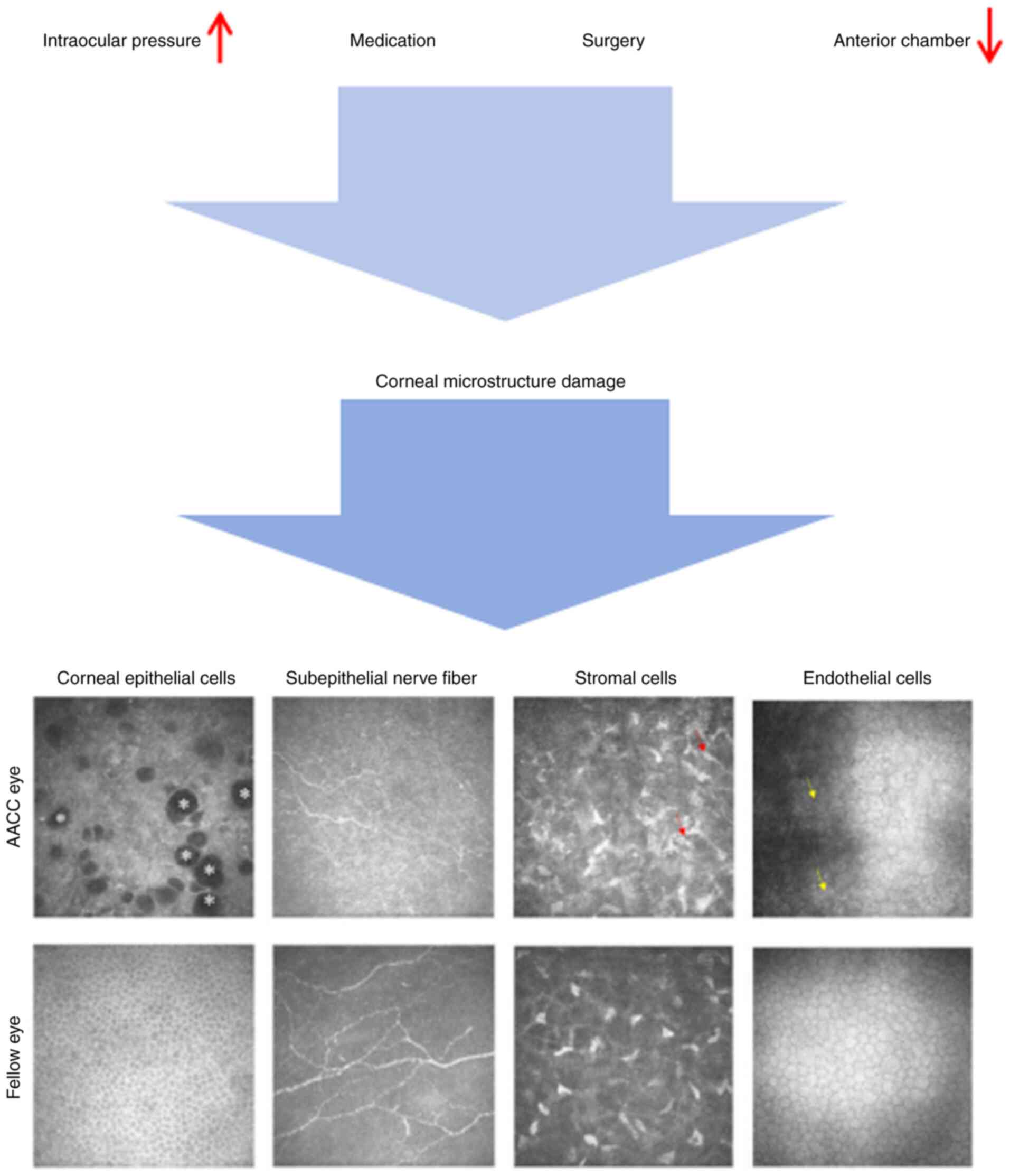
|
1
|
Kang JM and Tanna AP: Glaucoma. Med Clin
North Am. 105:493–510. 2021.PubMed/NCBI View Article : Google Scholar
|
|
2
|
He M, Jiang Y, Huang S, Chang DS, Munoz B,
Aung T, Foster PJ and Friedman DS: Laser peripheral iridotomy for
the prevention of angle closure: A single-centre, randomised
controlled trial. Lancet. 393:1609–1618. 2019.PubMed/NCBI View Article : Google Scholar
|
|
3
|
George R, Panda S and Vijaya L: Blindness
in glaucoma: primary open-angle glaucoma versus primary
angle-closure glaucoma-a meta-analysis. Eye (Lond). 36:2099–2105.
2022.PubMed/NCBI View Article : Google Scholar
|
|
4
|
Tuck H, Park M, Carnell M, Machet J,
Richardson A, Jukic M and Di Girolamo N: Neuronal-epithelial cell
alignment: A determinant of health and disease status of the
cornea. Ocul Surf. 21:257–270. 2021.PubMed/NCBI View Article : Google Scholar
|
|
5
|
Li EY, Mohamed S, Leung CK, Rao SK, Cheng
AC, Cheung CY and Lam DS: Agreement among 3 methods to measure
corneal thickness: Ultrasound pachymetry, Orbscan II, and Visante
anterior segment optical coherence tomography. Ophthalmology.
114:1842–1847. 2007.PubMed/NCBI View Article : Google Scholar
|
|
6
|
Heath MT, Mulpuri L, Kimiagarov E, Patel
RP, Murphy DA, Levine H, Tonk RS, Cooke DL and Riaz KM: Intraocular
lens power calculations in keratoconus eyes comparing keratometry,
total keratometry, and newer formulae. Am J Ophthalmol.
253:206–214. 2023.PubMed/NCBI View Article : Google Scholar
|
|
7
|
Sugar A: Clinical specular microscopy.
Surv Ophthalmol. 24:21–32. 1979.PubMed/NCBI View Article : Google Scholar
|
|
8
|
Badian RA, Ekman L, Pripp AH, Utheim TP,
Englund E, Dahlin LB, Rolandsson O and Lagali N: Comparison of
novel wide-field in vivo corneal confocal microscopy with skin
biopsy for assessing peripheral neuropathy in type 2 diabetes.
Diabetes. 72:908–917. 2023.PubMed/NCBI View Article : Google Scholar
|
|
9
|
Giannaccare G, Bernabei F, Pellegrini M,
Guaraldi F, Turchi F, Torrazza C, Senni C, Scotto R, Sindaco D, Di
Cello L, et al: Bilateral morphometric analysis of corneal
sub-basal nerve plexus in patients undergoing unilateral cataract
surgery: A preliminary in vivo confocal microscopy study. Br J
Ophthalmol. 105:174–179. 2021.PubMed/NCBI View Article : Google Scholar
|
|
10
|
Ren X, Chou Y, Wang Y, Jing D, Chen Y and
Li X: The utility of oral vitamin B1 and mecobalamin to improve
corneal nerves in dry eye disease: An in vivo confocal microscopy
study. Nutrients. 14(3750)2022.PubMed/NCBI View Article : Google Scholar
|
|
11
|
Misra SL, Slater JA, McGhee CNJ, Pradhan M
and Braatvedt GD: Corneal confocal microscopy in type 1 diabetes
mellitus: A six-year longitudinal study. Transl Vis Sci Technol.
11(17)2022.PubMed/NCBI View Article : Google Scholar
|
|
12
|
Posarelli M, Chirapapaisan C, Muller R,
Abbouda A, Pondelis N, Cruzat A, Cavalcanti BM, Cox SM, Jamali A,
Pavan-Langston D and Hamrah P: Corneal nerve regeneration is
affected by scar location in herpes simplex keratitis: A
longitudinal in vivo confocal microscopy study. Ocul Surf.
28:42–52. 2023.PubMed/NCBI View Article : Google Scholar
|
|
13
|
Wang W, Yang X, Yao Q, Xu Q, Liu W and Liu
J: Corneal confocal microscopic characteristics of acute
angle-closure crisis. BMC Ophthalmol. 22(21)2022.PubMed/NCBI View Article : Google Scholar
|
|
14
|
Sosuan GMN and Yap-Veloso MIR: Central
corneal thickness among filipino patients in an ambulatory eye
surgery center using anterior segment optical coherence tomography.
Clin Ophthalmol. 15:2653–2664. 2021.PubMed/NCBI View Article : Google Scholar
|
|
15
|
Park HM, Choi J, Lee WJ and Uhm KB: Rate
of central corneal thickness changes in primary angle closure eyes:
Long-term follow-up results. BMC Ophthalmol. 21(145)2021.PubMed/NCBI View Article : Google Scholar
|
|
16
|
Niu WR, Dong CQ, Zhang X, Feng YF and Yuan
F: Ocular biometric characteristics of chinese with history of
acute angle closure. J Ophthalmol. 2018(5835791)2018.PubMed/NCBI View Article : Google Scholar
|
|
17
|
Chen MJ, Liu CJ, Cheng CY and Lee SM:
Corneal status in primary angle-closure glaucoma with a history of
acute attack. J Glaucoma. 21:12–16. 2012.PubMed/NCBI View Article : Google Scholar
|
|
18
|
Sugumaran A, Devasena MA, Thomas M and
Periyathambi D: A cross sectional study on evaluating the corneal
endothelial cell density and central corneal thickness in eyes with
primary glaucoma. J Family Med Prim Care. 11:4650–4654.
2022.PubMed/NCBI View Article : Google Scholar
|
|
19
|
Radhakrishnan S, Chen PP, Junk AK,
Nouri-Mahdavi K and Chen TC: Laser peripheral iridotomy in primary
angle closure: A report by the American academy of ophthalmology.
Ophthalmology. 125:1110–1120. 2018.PubMed/NCBI View Article : Google Scholar
|
|
20
|
Unterlauft JD, Yafai Y and Wiedemann P:
Changes of anterior chamber architecture induced by laser
peripheral iridotomy in acute angle closure crisis. Int Ophthalmol.
35:549–556. 2015.PubMed/NCBI View Article : Google Scholar
|
|
21
|
Shon K, Sung KR and Yoon JY: Implications
of the relationship between refractive error and biometry in the
pathogenesis of primary angle closure. Invest Ophthalmol Vis Sci.
62(38)2021.PubMed/NCBI View Article : Google Scholar
|
|
22
|
Wang B, Cao K, Wang Z, Zhang Y, Congdon N
and Wang T: Analyzing anatomical factors contributing to angle
closure based on anterior segment optical coherence tomography
imaging. Curr Eye Res. 47:256–261. 2022.PubMed/NCBI View Article : Google Scholar
|
|
23
|
Zuo C, Gong R, Chen W, Chen C, Su J, Wei
K, Gao X, Lin M and Ge J: Investigation of corneal astigmatism in
chinese patients with primary angle closure disease. J Glaucoma.
27:1131–1135. 2018.PubMed/NCBI View Article : Google Scholar
|
|
24
|
Goldstein MH, Silva FQ, Blender N, Tran T
and Vantipalli S: Ocular benzalkonium chloride exposure: Problems
and solutions. Eye (Lond). 36:361–368. 2022.PubMed/NCBI View Article : Google Scholar
|
|
25
|
Hedengran A and Kolko M: The molecular
aspect of anti-glaucomatous eye drops - are we harming our
patients? Mol Aspects Med. 93(101195)2023.PubMed/NCBI View Article : Google Scholar
|
|
26
|
Thacker M, Sahoo A, Reddy AA, Bokara KK,
Singh S, Basu S and Singh V: Benzalkonium chloride-induced dry eye
disease animal models: Current understanding and potential for
translational research. Indian J Ophthalmol. 71:1256–1262.
2023.PubMed/NCBI View Article : Google Scholar
|
|
27
|
Valladales-Restrepo LF, Oyuela-Gutiérrez
MC, Delgado-Araujo AC and Machado-Alba JE: Use pattern of
ophthalmic antiglaucoma agents with and without preservatives: A
cross-sectional study. Pharmaceuticals (Basel).
16(753)2023.PubMed/NCBI View Article : Google Scholar
|
|
28
|
Güçlü H, Çınar AK, Çınar AC, Akaray İ,
Şambel Aykutlu M, Sakallıoğlu AK and Gürlü V: Corneal epithelium
and limbal region alterations due to glaucoma medications evaluated
by anterior segment optic coherence tomography: A case-control
study. Cutan Ocul Toxicol. 40:85–94. 2021.PubMed/NCBI View Article : Google Scholar
|
|
29
|
Mao J, Wang Y, Gao Y, Wan S, Jiang W, Pan
Y, Yan Y, Cong Y, Shi X, Huang L and Yang Y: Correlation between
anterior chamber angle status and limbal stem cell deficiency in
primary angle-closure glaucoma. Am J Ophthalmol. 262:178–185.
2024.PubMed/NCBI View Article : Google Scholar
|
|
30
|
Yu FX, Lee PSY, Yang L, Gao N, Zhang Y,
Ljubimov AV, Yang E, Zhou Q and Xie L: The impact of sensory
neuropathy and inflammation on epithelial wound healing in diabetic
corneas. Prog Retin Eye Res. 89(101039)2022.PubMed/NCBI View Article : Google Scholar
|
|
31
|
Patel S, Hwang J, Mehra D and Galor A:
Corneal nerve abnormalities in ocular and systemic diseases. Exp
Eye Res. 202(108284)2021.PubMed/NCBI View Article : Google Scholar
|
|
32
|
Rossi GCM, Scudeller L, Lumini C, Mirabile
AV, Picasso E, Bettio F, Pasinetti GM and Bianchi PE: An in vivo
confocal, prospective, masked, 36 months study on glaucoma patients
medically treated with preservative-free or preserved monotherapy.
Sci Rep. 9(4282)2019.PubMed/NCBI View Article : Google Scholar
|
|
33
|
Agnifili L, Brescia L, Villani E,
D'Onofrio G, Figus M, Oddone F, Nucci P and Mastropasqua R: In vivo
confocal microscopy of the corneal sub-basal nerve plexus in
medically controlled glaucoma. Microsc Microanal. 1–8.
2022.PubMed/NCBI View Article : Google Scholar : (Epub ahead of
print).
|
|
34
|
Graae Jensen P, Gundersen M, Nilsen C,
Gundersen KG, Potvin R, Gazerani P, Chen X, Utheim TP and Utheim
ØA: Prevalence of dry eye disease among individuals scheduled for
cataract surgery in a norwegian cataract clinic. Clin Ophthalmol.
17:1233–1243. 2023.PubMed/NCBI View Article : Google Scholar
|
|
35
|
Agnifili L, Brescia L, Oddone F, Sacchi M,
D'Ugo E, Di Marzio G, Perna F, Costagliola C and Mastropasqua R:
The ocular surface after successful glaucoma filtration surgery: A
clinical, in vivo confocal microscopy, and immune-cytology study.
Sci Rep. 9(11299)2019.PubMed/NCBI View Article : Google Scholar
|
|
36
|
Sebbag L, Crabtree EE, Sapienza JS, Kim K
and Rodriguez E: Corneal hypoesthesia, aqueous tear deficiency, and
neurotrophic keratopathy following micropulse transscleral
cyclophotocoagulation in dogs. Vet Ophthalmol. 23:171–180.
2020.PubMed/NCBI View Article : Google Scholar
|
|
37
|
Downie LE, Zhang X, Wu M, Karunaratne S,
Loi JK, Senthil K, Arshad S, Bertram K, Cunningham AL, Carnt N, et
al: Redefining the human corneal immune compartment using dynamic
intravital imaging. Proc Natl Acad Sci USA.
120(e2217795120)2023.PubMed/NCBI View Article : Google Scholar
|
|
38
|
Chen Q, Wang L, Zhang Y, Xu X, Wei Z,
Zhang Z, Wei Y, Pang J, Guo X, Cao K and Liang Q: Corneal
epithelial dendritic cells: An objective indicator for ocular
surface inflammation in patients with obstructive meibomian gland
dysfunction? Ocul Immunol Inflamm. 32:79–88. 2024.PubMed/NCBI View Article : Google Scholar
|
|
39
|
He W, Xu F, Chen L, Huang W, Jiang L, Tang
F, Yan W, Zhong S, Shen C, Huang H, et al: Association of
high-mobility group box-1 with inflammationrelated cytokines in the
aqueous humor with acute primary angle-closure eyes. Curr Mol Med.
21:237–245. 2021.PubMed/NCBI View Article : Google Scholar
|
|
40
|
Mastropasqua R, Agnifili L, Fasanella V,
Lappa A, Brescia L, Lanzini M, Oddone F, Perri P and Mastropasqua
L: In vivo distribution of corneal epithelial dendritic cells in
patients with glaucoma. Invest Ophthalmol Vis Sci. 57:5996–6002.
2016.PubMed/NCBI View Article : Google Scholar
|
|
41
|
Espana EM and Birk DE: Composition,
structure and function of the corneal stroma. Exp Eye Res.
198(108137)2020.PubMed/NCBI View Article : Google Scholar
|
|
42
|
Chen D, Wang L, Guo X, Zhang Z, Xu X, Jin
ZB and Liang Q: Evaluation of limbal stem cells in patients with
type 2 diabetes: An in vivo confocal microscopy study. Cornea.
43:67–75. 2024.PubMed/NCBI View Article : Google Scholar
|
|
43
|
Cagini C, Di Lascio G, Torroni G,
Mariniello M, Meschini G, Lupidi M and Messina M: Dry eye and
inflammation of the ocular surface after cataract surgery:
Effectiveness of a tear film substitute based on
trehalose/hyaluronic acid vs hyaluronic acid to resolve signs and
symptoms. J Cataract Refract Surg. 47:1430–1435. 2021.PubMed/NCBI View Article : Google Scholar
|
|
44
|
Yuan XL, Wen Q, Zhang MY and Fan TJ:
Cytotoxicity of pilocarpine to human corneal stromal cells and its
underlying cytotoxic mechanisms. Int J Ophthalmol. 9:505–511.
2016.PubMed/NCBI View Article : Google Scholar
|
|
45
|
Vallabh NA, Kennedy S, Vinciguerra R,
McLean K, Levis H, Borroni D, Romano V and Willoughby CE: Corneal
endothelial cell loss in glaucoma and glaucoma surgery and the
utility of management with descemet membrane endothelial
keratoplasty (DMEK). J Ophthalmol. 2022(1315299)2022.PubMed/NCBI View Article : Google Scholar
|
|
46
|
Wenzel DA, Schultheiss C, Druchkiv V,
Hellwinkel OJC, Spitzer MS, Schultheiss M, Casagrande M and
Steinhorst NA: Effect of elevated irrigation bottle height during
cataract surgery on corneal endothelial cells in porcine eyes. BMC
Ophthalmol. 23(211)2023.PubMed/NCBI View Article : Google Scholar
|
|
47
|
Li X, Zhang Z, Ye L, Meng J, Zhao Z, Liu Z
and Hu J: Acute ocular hypertension disrupts barrier integrity and
pump function in rat corneal endothelial cells. Sci Rep.
7(6951)2017.PubMed/NCBI View Article : Google Scholar
|
|
48
|
Li Z, Fan N, Cheng Y, Xiang F, Pan X, Cao
K, Zhang Y, Zhang Q and Li S: Factors associated with severe
corneal endothelial damage following acute primary angle closure in
Chinese subjects. Graefes Arch Clin Exp Ophthalmol. 261:2927–2934.
2023.PubMed/NCBI View Article : Google Scholar
|
|
49
|
Tham CC, Kwong YY, Lai JS and Lam DS:
Effect of a previous acute angle closure attack on the corneal
endothelial cell density in chronic angle closure glaucoma
patients. J Glaucoma. 15:482–485. 2006.PubMed/NCBI View Article : Google Scholar
|
|
50
|
Yeom H, Hong EH, Shin YU, Kang MH, Cho HY
and Seong M: Corneal endothelial cell loss after
phacoemulsification in eyes with a prior acute angle-closure
attack. Korean J Ophthalmol. 34:432–438. 2020.PubMed/NCBI View Article : Google Scholar
|
|
51
|
Verma S, Nongpiur ME, Husain R, Wong TT,
Boey PY, Quek D, Perera SA and Aung T: Characteristics of the
corneal endothelium across the primary angle closure disease
spectrum. Invest Ophthalmol Vis Sci. 59:4525–4530. 2018.PubMed/NCBI View Article : Google Scholar
|
|
52
|
Imai K, Sawada H, Hatase T and Fukuchi T:
Iridocorneal contact as a potential cause of corneal decompensation
following laser peripheral iridotomy. Jpn J Ophthalmol. 65:460–471.
2021.PubMed/NCBI View Article : Google Scholar
|
|
53
|
Yuan Y, Wang W, Xiong R, Zhang J, Li C,
Yang S, Friedman DS, Foster PJ and He M: Fourteen-year outcome of
angle-closure prevention with laser iridotomy in the zhongshan
angle-closure prevention study: Extended follow-up of a randomized
controlled trial. Ophthalmology. 130:786–794. 2023.PubMed/NCBI View Article : Google Scholar
|
|
54
|
Chen HC, Lee CY, Liu CF, Hsueh YJ, Meir
YJ, Cheng CM and Wu WC: Corneal endothelial changes following early
capsulotomy using neodymium:yttrium-aluminum-garnet laser.
Diagnostics (Basel). 12(150)2022.PubMed/NCBI View Article : Google Scholar
|
|
55
|
Higashihara H, Sotozono C, Yokoi N,
Inatomi T and Kinoshita S: The blood-aqueous barrier breakdown in
eyes with endothelial decompensation after argon laser iridotomy.
Br J Ophthalmol. 95:1032–1034. 2011.PubMed/NCBI View Article : Google Scholar
|
|
56
|
Liao C, Zhang J, Jiang Y, Huang S, Aung T,
Foster PJ, Friedman D and He M: Long-term effect of YAG laser
iridotomy on corneal endothelium in primary angle closure suspects:
A 72-month randomised controlled study. Br J Ophthalmol.
105:348–353. 2021.PubMed/NCBI View Article : Google Scholar
|
|
57
|
Ono T, Iida M, Sakisaka T, Minami K and
Miyata K: Effect of laser peripheral iridotomy using argon and
neodymium-YAG lasers on corneal endothelial cell density: 7-year
longitudinal evaluation. Jpn J Ophthalmol. 62:216–220.
2018.PubMed/NCBI View Article : Google Scholar
|


