Open Access

Considerations for two inoculation methods of rabbit hepatic tumors: Pathology and image features

  • Authors:
    • Jun-Hui Sun
    • Yue-Lin Zhang
    • Chun-Hui Nie
    • Xiao-Bo  Yu
    • Hai‑Yang Xie
    • Lin Zhou
    • Shu-Sen Zheng
  • View Affiliations

  • Published online on: December 28, 2011     https://doi.org/10.3892/etm.2011.435
  • Pages: 386-390
Metrics: Total Views: 0 (Spandidos Publications: | PMC Statistics: )
Total PDF Downloads: 0 (Spandidos Publications: | PMC Statistics: )


Abstract

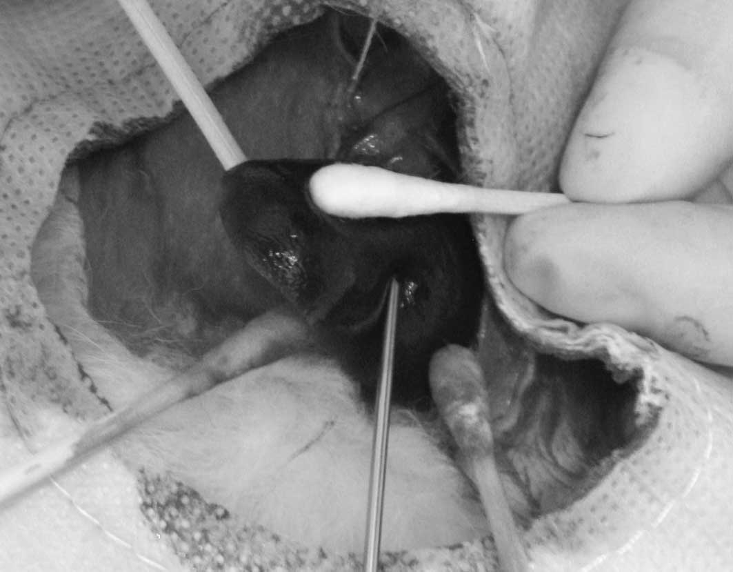
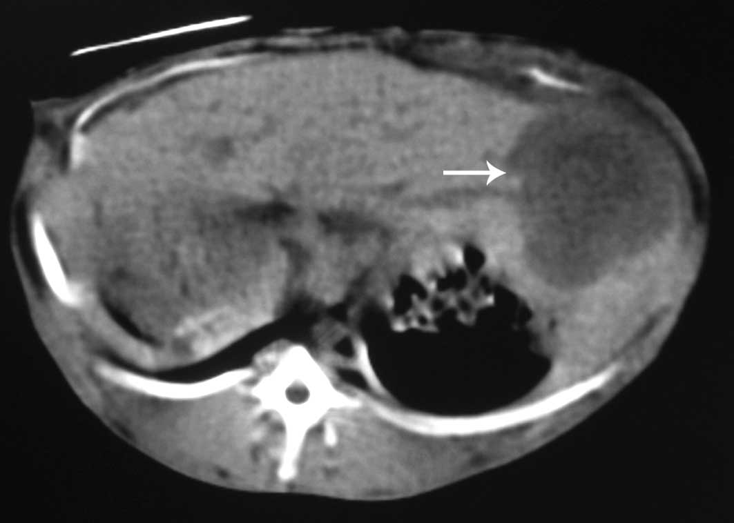
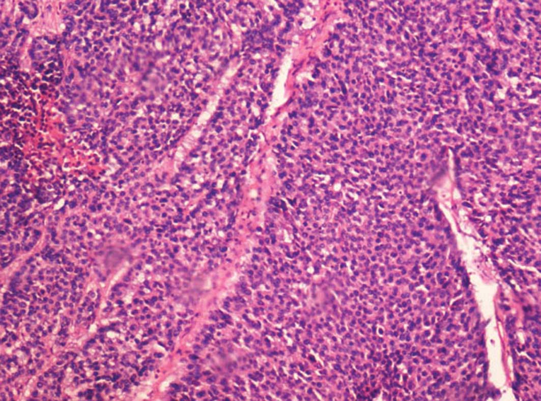
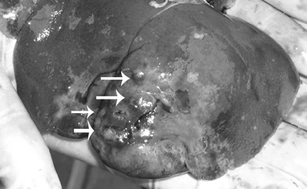
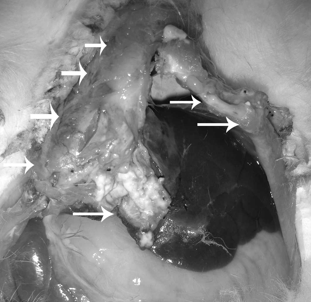
The rabbit VX2 hepatic tumor model has been widely used for the investigation of liver cancer. The present study was designed to compare two inoculation methods in order to identify an improved modality for establishing rabbit hepatic tumors. A total of 44 rabbits were randomly divided into 2 groups. All inoculations were performed by open laparotomy. In group 1, 22 rabbits received a VX2 tumor cell suspension in a volume of 0.2 ml, which was slowly injected into the rabbit liver parenchyma using an 18-gauge needle. In group 2, a 0.2-ml suspension of small, minced tumor fragments of a VX2 carcinoma, inserted into an 18-gauge needle, was implanted into the liver parenchyma. The tumor survival ratio, size and extrahepatic metastases were evaluated and compared using computed tomography (CT)-scan, angiography and pathological specimens. At 2 weeks following implantation, successful liver tumor growth was achieved in 14 out of the 22 rabbits in group 1 (63.6%) and 21 out of the 22 rabbits in group 2 (95.5%) (P<0.05). Further sub-analysis revealed that single nodular tumors in the liver were observed in 4 out of the 14 rabbits in group 1 (28.6%) and 14 out of the 21 rabbits in group 2 (66.7%), and that multinodular tumors in the liver were observed in 10 out of the 14 rabbits in group 1 (71.4%) and 7 out of the 21 rabbits in group 2 (33.3%) (P<0.05). Extrahepatic metastases were observed in 5 out of the 14 rabbits in group 1 (35.7%) and 1 of the 21 rabbits in group 2 (4.8%) (P<0.05). Imaging findings of hepatic tumors were in concordance with histopathology. In conclusion, direct implantation of VX2 tumor fragments into the liver using a fine needle achieved a higher success rate than injecting VX2 tumor cells. The rabbit hepatic tumors inoculated by this method may therefore be a more suitable animal model for investigating liver cancer.
View Figures
View References

Related Articles

Journal Cover

March 2012
Volume 3 Issue 3

Print ISSN: 1792-0981
Online ISSN:1792-1015

Sign up for eToc alerts

Recommend to Library

Copy and paste a formatted citation
x
Spandidos Publications style
Sun J, Zhang Y, Nie C, Yu X, Xie HY, Zhou L and Zheng S: Considerations for two inoculation methods of rabbit hepatic tumors: Pathology and image features. Exp Ther Med 3: 386-390, 2012.
APA
Sun, J., Zhang, Y., Nie, C., Yu, X., Xie, H., Zhou, L., & Zheng, S. (2012). Considerations for two inoculation methods of rabbit hepatic tumors: Pathology and image features. Experimental and Therapeutic Medicine, 3, 386-390. https://doi.org/10.3892/etm.2011.435
MLA
Sun, J., Zhang, Y., Nie, C., Yu, X., Xie, H., Zhou, L., Zheng, S."Considerations for two inoculation methods of rabbit hepatic tumors: Pathology and image features". Experimental and Therapeutic Medicine 3.3 (2012): 386-390.
Chicago
Sun, J., Zhang, Y., Nie, C., Yu, X., Xie, H., Zhou, L., Zheng, S."Considerations for two inoculation methods of rabbit hepatic tumors: Pathology and image features". Experimental and Therapeutic Medicine 3, no. 3 (2012): 386-390. https://doi.org/10.3892/etm.2011.435