Dual‑source CT coronary angiographic evaluation of coronary artery fistulas
- Authors:
- Min Liu
- Qing Hou
- Xiaojuan Guo
- Shuangkun Wang
- Zhanhong Ma
View Affiliations
Affiliations: Department of Radiology, Beijing Chaoyang Hospital, Capital Medical University, Beijing 100020, P.R. China, Department of Radiology, Beijing Puren Hospital, Beijing 100069, P.R. China
- Published online on: March 4, 2014 https://doi.org/10.3892/etm.2014.1602
-
Pages:
1155-1159
Metrics: Total
Views: 0 (Spandidos Publications: | PMC Statistics: )
Metrics: Total PDF Downloads: 0 (Spandidos Publications: | PMC Statistics: )
This article is mentioned in:
Abstract
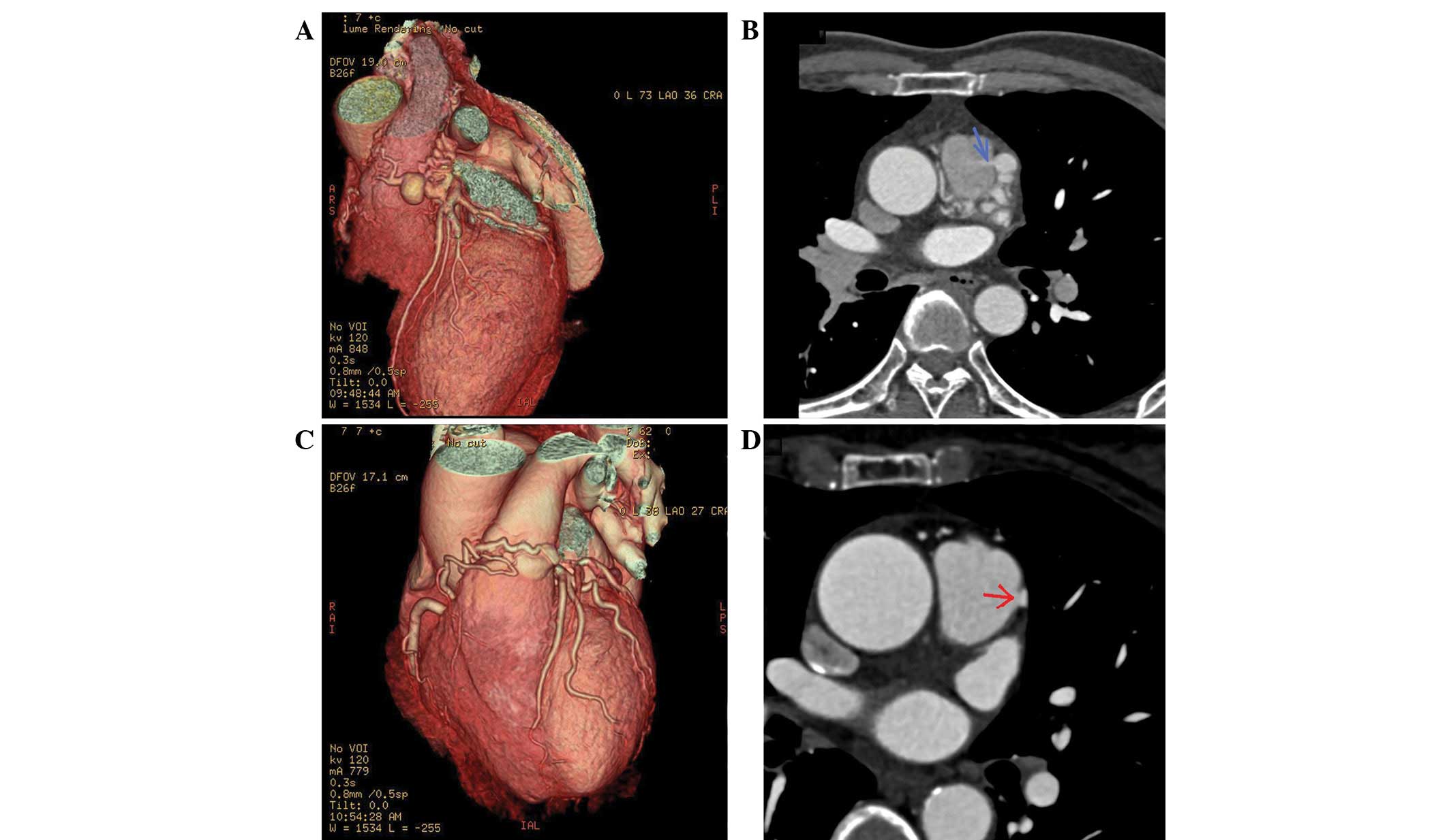
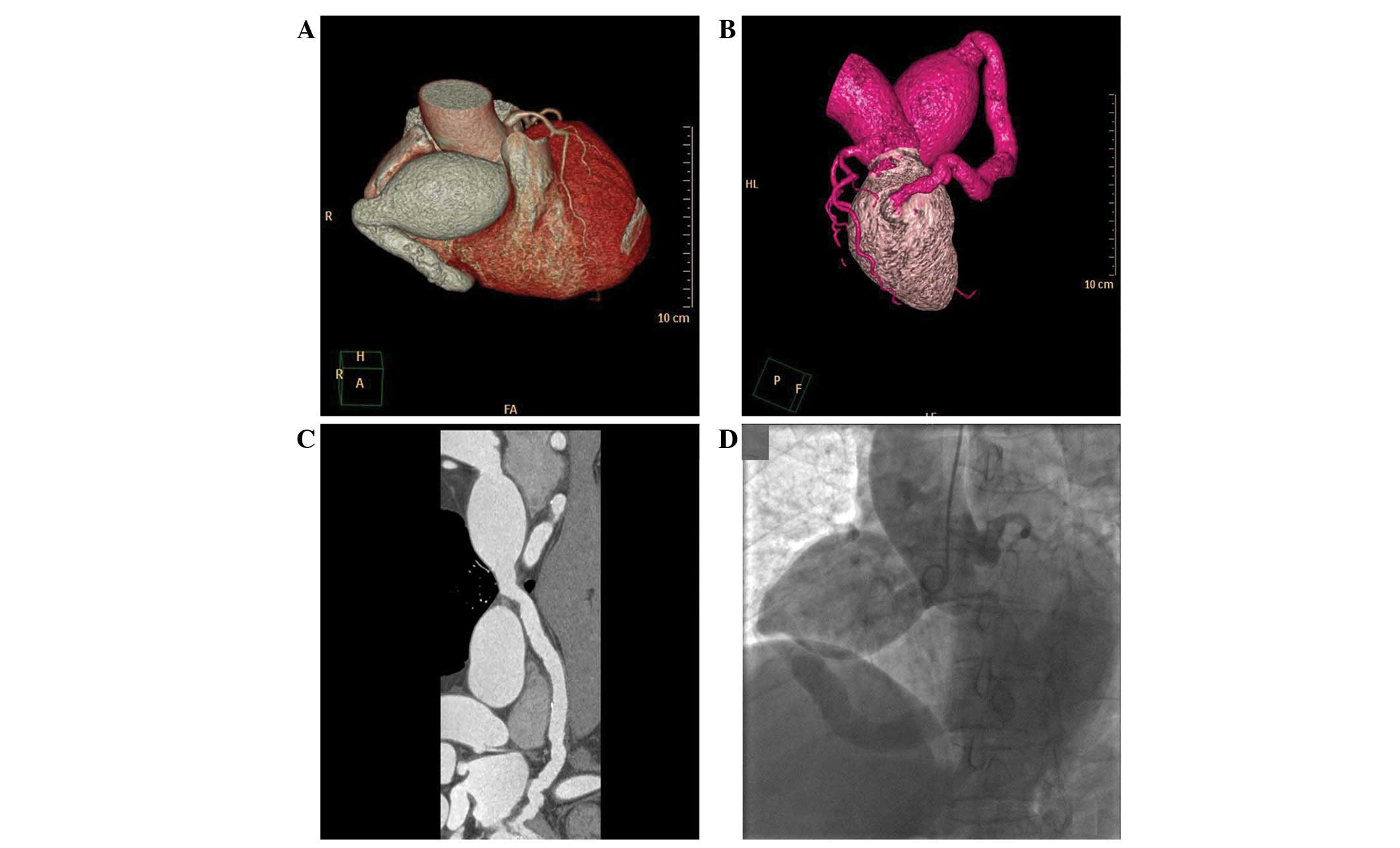
The aim of the present study was to retrospectively evaluate the incidence and morphological features of coronary artery fistulas (CAFs) detected by dual‑source computed tomography coronary angiography (DS‑CTCA). Between January 2011 and January 2013, 19,584 consecutive patients that had undergone electrocardiogram‑triggering DS‑CTCA were retrospectively reviewed. Image reconstructions were performed and image quality was evaluated. The medical information of the patients with CAF was reviewed from the medical records. Among the 19,584 patients, 66 patients were diagnosed with CAFs by CTCA, including 60 patients with coronary pulmonary artery fistulas (CPAFs) and six with coronary left ventricular fistulas. Therefore, the incidence of CAFs was 0.34%. Image quality was considered to be excellent in 61 patients and moderate in five cases. CPAFs were identified as small and tortuous vessels in 24 patients and dilated vessels close to the surface of the pulmonary artery (PA) in 36 patients. The coronary left ventricular fistulas were identified as dilated vessels that were draining into the posterior wall of the left ventricle. Among the 66 patients, 54 patients had one traceable fistula and the remaining 12 patients were shown to have two fistula vessels. The average diameter of the detected fistulas, measured with CTCA, was 3.1±1.9 mm. A high‑density flow jet of contrast agent shunting from the fistula into the low density PA was observed in 46 patients with CPAF. The results indicate that DS‑CTCA is a reliable noninvasive tool that allows the accurate delineation of CAFs.
View References
1
|
Meyer J, Reul GJ, Mullins CE, McCoy J,
Hallman GL and Cooley DA: Congenital fistulae of the coronary
arteries. Clinical considerations and surgical management in 23
patients. J Cardiovasc Surg (Torino). 16:506–511. 1975.PubMed/NCBI
|
2
|
Kim MS, Jung JI and Chun HJ: Coronary to
pulmonary artery fistula: morphologic features at multidetector CT.
Int J Cardiovasc Imaging. 26(Suppl 2): 273–280. 2010. View Article : Google Scholar : PubMed/NCBI
|
3
|
Sabarudin A, Md Yusof AK, Tay MF, Ng KH
and Sun Z: Dual-source CT coronary angiography: effectiveness of
radiation dose reduction with lower tube voltage. Radiat Prot
Dosimetry. 153:441–447. 2013.PubMed/NCBI
|
4
|
Sherwood MC, Rockenmacher S, Colan SD and
Geva T: Prognostic significance of clinically silent coronary
artery fistulas. Am J Cardiol. 83:407–411. 1999. View Article : Google Scholar : PubMed/NCBI
|
5
|
Lau G: Sudden death arising from a
congenital coronary artery fistula. Forensic Sci Int. 73:125–130.
1995. View Article : Google Scholar : PubMed/NCBI
|
6
|
Gowda RM, Vasavada BC and Khan IA:
Coronary artery fistulas: clinical and therapeutic considerations.
Int J Cardiol. 107:7–10. 2006. View Article : Google Scholar : PubMed/NCBI
|
7
|
Cebi N, Schulze-Waltrup N, Frömke J,
Scheffold T and Heuer H: Congenital coronary artery fistulas in
adults: concomitant pathologies and treatment. Int J Cardiovasc
Imaging. 24:349–355. 2008. View Article : Google Scholar : PubMed/NCBI
|
8
|
Lin FC, Chang HJ, Chern MS, Wen MS, Yeh SJ
and Wu D: Multiplane transesophageal echocardiography in the
diagnosis of congenital coronary artery fistula. Am Heart J.
130:1236–1244. 1995. View Article : Google Scholar : PubMed/NCBI
|
9
|
Dodge-Khatami A, Mavroudis C and Backer
CL: Congenital heart surgery nomenclature and database project:
anomalies of the coronary arteries. Ann Thorac Surg. 69(4 Suppl):
S270–S297. 2000. View Article : Google Scholar : PubMed/NCBI
|
10
|
Yun H, Zeng MS, Yang S, Jin H and Yang X:
Congenital coronary artery fistulas: dual-source CT findings from
consecutive 6,624 patients with suspected or confirmed coronary
artery disease. Chin Med J (Engl). 124:4172–4177. 2011.PubMed/NCBI
|
11
|
Nakamura M, Matsuoka H, Kawakami H, et al:
Giant congenital coronary artery fistula to left brachial vein
clearly detected by multidetector computed tomography. Circ J.
70:796–799. 2006. View Article : Google Scholar : PubMed/NCBI
|
12
|
Liberthson RR, Sagar K, Berkoben JP,
Weintraub RM and Levine FH: Congenital coronary arteriovenous
fistula. Report of 13 patients, review of the literature and
delineation of management. Circulation. 59:849–854. 1979.
View Article : Google Scholar : PubMed/NCBI
|