Calcifying aponeurotic fibroma of the finger in an elderly patient: CT and MRI findings with pathologic correlation
- Authors:
- Jun Nishio
- Hideaki Inamitsu
- Hiroshi Iwasaki
- Hiroyuki Hayashi
- Masatoshi Naito
View Affiliations
Affiliations: Department of Orthopaedic Surgery, Faculty of Medicine, Fukuoka University, Fukuoka 814‑0180, Japan, Department of Pathology, Faculty of Medicine, Fukuoka University, Fukuoka 814‑0180, Japan
- Published online on: July 11, 2014 https://doi.org/10.3892/etm.2014.1838
-
Pages:
841-843
Metrics: Total
Views: 0 (Spandidos Publications: | PMC Statistics: )
Metrics: Total PDF Downloads: 0 (Spandidos Publications: | PMC Statistics: )
This article is mentioned in:
Abstract
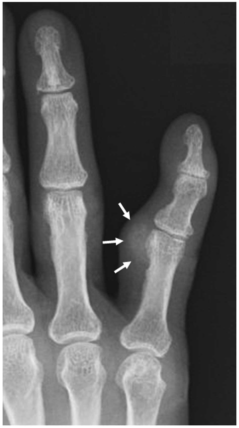
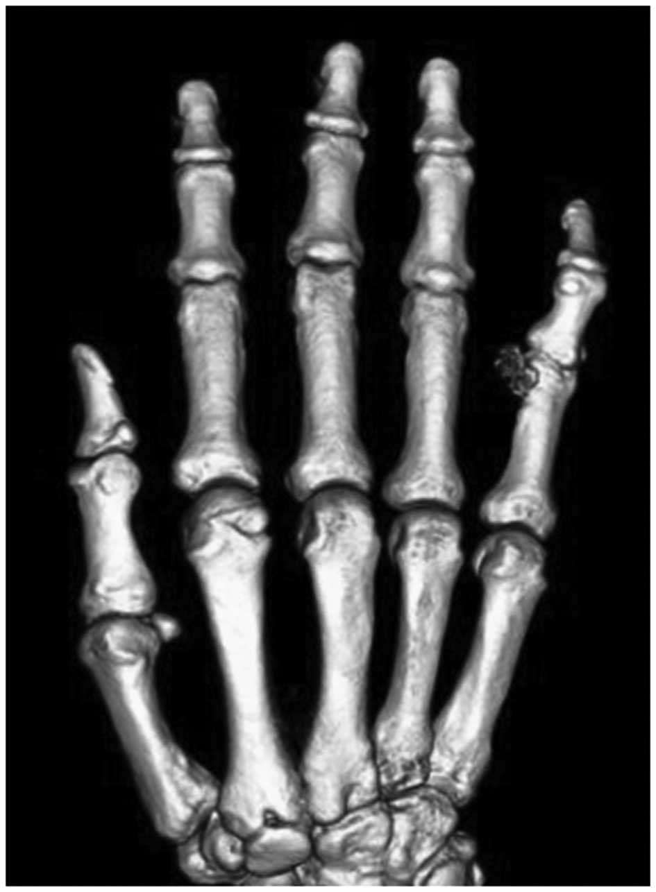
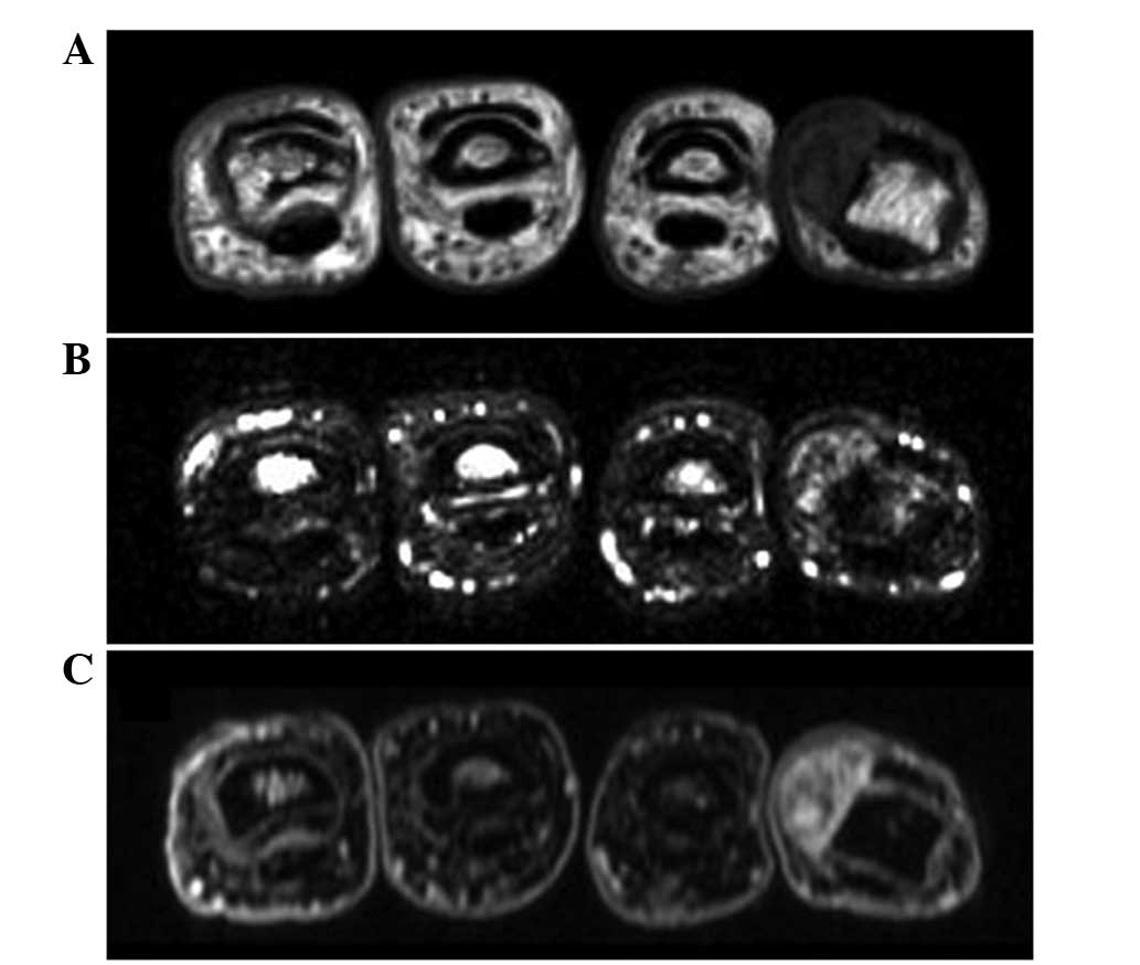
Calcifying aponeurotic fibroma (CAF) is a rare, locally aggressive fibroblastic lesion that occurs predominantly in the distal extremities of children and adolescents. In the present study, a case of pathologically proven CAF arising in the right little finger of a 69‑year‑old woman is presented. Physical examination revealed a firm, immobile, non‑tender mass. Plain radiographs showed a faintly calcified soft tissue mass without bone involvement and computed tomography scans clearly demonstrated the presence of the lesion. Magnetic resonance imaging revealed that the lesion exhibited low to intermediate signal intensity on T1‑weighted images and heterogeneous high signal intensity with small foci of low signal intensity on T2‑weighted spectral presaturation with inversion recovery images. Contrast‑enhanced fat‑suppressed T1‑weighted images demonstrated intense heterogeneous enhancement throughout the mass. The patient underwent an excisional biopsy. Histologically, the tumor showed a biphasic pattern, composed of a moderately cellular fibromatosis‑like component and irregular calcified areas with polygonal epithelioid cells. There has been no evidence of local recurrence four months following surgery. To the best of our knowledge, this case report describes the oldest patient with this condition.
View References
1
|
Kilpatrick SE: Calcifying aponeurotic
fibroma. World Health Organization Classification of Tumours of
Soft Tissue and Bone. Fletcher CDM, Bridge JA, Hogendoorn PCW and
Mertens F: 4th edition. IARC Press; Lyon: pp. 632013
|
2
|
Lafferty KA, Nelson EL, Demuth RJ, Miller
SH and Harrison MW: Juvenile aponeurotic fibroma with disseminated
fibrosarcoma. J Hand Surg Am. 11:737–740. 1986. View Article : Google Scholar : PubMed/NCBI
|
3
|
Fetsch JF and Miettinen M: Calcifying
aponeurotic fibroma: a clinicopathologic study of 22 cases arising
in uncommon sites. Hum Pathol. 29:1504–1510. 1998. View Article : Google Scholar : PubMed/NCBI
|
4
|
Weiss SW and Goldblum JR: Enzinger and
Weiss’s Soft Tissue Tumors. Calcifying aponeurotic fibroma. 5th
edition. Mosby Elsevier; Philadelphia, USA: pp. 289–293. 2008
|
5
|
Murphey MD, Ruble CM, Tyszko SM,
Zbojniewicz AM, Potter BK and Miettinen M: From the archives of the
AFIP: musculoskeletal fibromatoses: radiologic-pathologic
correlation. Radiographics. 29:2143–2173. 2009. View Article : Google Scholar : PubMed/NCBI
|
6
|
Morii T, Yoshiyama A, Morioka H, Anazawa
U, Mochizuki K and Yabe H: Clinical significance of magnetic
resonance imaging in the preoperative differential diagnosis of
calcifying aponeurotic fibroma. J Orthop Sci. 13:180–186. 2008.
View Article : Google Scholar
|
7
|
Rosenberg AE and Mandahl N: Soft-tissue
chondroma. World Health Organization Classification of Tumours of
Soft Tissue and Bone. Fletcher CDM, Bridge JA, Hogendoorn PCW and
Mertens F: 4th edition. IARC Press; Lyon: pp. 160–161. 2013
|
8
|
Hondar Wu HT, Chen W, Lee O and Chang CY:
Imaging and pathological correlation of soft-tissue chondroma: a
serial five-case study and literature review. Clin Imaging.
30:32–36. 2006.PubMed/NCBI
|
9
|
Tai LH, Johnston JO, Klein HZ, Rowland J
and Sudilovsky D: Calcifying aponeurotic fibroma features seen on
fine-needle aspiration biopsy: case report and brief review of the
literature. Diagn Cytopathol. 24:336–339. 2001. View Article : Google Scholar : PubMed/NCBI
|