Analgesic mechanism of electroacupuncture in a rat L5 spinal nerve ligation model

  • Authors:
    • Chunchun Xue
    • Lei Xie
    • Xia Li
    • Jianfeng Cai
    • Zhen Gu
    • Kaiqiang Wang
  • View Affiliations

  • Published online on: January 2, 2015     https://doi.org/10.3892/etm.2015.2165
  • Pages: 987-991
Metrics: Total Views: 0 (Spandidos Publications: | PMC Statistics: )
Total PDF Downloads: 0 (Spandidos Publications: | PMC Statistics: )


Abstract

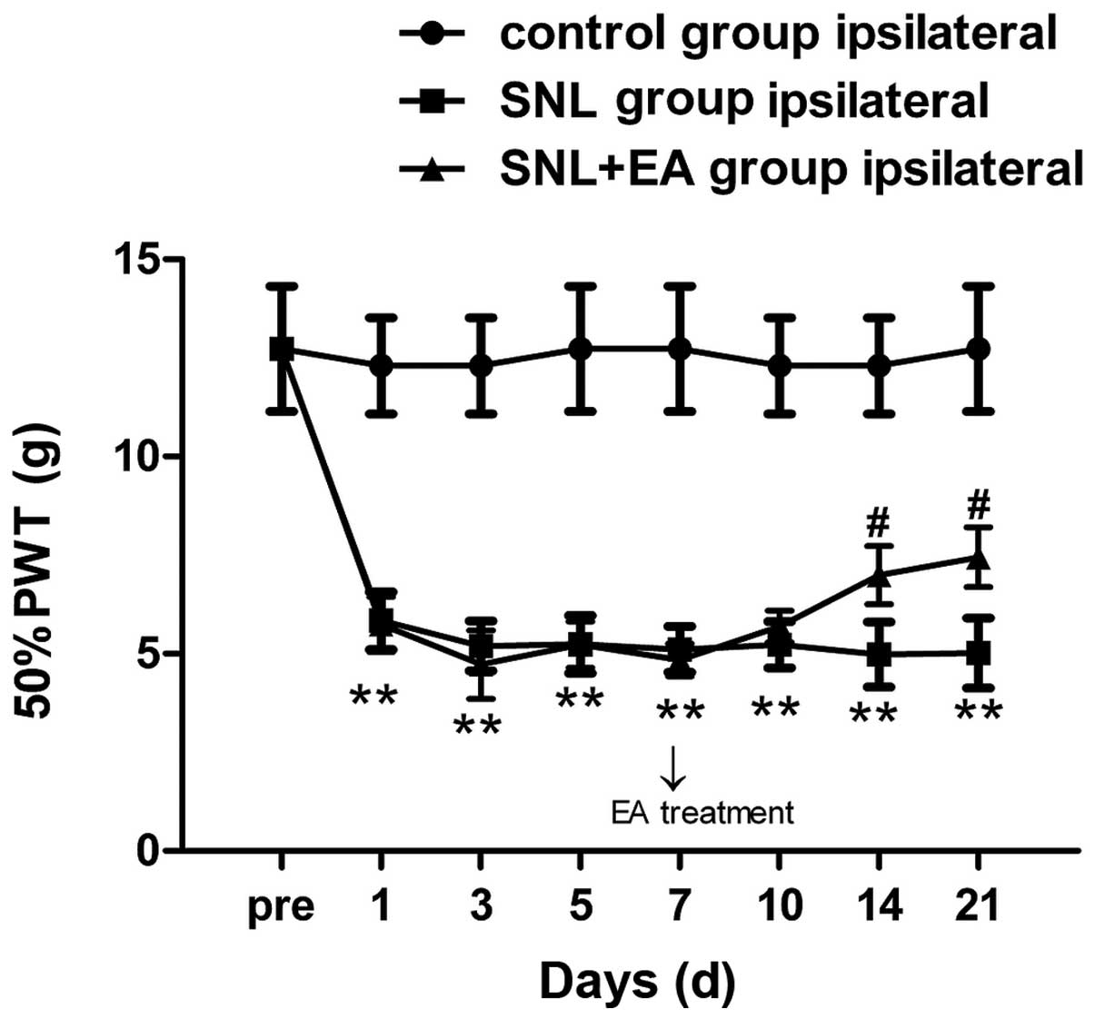
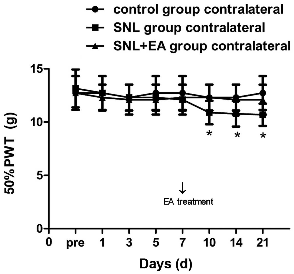
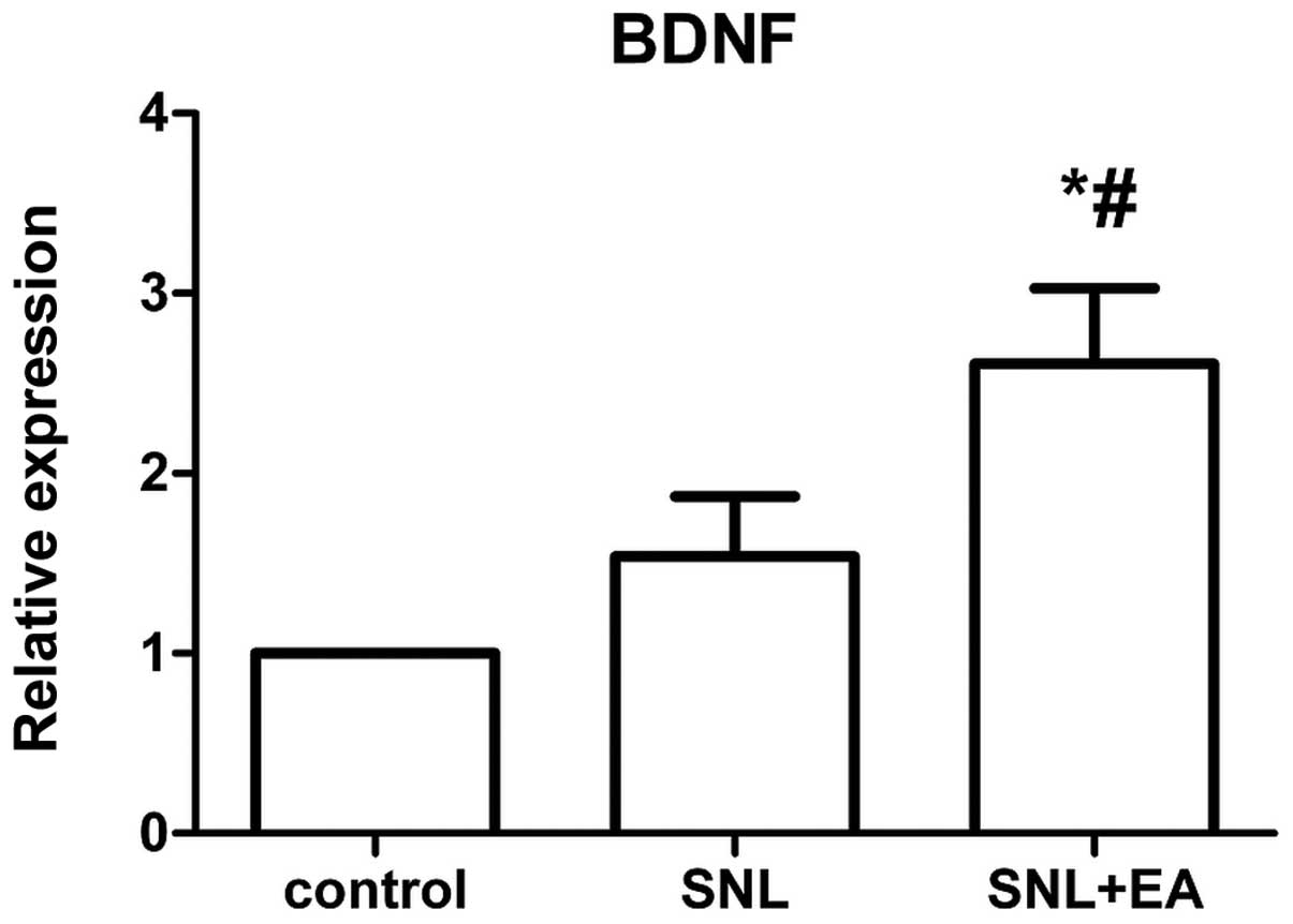
The aim of this study was to investigate the analgesic mechanism of electroacupuncture (EA) in the treatment of neuropathological pain. A total of 60 Sprague‑Dawley rats were randomly divided into three groups, namely the spinal nerve ligation (SNL), electroacupuncture (SNL + EA) and normal control groups, with 20 rats in each group. The up-down method was used to determine the bipedal 50% mechanical paw withdrawal threshold (PWT). The ultrastructure of the injured‑side L5 nerve root (n=6) was observed by electron microscopy. The mRNA levels of brain-derived neurotrophic factor (BDNF) and purinergic receptor P2X, ligand-gated ion channel 4 (P2X4) in the spinal cord (n=14) were determined by reverse transcription-quantitative polymerase chain reaction (RT-qPCR). The postoperative PWT of the injured‑side hindpaw in the SNL group at each time point was lower than that in the control group (P<0.01); there were differences of statistical significance between the PWT values of the SNL + EA and SNL groups on postoperative days 14 and 21 (P<0.05). Postoperatively, the PWT of the hindpaw on the uninjured-side was significantly lower in the SNL group when compared with that of the control group on days 10, 14 and 21 (P<0.05). Following the EA treatment, axonal demyelination was reduced and vascular proliferation was observed within the visual field. In addition, following the EA treatment, BDNF expression levels in the spinal dorsal horn increased (P<0.05), while the expression of P2X4 was not different from that in the SNL group. EA exerted an analgesic effect on the SNL model in a time-dependent manner, and improved the blood supply to the nerve root. Following the EA treatment, the expression of P2X4 did not change significantly compared with that in the SNL group, whereas the spinal secretion of BDNF increased. However, the exact mechanism requires further study.
View Figures
View References

Related Articles

Journal Cover

March-2015
Volume 9 Issue 3

Print ISSN: 1792-0981
Online ISSN:1792-1015

Sign up for eToc alerts

Recommend to Library

Copy and paste a formatted citation
x
Spandidos Publications style
Xue C, Xie L, Li X, Cai J, Gu Z and Wang K: Analgesic mechanism of electroacupuncture in a rat L5 spinal nerve ligation model. Exp Ther Med 9: 987-991, 2015.
APA
Xue, C., Xie, L., Li, X., Cai, J., Gu, Z., & Wang, K. (2015). Analgesic mechanism of electroacupuncture in a rat L5 spinal nerve ligation model. Experimental and Therapeutic Medicine, 9, 987-991. https://doi.org/10.3892/etm.2015.2165
MLA
Xue, C., Xie, L., Li, X., Cai, J., Gu, Z., Wang, K."Analgesic mechanism of electroacupuncture in a rat L5 spinal nerve ligation model". Experimental and Therapeutic Medicine 9.3 (2015): 987-991.
Chicago
Xue, C., Xie, L., Li, X., Cai, J., Gu, Z., Wang, K."Analgesic mechanism of electroacupuncture in a rat L5 spinal nerve ligation model". Experimental and Therapeutic Medicine 9, no. 3 (2015): 987-991. https://doi.org/10.3892/etm.2015.2165