Introduction
Osteoarthritis (OA) is a common chronic joint
disease, which is characterized by cartilage degradation,
synovitis, subchondral bone sclerosis and osteophyte formation
(1). Worldwide, the World Health
Organization estimates that ~10% of men and 18% of women aged
>60 years have symptomatic OA, 80% of which suffer from movement
limitations (2). OA imposes a
substantial burden of disease at the global, regional and
individual levels, and this is likely to increase with time due to
an aging world population with increasing rates of obesity
(3–5). Although previous clinical studies on OA
have been performed, the etiology of this disease is yet to be
elucidated (6). Several biochemical
and biomechanical factors are attributable to the pathogenesis of
OA.
Wnt inhibitory factor-1 (WIF-1) is a secreted
protein that antagonizes Wnt signaling (7). WIF-1 was initially detected in the
human retina and highly conserved homologs have previously been
described in various vertebrates (8). WIF-1 mRNA expression has been detected
in numerous murine and human tissues, and WIF-1 expression was
demonstrated to be most abundant in the brain, lung, retina and
cartilage (8–12). Surmann-Schmitt et al (11) demonstrated that WIF-1 is capable of
disrupting the CTGF-dependent induction of Acan and COL2A1 gene
expression in primary murine chondrocytes. These results indicated
that WIF-1 may be a multifunctional modulator of signalling
pathways in the cartilage compartment. Stock et al (12) demonstrated that WIF-1 promoted local
bone erosion and systemic bone loss whilst protecting against
arthritis-triggered cartilage degradation, indicating that WIF-1
may function to rescue the articular surface from excessive Wnt
signals.
To the best of our knowledge, there have been no
detailed studies investigating WIF-1 expression levels in the
articular cartilage of patients with various stages of OA of the
knee. Whether WIF-1 expression levels in articular cartilage are
associated with the disease severity of OA of the knee remains
unclear. We hypothesized that WIF-1 in articular cartilage may be
associated with disease severity in patients with OA of the knee.
The aim of the present study was to investigate the expression
levels of WIF-1 in the articular cartilage of patients with primary
OA of the knee, and identify the possible correlations with the
Modified Mankin score (MMS) of OA of the knee, which may serve as a
useful tool for indicating the disease severity and progression of
OA of the knee.
Materials and methods
Patients and preparation of
samples
The protocol of the present study was approved by
the Human Ethics Committee of Xiangya Hospital (Changsha, China).
According to the criteria outlined by the American College of
Rheumatology (13), 40 patients aged
between 48 and 79 years with no history of any form of secondary OA
or inflammatory joint diseases, including rheumatoid arthritis, were
eligible for enrollment in the present study. A total of 40 human
articular cartilage samples were harvested from the lateral and
medial sides of the tibia plateau of the 40 knees of these patients
who had undergone total knee arthroplasty (TKA). Radiographs of the
knee were captured and evaluated using the Kellgren-Lawrence
grading scale (14). Control
experiments were performed on 10 samples which were harvested from
the disease-free cartilage of patients who had undergone above knee
amputation due to severe trauma. Control subjects had no history of
secondary OA, knee injury, intra-articular fracture, OA in other
joints, osteoporosis, rheumatoid arthritis or tuberculous
arthritis, and had not received steroid injection within the
preceeding 3 months. Control subjects were matched according to
age, gender and body mass index (BMI) with the OA group. Causes of
amputation included: Femoral fractures (20%), open tibial-fibular
fractures (50%) and multiple fractures to the lower leg (30%). All
fractures were complicated with vascular disruptions or destructive
soft tissue injuries.
Histology
Biopsies of cartilage and bone were obtained from
the lateral and medial sides of the tibia plateau including the
loading zone and the margin zone whenever possible. All cartilage
samples underwent decalcification with 15%
ethylenediaminetetraacetic acid solution (Beyotime Institute of
Biotechnology, Haimen, China) for 3 weeks. Specimens, which were
1.0 cm-thick with a cartilage surface of ~2.0×0.5 cm, were
incubated in freshly prepared paraformaldehyde (Tianjin Kemiou
Chemical Reagent Co., Ltd., Tianjin, China) prior to dehydration
with a graded concentration of ethanol and xylene and paraffin
embedding (all Sigma-Aldrich, St. Louis, MO, USA). Subsequently,
sagittal 5-µm thick sections were cut and stained with hematoxylin
and eosin and safranin-O fast green (both Sigma-Aldrich). The
severity of cartilage damage was assessed according to the MMS
(15): Normal (MMS, 0–1; n=10), mild
lesions (MMS, 2–5; n=9), moderate lesions (MMS, 6–9; n=14) and
severe lesions (MMS, 10–14; n=15). Two samples were excluded from
the present study as the specimens were destroyed.
Immunohistochemistry
Paraffin-fixed samples were deparaffinized in xylene
and ethanol prior to rehydration in water. Subsequently, the
sections were incubated with trypsin (Beyotime Institute of
Biotechnology) for 30 min at 37°C and incubated with citrate sodium
(Sigma-Aldrich) in a water bath for 10 min. Following washing with
phosphate-buffered saline (PBS), sections were blocked with 0.5%
bovine serum albumin (diluted in PBS; both Sigma-Aldrich) for 30
min at room temperature. The sections were subsequently incubated
with diluted WIF-1 antibody (1:100; CB52917240 Beijing Biosynthesis
Biotechnology Co., Ltd., Beijing, China) for 1 h. Following rinsing
with PBS, the sections were incubated with horseradish
peroxidase/Fab polymer conjugate (Zymed Picure-Plus kit; Thermo
Fisher Scientific, Inc., Waltham, MA, USA) for 30 min. Finally, the
sections were incubated with 3,3′-diaminobenzidine (Sigma-Aldrich)
for 5 min in order to develop the signals. A negative control was
included by omitting the primary antibody. All the sections were
examined by an independent pathologist who was unaware of the
clinical characteristics of the samples included in the present
study. All the sections were examined using a Motic BA210
microscope (Motic Instruments, Xiamen, China) at ×100
magnification. The relative WIF-1 distribution of cartilage tissue
was visualized and quantified using optical density (OD) values
(16,17). Positive WIF-1 immunostaining was
defined as detectable immunoreactivity in the perinuclear and/or
other cytoplasmic regions in chondrocytes. Semiquantitative
assessment of the average optical density of WIF-1 expression
levels was performed on scanned autoradiograms using MIAS-4400
ImageJ (National Institutes of Health, Bethesda, MA, USA) (1). All densities were normalized against
PBS and the experiment was repeated in triplicate. The coefficient
of variation (CV) of WIF-1 expression in articular cartilage was
<2%.
Statistical analysis
Statistical analyses were performed using SPSS
software 13.0 (SPSS, Inc., Chicago, IL, USA). One-way analysis of
variance was performed in the present study in order to determine
between-group differences. Associations between WIF-1 expression
levels in the articular cartilage, as determined by OD values, and
the MMSs of OA were analyzed using Pearson's correlation analysis.
Data were expressed as the mean ± standard deviation. P<0.05 was
considered to indicate a statistically significant difference.
Results
Patient characteristics
A total of 48 knee samples were analyzed from 38
patients with OA of the knee and 10 control subjects. Baseline
clinical characteristics of both groups are presented in Table I. No significant differences were
detected in age, gender and BMI indices between the two groups.
Kellgren-Lawrence radiological scoring confirmed that the majority
of patients with OA of the knee presented with severe OA, which was
appropriate for total knee replacement (Table II).
 | Table I.Characteristics of patients with
osteoarthritis of the knee (n=38) and control subjects (n=10). |
Table I.
Characteristics of patients with
osteoarthritis of the knee (n=38) and control subjects (n=10).
| Parameter | Patients | Controls | P-value |
|---|
| Age (years) |
62.0±13.6 |
56.2±18.9 | 0.66 |
| Gender (%
female) | 45 | 40 | 0.32 |
| BMI
(kg/m2) | 28.08±0.49 | 26.34±0.56 | 0.13 |
 | Table II.Kellgren-Lawrence radiological scores
in patients with osteoarthritis of the knee (n=40). |
Table II.
Kellgren-Lawrence radiological scores
in patients with osteoarthritis of the knee (n=40).
| Kellgren-Lawrence
score | Patients (n) |
|---|
| Grade 1 | 0 |
| Grade 2 | 0 |
| Grade 3 | 9 |
| Grade 4 | 31 |
WIF-1 expression levels
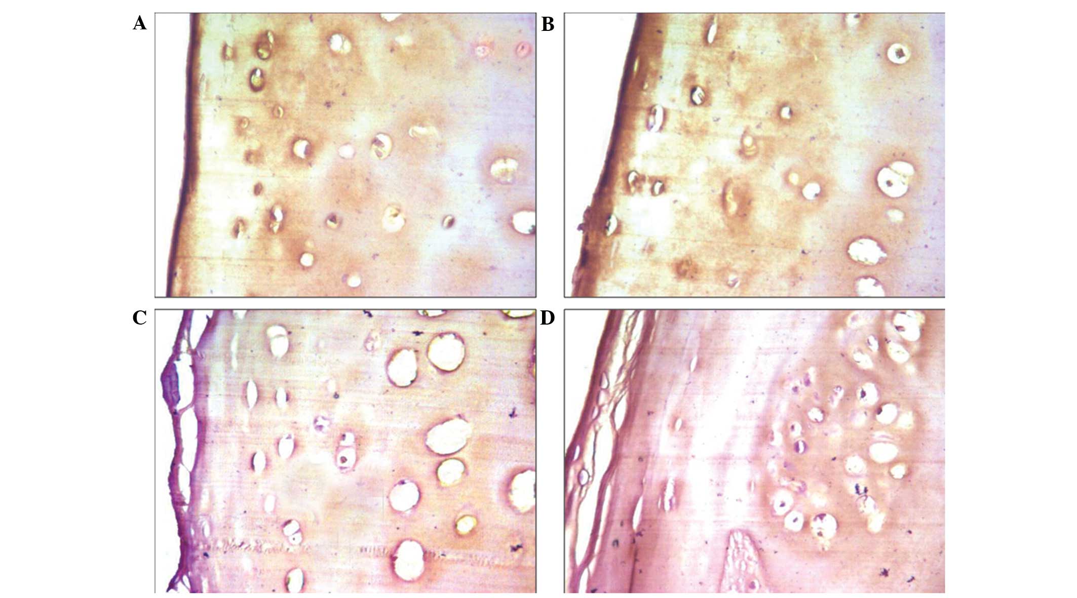
MMSs of each group are shown in Table III. WIF-1 expression levels were
detected in the articular cartilage of all four groups. In both the
control and mild lesion groups, WIF-1 expression was predominantly
detected in the superficial layers of the articular cartilage
(Fig. 1); whereas faint staining was
detected in the territorial matrix of the deep zone of articular
cartilage in the moderate and severe lesions (Fig. 1).
 | Table III.Modified Mankin scores in the various
groups. |
Table III.
Modified Mankin scores in the various
groups.
| Group | Samples (n) | Modified Mankin
score |
|---|
| Normal | 10 |
0.60±0.52 |
| Mild lesions | 9 |
3.11±1.05 |
| Moderate lesions | 14 |
7.21±1.12 |
| Severe lesions | 15 | 12.07±1.33 |
OD values
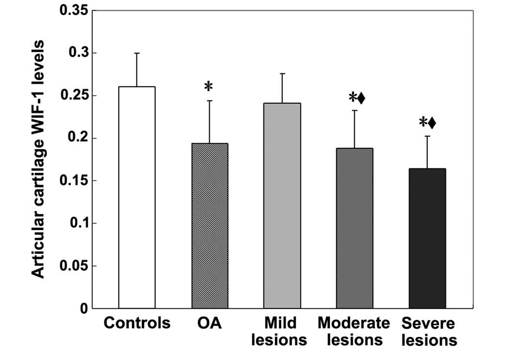
Average OD values of WIF-1 in the articular
cartilage was 0.26±0.04 in the control patients, 0.19±0.05 in
patients with OA of the knee and 0.24±0.03, 0.19±0.04 and 0.16±0.04
in the mild, moderate and severe lesions, respectively. Patients
with OA demonstrated significantly reduced WIF-1 expression levels
in the articular cartilage, as compared to the controls (P<0.01)
(Fig. 2). Furthermore, WIF-1
expression levels in the moderate and severe lesions of articular
cartilage were significantly reduced, as compared with the control
(P<0.01) and mild lesion (P<0.05) groups (Fig. 2). The MMSs (r=−0.896; P<0.001)
indicate that WIF-1 expression in articular cartilage was
negatively correlated with severity of disease (Fig. 3).
Discussion
To the best of our knowledge, the present study is
the first to investigate the WIF-1 expression levels of articular
cartilage in patients with various stages of OA of the knee and its
correlation with OA disease severity. The results of the present
study demonstrated that WIF-1 expression is predominantly detected
in the superficial layers of articular cartilage. These results are
consistent with previous studies which have demonstrated that WIF-1
is highly expressed in the superficial layers of epiphyseal
cartilage in bone and tendons (11,12,18).
Since WIF-1 is predominantly expressed in articular cartilage, and
in bone to a lesser extent (12,18), the
present study aimed to investigate whether WIF-1 is upregulated in
patients with OA. Notably, the present study demonstrated a marked
reduction in WIF-1 expression levels in the articular cartilage of
patients with OA of the knee, as compared with the controls.
Cartilage damage is one of the predominant
pathological changes detected in patients with OA. According to the
MMS, the results of the present study demonstrated that WIF-1
expression levels in articular cartilage were negatively correlated
with the severity of disease. These results suggested that the loss
of WIF-1 expression may be an early event in the OA disease
progress and indicated that WIF-1 may be associated with the
pathogenesis of OA. A previous study demonstrated specific binding
of WIF-1 to Wnt3a, Wnt4, Wnt5a, Wnt7a, Wnt9a and Wnt11 (18). Furthermore, it has been demonstrated
that WIF-1 is capable of blocking Wnt3a-mediated activation of the
canonical Wnt signalling pathway, which is associated with
cartilage degeneration (18). The
results of another previous study demonstrated that WIF-1 was
capable of attenuating the CTGF-dependent induction of cartilage
matrix gene expression, including aggrecan and COL2A1, in primary
murine chondrocytes (11).
Therefore, this mechanism, which is mediated by WIF-1, may have a
key role in OA.
Stock et al (12) demonstrated that WIF-1 expression
levels were repressed by tumor necrosis factor (TNF)α in
chondrocytes and osteoblasts, and were downregulated in
inflammatory arthritis. WIF-1 deficiency partially protected
TNF-transgenic mice against bone loss and reduced arthritis-related
increases in osteoclast numbers. Enhanced cartilage destruction was
detected during WIF-1 deficiency and systemic overexpression of
WIF-1 was demonstrated to partially protect against
arthritis-related cartilage loss, which suggested that WIF-1 may
have a protective role in cartilage destruction in patients with
arthritis. Furthermore, in chondrocytes, TNFα induced canonical Wnt
signaling, which was successfully blocked by WIF-1, indicating that
TNFα and WIF-1 may have a direct effect on Wnt signaling in this
system. These data suggested that WIF-1 may be associated with the
fine-tuning of cartilage and bone turnover, thus promoting the
balance between cartilage and bone anabolism.
Aberrant methylation of regulatory regions that
silence the transcription of genes has been postulated to be a
mechanism for the inactivation of tumor suppressor genes in human
cancer (19–22). Mazieres et al (23) demonstrated that methylation silencing
of WIF-1 was a common mechanism of aberrant activation of the Wnt
signaling pathway in lung cancer pathogenesis. Furthermore, Urakami
et al (21) previously
demonstrated that, independent of histological grade or stage,
bladder tumor samples exhibited decreased WIF-1 expression levels
and enhanced WIF-1 promoter methylation, as compared with normal
bladder mucosa samples. Therefore, it is possible that dysregulated
WIF-1 expression, induced by promoter hypermethylation, may
activate the Wnt/β-catenin signaling pathway via the nuclear
translocation of β-catenin in the pathogenesis of OA.
The present study had various limitations. Firstly,
the present study had a small sample size and was a single-center
study. Secondly, only patients with OA of the knee who attended
Xiangya Hospital were enrolled in the study. Thirdly, the
associations between WIF-1 expression levels in the serum, synovial
fluid and articular cartilage, which were determined via
radiographic grading of OA, were not analyzed. Therefore, in order
to determine whether WIF-1 expression levels are capable of
predicting clinical prognosis and outcomes in patients with OA of
the knee, a cross-sectional study is required.
In conclusion, the results of the present study
demonstrated that patients OA of the knee exhibited reduced WIF-1
expression levels, as compared with healthy control subjects.
Furthermore, WIF-1 expression levels in articular cartilage were
demonstrated to be negatively associated with the MMS, indicating
that WIF-1 expression may be a potential indictor of disease
severity of OA of the knee. Further investigations are being
performed by the authors of the present study in order to define
WIF-1 expression levels in the synovial fluid or serum, the
signaling events induced by WIF-1 and the potential utility of
WIF-1 as a therapeutic target to inhibit the progression of OA.
Measurements of WIF-1 expression levels in the synovial fluid or
serum may have predictive value for the progression of OA of the
knee. Subsequent longitudinal studies may further elucidate the
therapeutic value of WIF-1 as a potential marker to monitor the
disease severity and progression of OA of the knee.
Acknowledgements
The present study was supported by the National
Natural Science Foundation of China (grant nos. 81201420 and
81272034), the Provincial Science Foundation of Hunan (grant no.
14JJ3032), the Scientific Research Project of the Development and
Reform Commission of Hunan Province (grant no. 20131199), the
Scientific Research Project of Science and Technology Office of
Hunan Province (grant no. 2013SK2018), the Doctoral Scientific Fund
Project of the Ministry of Education of China (grant no.
20120162110036) and the Clinical Research Project of Xingya
Hospital (grant no 2015101).
References
|
1
|
Arden NK and Leyland KM: Osteoarthritis
year 2013 in review: Clinical. Osteoarthritis Cartilage.
21:1409–1413. 2013. View Article : Google Scholar : PubMed/NCBI
|
|
2
|
Gao SG, Li KH, Zeng KB, Tu M, Xu M and Lei
GH: Elevated osteopontin level of synovial fluid and articular
cartilage is associated with disease severity in knee
osteoarthritis patients. Osteoarthritis Cartilage. 18:82–87. 2010.
View Article : Google Scholar : PubMed/NCBI
|
|
3
|
Lawrence RC, Felson DT, Helmick CG, Arnold
LM, Choi H, Deyo RA, Gabriel S, Hirsch R, Hochberg MC, Hunder GG,
et al: Estimates of the prevalence of arthritis and other rheumatic
conditions in the United States. Part II. Arthritis Rheum.
58:26–35. 2008. View Article : Google Scholar : PubMed/NCBI
|
|
4
|
Liu M and Hu C: Association of MIF in
serum and synovial fluid with severity of knee osteoarthritis. Clin
Biochem. 45:737–739. 2012. View Article : Google Scholar : PubMed/NCBI
|
|
5
|
Holt HL, Katz JN, Reichmann WM, Gerlovin
H, Wright EA, Hunter DJ, Jordan JM, Kessler CL and Losina E:
Forecasting the burden of advanced knee osteoarthritis over a
10-year period in a cohort of 60–64 year-old US adults.
Osteoarthritis Cartilage. 19:44–50. 2011. View Article : Google Scholar : PubMed/NCBI
|
|
6
|
Bay-Jensen AC, Reker D, Kjelgaard-Petersen
CF, Mobasheri A, Karsdal MA, Ladel C, Henrotin Y and Thudium CS:
Osteoarthritis year in review: 2015 soluble biomarkers and the
BIPED criteria. Osteoarthritis Cartilage. 24:9–20. 2016. View Article : Google Scholar : PubMed/NCBI
|
|
7
|
Ai L, Tao Q, Zhong S, Fields CR, Kim WJ,
Lee MW, Cui Y, Brown KD and Robertson KD: Inactivation of Wnt
inhibitory factor-1 (WIF1) expression by epigenetic silencing is a
common event in breast cancer. Carcinogenesis. 27:1341–1348. 2006.
View Article : Google Scholar : PubMed/NCBI
|
|
8
|
Hsieh JC, Kodjabachian L, Rebbert ML,
Rattner A, Smallwood PM, Samos CH, Nusse R, Dawid IB and Nathans J:
A new secreted protein that binds to Wnt proteins and inhibits
their activities. Nature. 398:431–436. 1999. View Article : Google Scholar : PubMed/NCBI
|
|
9
|
Hu YA, Gu X, Liu J, Yang Y, Yan Y and Zhao
C: Expression pattern of Wnt inhibitor factor 1(Wif1) during the
development in mouse CNS. Gene Expr Patterns. 8:515–522. 2008.
View Article : Google Scholar : PubMed/NCBI
|
|
10
|
Hunter DD, Zhang M, Ferguson JW, Koch M
and Brunken WJ: The extracellular matrix component WIF-1 is
expressed during and can modulate, retinal development. Mol Cell
Neurosci. 27:477–488. 2004. View Article : Google Scholar : PubMed/NCBI
|
|
11
|
Surmann-Schmitt C, Sasaki T, Hattori T,
Eitzinger N, Schett G, von der Mark K and Stock M: The Wnt
antagonist Wif-1 interacts with CTGF and inhibits CTGF activity. J
Cell Physiol. 227:2207–2216. 2012. View Article : Google Scholar : PubMed/NCBI
|
|
12
|
Stock M, Böhm C, Scholtysek C, Englbrecht
M, Fürnrohr BG, Klinger P, Gelse K, Gayetskyy S, Engelke K,
Billmeier U, et al: Wnt inhibitory factor 1 deficiency uncouples
cartilage and bone destruction in tumor necrosis factor α-mediated
experimental arthritis. Arthritis Rheum. 65:2310–2322. 2013.
View Article : Google Scholar : PubMed/NCBI
|
|
13
|
Altman R, Asch E, Bloch D, Bole G,
Borenstein D, Brandt K, Christy W, Cooke TD, Greenwald R and
Hochberg M: Development of criteria for the classification and
reporting of osteoarthritis: Classification of osteoarthritis of
the knee Diagnostic and Therapeutic Criteria of the American
Rheumatism Association. Arthritis Rheum. 29:1039–1049. 1986.
View Article : Google Scholar : PubMed/NCBI
|
|
14
|
Kellgren JH and Lawrence JS: Radiological
assessment of osteo-arthrosis. Ann Rheum Dis. 16:494–502. 1957.
View Article : Google Scholar : PubMed/NCBI
|
|
15
|
van der Sluijs JA, Geesink RG, van der
Linden AJ, Bulstra SK, Kuyer R and Drukker J: The reliability of
the Mankin score for osteoarthritis. J Orthop Res. 10:58–61. 1992.
View Article : Google Scholar : PubMed/NCBI
|
|
16
|
Jiang W, Gao SG, Chen XG, Xu XC, Xu M, Luo
W, Tu M, Zhang FJ, Zeng C and Lei GH: Expression of synovial fluid
and articular cartilage VIP in human osteoarthritic knee: A new
indicator of disease severity? Clin Biochem. 45:1607–1612. 2012.
View Article : Google Scholar : PubMed/NCBI
|
|
17
|
Zhang FJ, Luo W, Gao SG, Su DZ, Li YS,
Zeng C and Lei GH: Expression of CD44 in articular cartilage is
associated with disease severity in knee osteoarthritis. Mod
Rheumatol. 23:1186–1191. 2013. View Article : Google Scholar : PubMed/NCBI
|
|
18
|
Surmann-Schmitt C, Widmann N, Dietz U,
Saeger B, Eitzinger N, Nakamura Y, Rattel M, Latham R, Hartmann C,
von der Mark H, et al: Wif-1 is expressed at cartilage-mesenchyme
interfaces and impedes Wnt3a-mediated inhibition of chondrogenesis.
J Cell Sci. 122:3627–3637. 2009. View Article : Google Scholar : PubMed/NCBI
|
|
19
|
Herman JG and Baylin SB: Gene silencing in
cancer in association with promoter hypermethylation. N Engl J Med.
349:2042–2054. 2003. View Article : Google Scholar : PubMed/NCBI
|
|
20
|
Jones PA and Baylin SB: The fundamental
role of epigenetic events in cancer. Nat Rev Genet. 3:415–428.
2002.PubMed/NCBI
|
|
21
|
Urakami S, Shiina H, Enokida H, Kawakami
T, Tokizane T, Ogishima T, Tanaka Y, Li LC, Ribeiro-Filho LA,
Terashima M, et al: Epigenetic inactivation of Wnt inhibitory
factor-1 plays an important role in bladder cancer through aberrant
canonical Wnt/beta-catenin signaling pathway. Clin Cancer Res.
12:383–391. 2006. View Article : Google Scholar : PubMed/NCBI
|
|
22
|
Yang Z and Wang Y, Fang J, Chen F, Liu J,
Wu J and Wang Y: Expression and aberrant promoter methylation of
Wnt inhibitory factor-1 in human astrocytomas. J Exp Clin Cancer
Res. 29:262010. View Article : Google Scholar : PubMed/NCBI
|
|
23
|
Mazieres J, He B, You L, Xu Z, Lee AY,
Mikami I, Reguart N, Rosell R, McCormick F and Jablons DM: Wnt
inhibitory factor-1 is silenced by promoter hypermethylation in
human lung cancer. Cancer Res. 64:4717–4720. 2004. View Article : Google Scholar : PubMed/NCBI
|


