Chronic myelocytic leukemic fundus lesion: A case report
- Authors:
- Le Wen Xu
- Yinghua Zhang
- Yan Liu
- Yan Ding
View Affiliations
Affiliations: Department of Ophthalmology, Rizhao People's Hospital, Rizhao, Shandong 276800, P.R. China
- Published online on: August 5, 2016 https://doi.org/10.3892/etm.2016.3577
-
Pages:
2253-2256
Metrics: Total
Views: 0 (Spandidos Publications: | PMC Statistics: )
Metrics: Total PDF Downloads: 0 (Spandidos Publications: | PMC Statistics: )
This article is mentioned in:
Abstract
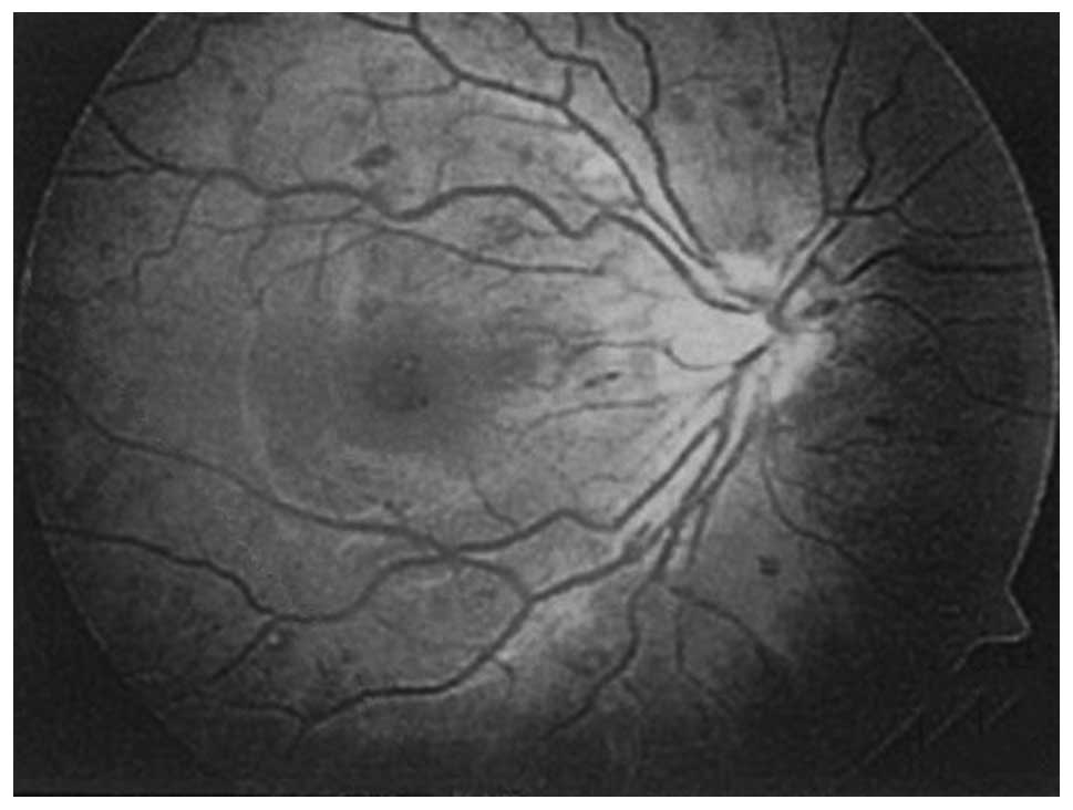
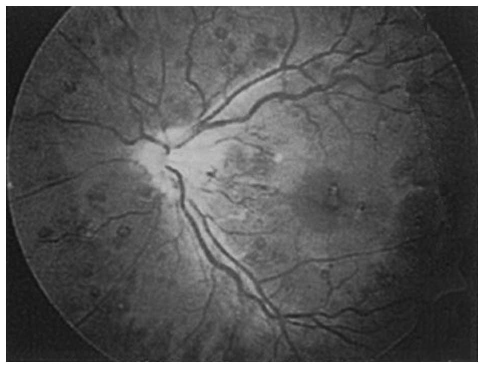
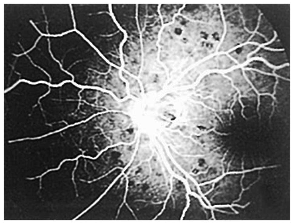
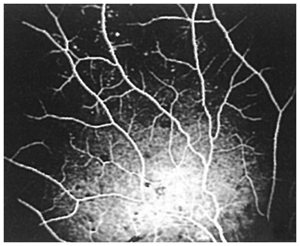
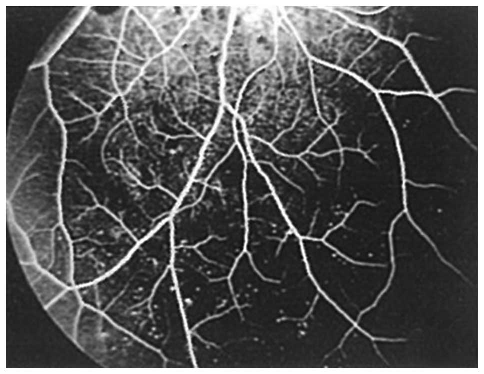
The current study reports a case of a patient with a chronic myelocytic leukemic fundus lesion, initially diagnosed in the Department of Ophthalmology, Rizhao People's Hospital (Rizhao, China). A male, 23‑years‑of‑age, presented with a dark shadow in the front of the right eye, accompanied with blurred vision for 3 days (visual acuity of right eye, 0.4; visual acuity of left eye, 0.6). In addition, the patient had experienced gingival bleeding for 2 years, and recurrent upper respiratory infections for 1 year. A fundus examination revealed mild binocular papillary edema, dilated and tortuous veins, and the retina exhibited large quantities of scattered and dark red bleeding spots. The bleeding spots had white spots in the center, which exhibited typical Roth spots, whilst routine blood examination, abdominal ultrasound, marrow biopsy and other laboratory tests confirmed the diagnosis of chronic myelogenous leukemia. Patients with leukemia typically present with initial symptoms that include fever, fatigue, anemia and hepatosplenomegaly, and a diagnosis as a result of eye‑related symptoms is rare, rendering the present case unique.
View Figures |
Figure 1
|
|
Figure 2
|
|
Figure 3
|
|
Figure 4
|
|
Figure 5
|
|
Figure 6
|
View References
1
|
Howlader N, Noone AM, Krapcho M, Neyman N,
Aminou R, Waldron W, Altekruse SF, Kosary CL, Ruhl J, Tatalovich Z,
et al: SEER Cancer Statistics Review, 1975-2009 (Vintage 2009
Populations). National Cancer Institute; Bethesda, MD:
(seer.cancer.gov/csr/1975_2009_pops09/). Accessed. September.
2013
|
2
|
Baccarani M, Cortes J, Pane F,
Niederwieser D, Saglio G, Apperley J, Cervantes F, Deininger M,
Gratwohl A, Guilhot F, et al: Chronic myeloid leukemia: An update
of concepts and management recommendations of European Leukemia
Net. J Clin Oncol. 27:6041–6051. 2009. View Article : Google Scholar : PubMed/NCBI
|
3
|
Quintás-Cardama A and Cortes J: Molecular
biology of bcr-abl1-positive chronic myeloid leukemia. Blood.
113:1619–1630. 2009. View Article : Google Scholar : PubMed/NCBI
|
4
|
Cortes J: Natural history and staging of
chronic myelogenous leukemia. Hematol Oncol Clin North Am.
18:569–584. 2004. View Article : Google Scholar : PubMed/NCBI
|
5
|
GambacortiPasserini C, Antolini L, Mahon
FX, Guilhot F, Deininger M, Fava C, Nagler A, Della Casa CM, Morra
E, Abruzzese E, et al: Multicenter independent assessment of
outcomes in chronic myeloid leukemia patients treated with
imatinib. J Natl Cancer Inst. 103:553–561. 2011. View Article : Google Scholar : PubMed/NCBI
|
6
|
Huang PK and Sanjay S: Visual disturbance
as the first symptom of chronic myeloid leukemia. Middle East Afr J
Ophthalmol. 18:336–338. 2011. View Article : Google Scholar : PubMed/NCBI
|
7
|
Vrcek I, Finnerty K, Ford P, Hogan RN and
Mancini R: Relapsing acute lymphoblastic leukemia presenting with a
rapidly enlarging and vision-threatening orbital mass. Int
Ophthalmol. 35:257–260. 2015. View Article : Google Scholar : PubMed/NCBI
|
8
|
Abuel-Asrar AM, Al-Momen AK, Kangave D,
Harakati MS and Ajarim DS: Correlation of fundus lesions and
hematologic findings in leukemic retinopathy. Eur J Ophthalmol.
6:167–172. 1996.PubMed/NCBI
|
9
|
Abuel-Asrar AM, Al-Momen AK, Kangave D and
Harakati MS: Prognostic importance of retinopathy in acute
leukemia. Doc Ophthalmol. 91:273–281. 1995-1996. View Article : Google Scholar
|
10
|
AlAbdulla NA, Thompson JT and La Borwit
SE: Simultaneous bilateral central retinal vein occlusion
associated with anticardiolipin antibodies in leukemia. Am J
Ophthalmol. 132:266–268. 2001. View Article : Google Scholar : PubMed/NCBI
|
11
|
Fred HL: Little black bags, ophthalmoscopy
and the Roth spot. Tex Heart Inst J. 40:115–116. 2013.PubMed/NCBI
|
12
|
Ling R and James B: White centered retinal
haemorrhages (Roth spots). Postgrad Med J. 74:581–582. 1998.
View Article : Google Scholar : PubMed/NCBI
|
13
|
Falcone PM and Larrison WI: Roth spots
seen on ophthalmoscopy: Diseases with which they may be associated.
Conn Med. 59:271–273. 1995.PubMed/NCBI
|
14
|
Reddy SC and Jackson N: Retinopathy in
acute leukaemia at initial diagnosis: Correlation of fundus lesions
and haematological parameters. Acta Ophthalmol Scand. 82:81–85.
2004. View Article : Google Scholar : PubMed/NCBI
|
15
|
Specchia G, Albano F, Guerriero S,
Buquicchio C, Pomes L, Pastore D, Carluccio P, Delle Noci N and
Liso V: Retinal abnormalities in newly diagnosed adult acute
myeloid leukemia. Acta Haematol. 105:197–203. 2001. View Article : Google Scholar : PubMed/NCBI
|