Open Access

A micro-CT study of microstructure change of alveolar bone during orthodontic tooth movement under different force magnitudes in rats

  • Authors:
    • Jingtao An
    • Ying Li
    • Zhongshuang Liu
    • Rui Wang
    • Bin Zhang
  • View Affiliations

  • Published online on: March 6, 2017     https://doi.org/10.3892/etm.2017.4186
  • Pages: 1793-1798
  • Copyright: © An et al. This is an open access article distributed under the terms of Creative Commons Attribution License.

Metrics: Total Views: 0 (Spandidos Publications: | PMC Statistics: )
Total PDF Downloads: 0 (Spandidos Publications: | PMC Statistics: )


Abstract

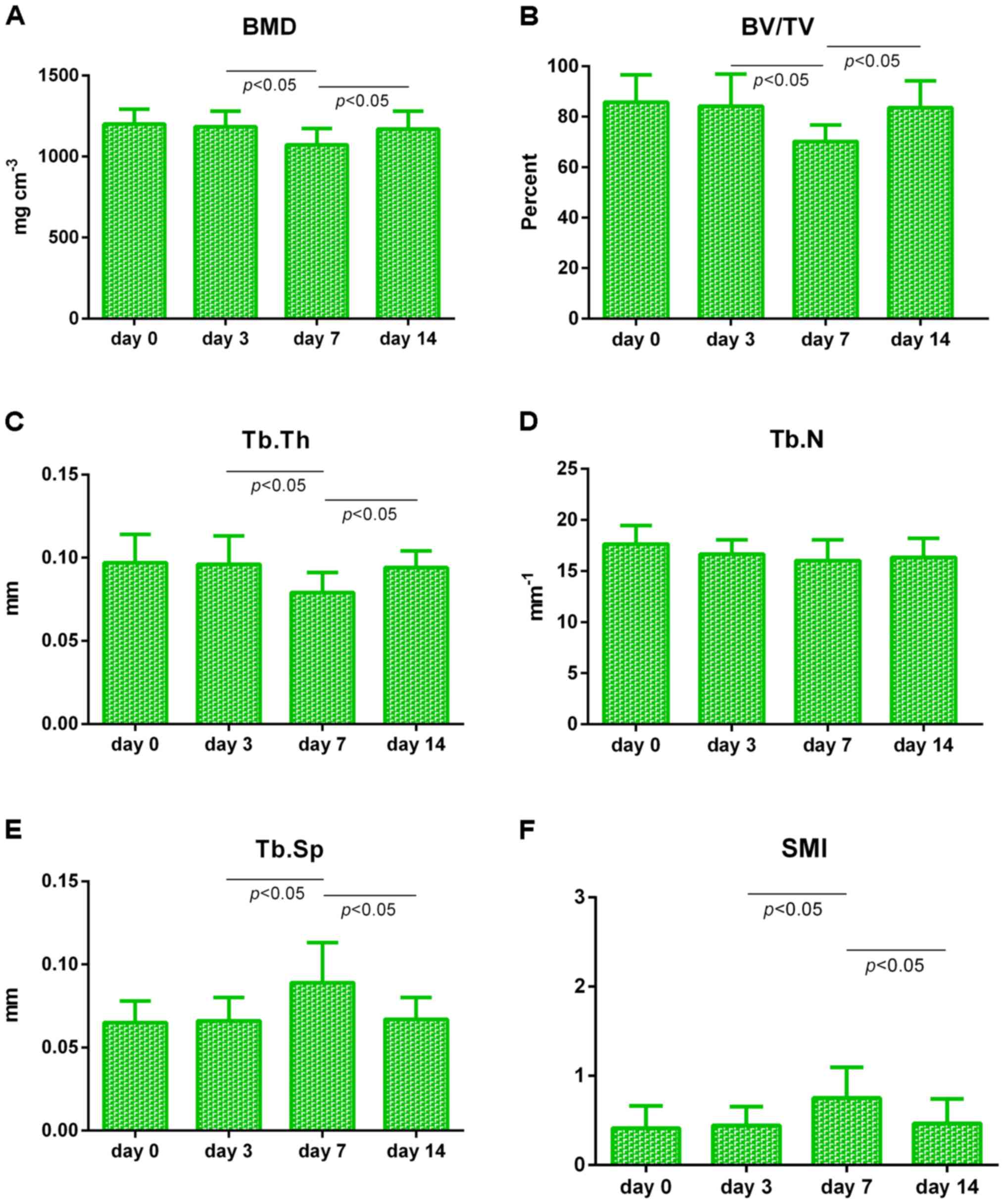
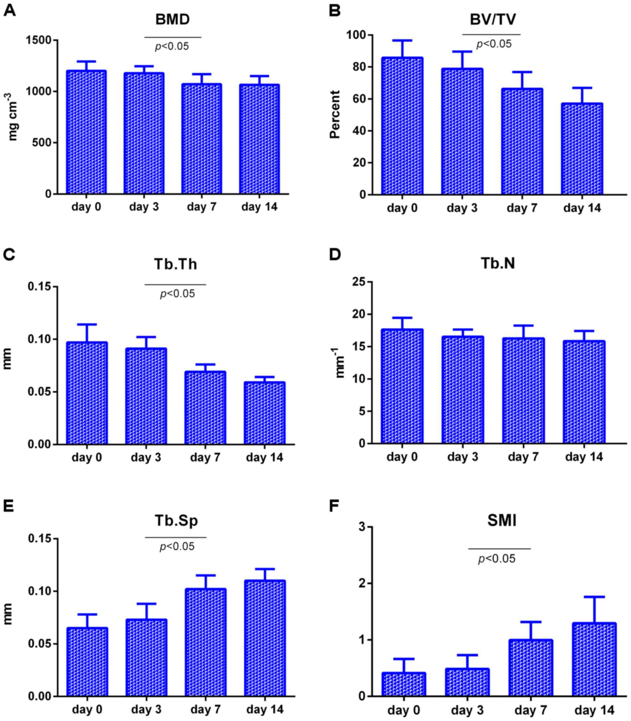
The dynamic changes of the microstructure of alveolar bone during orthodontic tooth movement in rats was explored by employing micro-computed tomography (micro-CT) system and to provide theoretical reference for clinical orthodontic treatment. Ten rats were selected randomly as control among 70 adult female Wistar rats, and the other 60 rats were divided into 25-g and 75-g groups of equal number. Orthodontic appliance with force of 25 g and 75 g was installed to perform the molar mesial movement. Microstructural parameters for trabecular bone mesial to the distobuccal root were evaluated at different time points using micro‑CT system. Moreover, distance for mesial movement of the molar were measured. Microstructural parameters for trabecular bone of two groups showed no significant changes from day 0 to day 3 (P>0.05); from day 3 to day 7, bone mineral density (BMD), bone volume/total volume (BV/TV) and trabecular thickness (Tb.Th) decreased significantly (P<0.05), whereas trabecular separation (Tb.Sp) and structure model index (SMI) increased significantly (P<0.05); from day 7 to day 14, in 25‑g group, BMD, BV/TV and Tb.Th increased significantly (P<0.05), while Tb.Sp and SMI decreased significantly (P<0.05). Correspondingly, in 75‑g group, changes of parameters did not carry any statistical significance (P>0.05). Furthermore, the 75‑g group showed larger distance than 25-g group only at day 14 (P<0.05). In conclusion, in order to maintain the health of periodontal tissues, adequate time for repair and recovery is needed to ensure reasonable remolding of alveolar bone and healthy movement of the orthodontic tooth.
View Figures
View References

Related Articles

Journal Cover

May-2017
Volume 13 Issue 5

Print ISSN: 1792-0981
Online ISSN:1792-1015

Sign up for eToc alerts

Recommend to Library

Copy and paste a formatted citation
x
Spandidos Publications style
An J, Li Y, Liu Z, Wang R and Zhang B: A micro-CT study of microstructure change of alveolar bone during orthodontic tooth movement under different force magnitudes in rats. Exp Ther Med 13: 1793-1798, 2017.
APA
An, J., Li, Y., Liu, Z., Wang, R., & Zhang, B. (2017). A micro-CT study of microstructure change of alveolar bone during orthodontic tooth movement under different force magnitudes in rats. Experimental and Therapeutic Medicine, 13, 1793-1798. https://doi.org/10.3892/etm.2017.4186
MLA
An, J., Li, Y., Liu, Z., Wang, R., Zhang, B."A micro-CT study of microstructure change of alveolar bone during orthodontic tooth movement under different force magnitudes in rats". Experimental and Therapeutic Medicine 13.5 (2017): 1793-1798.
Chicago
An, J., Li, Y., Liu, Z., Wang, R., Zhang, B."A micro-CT study of microstructure change of alveolar bone during orthodontic tooth movement under different force magnitudes in rats". Experimental and Therapeutic Medicine 13, no. 5 (2017): 1793-1798. https://doi.org/10.3892/etm.2017.4186