|
1
|
Nagy Z and Nardai S: Cerebral
ischemia/repefusion injury: From bench space to bedside. Brain Res
Bull. 134:30–37. 2017. View Article : Google Scholar : PubMed/NCBI
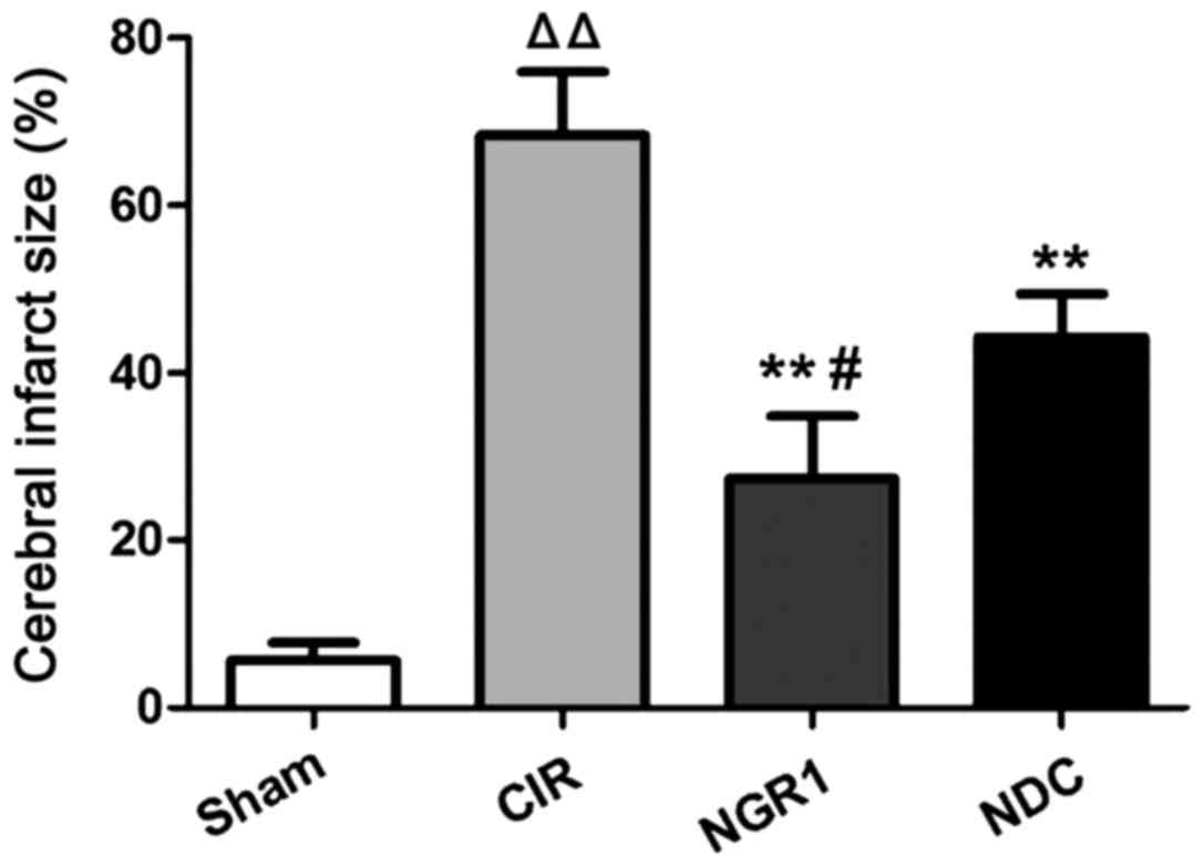
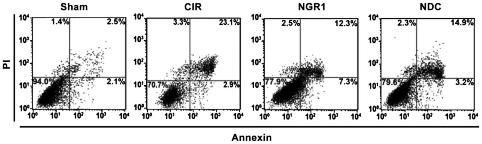
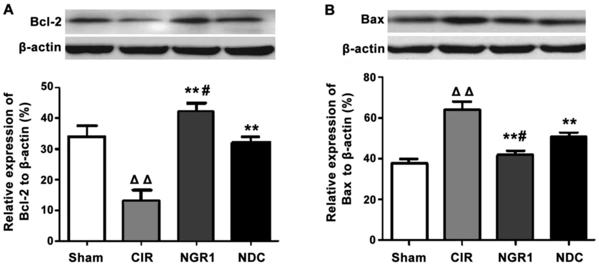
|
|
2
|
Broughton BR, Reutens DC and Sobey CG:
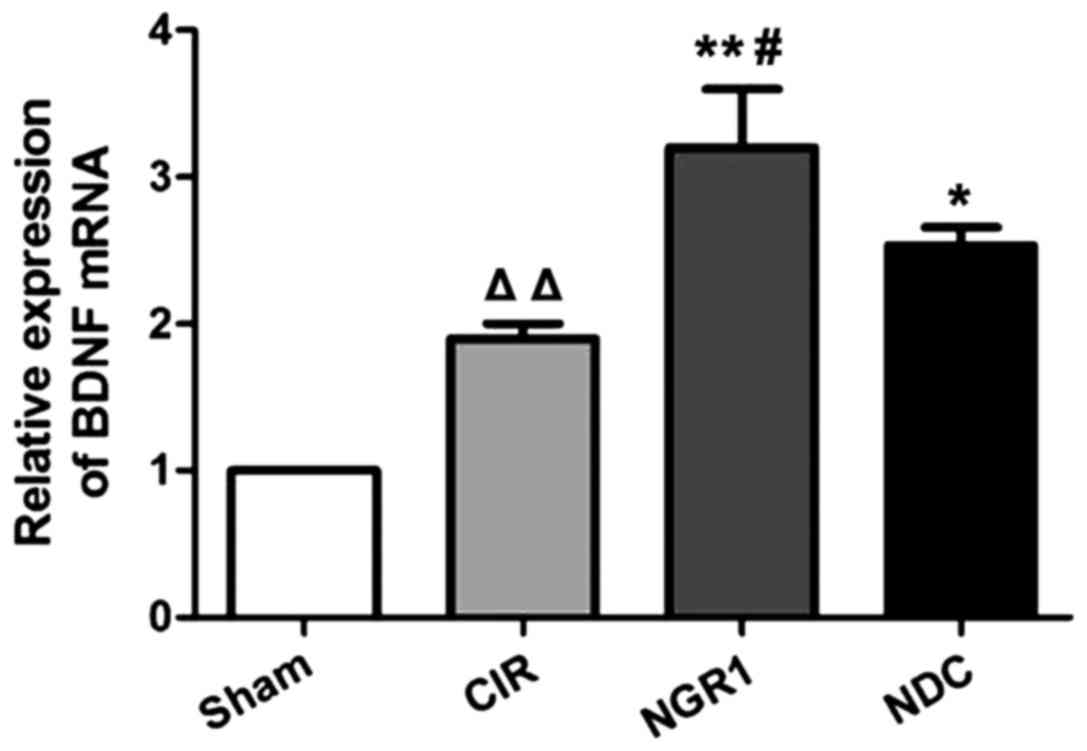
Apoptotic mechanisms after cerebral ischemia. Stroke. 40:e331–e339.
2009. View Article : Google Scholar : PubMed/NCBI
|
|
3
|
Iadecola C and Alexander M: Cerebral
ischemia and inflammation. Curr Opin Neurol. 14:89–94. 2001.
View Article : Google Scholar : PubMed/NCBI
|
|
4
|
Allen CL and Bayraktutan U: Oxidative
stress and its role in the pathogenesis of ischaemic stroke. Int J
Stroke. 4:461–470. 2009. View Article : Google Scholar : PubMed/NCBI
|
|
5
|
O'Collins VE, Macleod MR, Donnan GA, Horky
LL, van der Worp BH and Howells DW: 1,026 experimental treatments
in acute stroke. Ann Neurol. 59:467–477. 2006. View Article : Google Scholar : PubMed/NCBI
|
|
6
|
Zhan HQ, Zhang WX, Yan FL, Liu FQ and
Zhang XY: Effects of notoginsenoside-Rg1 on the expression of
apoptosis factors in brain tissue after ischemia injury. Guangdong
Yixue. 2014.(In Chinese).
|
|
7
|
Li J, Zhu P, Si Y, Xu Hong and Wu H:
Research on the effects of the total Saponin from Sanqi (the dried
root of Panax Notoginseng) on proapoptotic caspase-3 in the
forebrain in the rats with intracerebral hemorrhage. J Beijing Univ
Tradit Chin Med. 26:22–25. 2003.
|
|
8
|
Lu T, Jiang Y, Zhou Z, Yue X, Wei N, Chen
Z, Ma M, Xu G and Liu X: Intranasal ginsenoside Rb1 targets the
brain and ameliorates cerebral ischemia/reperfusion injury in rats.
Biol Pharm Bull. 34:1319–1324. 2011. View Article : Google Scholar : PubMed/NCBI
|
|
9
|
Zhang HS and Wang SQ: Notoginsenoside R1
inhibits TNF-alpha-induced fibronectin production in smooth muscle
cells via the ROS/ERK pathway. Free Radic Biol Med. 40:1664–1674.
2006. View Article : Google Scholar : PubMed/NCBI
|
|
10
|
Ferrer I, Ballabriga J, Martí E, Pérez E,
Alberch J and Arenas E: BDNF up-regulates TrkB protein and prevents
the death of CA1 neurons following transient forebrain ischemia.
Brain Pathol. 8:253–261. 1998. View Article : Google Scholar : PubMed/NCBI
|
|
11
|
Yang KH, Ge SX, Xu BY, Yan JL and Wu LO:
Variation of BDNF mRNA on focalcerebral ischemia reperfusion injury
in rats with notogisenoside-Rg1. Zhong Yao Cai. 30:313–316.
2007.(In Chinese). PubMed/NCBI
|
|
12
|
Schmidt-Kastner R, Aguirre-Chen C, Saul I,
Yick L, Hamasaki D, Busto R and Ginsberg MD: Astrocytes react to
oligemia in the forebrain induced by chronic bilateral common
carotid artery occlusion in rats. Brain Res. 1052:28–39. 2005.
View Article : Google Scholar : PubMed/NCBI
|
|
13
|
Chan PH: Reactive oxygen radicals in
signaling and damage in the ischemic brain. J Cereb Blood Flow
Metab. 21:2–14. 2001. View Article : Google Scholar : PubMed/NCBI
|
|
14
|
Erfani S, Khaksari M, Oryan S, Shamsaei N,
Aboutaleb N, Nikbakht F, Jamali-Raeufy N and Gorjipour F: Visfatin
reduces hippocampal CA1 cells death and improves learning and
memory deficits after transient global ischemia/reperfusion.
Neuropeptides. 49:63–68. 2015. View Article : Google Scholar : PubMed/NCBI
|
|
15
|
Endres M, Fan G, Hirt L, Fujii M,
Matsushita K, Liu X, Jaenisch R and Moskowitz MA: Ischemic brain
damage in mice after selectively modifying BDNF or NT4 gene
expression. J Cereb Blood Flow Metab. 20:139–144. 2000. View Article : Google Scholar : PubMed/NCBI
|
|
16
|
Kiprianova I, Sandkühler J, Schwab S,
Hoyer S and Spranger M: Brain-derived neurotrophic factor improves
long-term potentiation and cognitive functions after transient
forebrain ischemia in the rat. Exp Neurol. 159:511–519. 1999.
View Article : Google Scholar : PubMed/NCBI
|
|
17
|
Huang XP, Ding H, Lu JD, Tang YH, Deng BX
and Deng CQ: Study on protective effect of Panax notoginseng
saponins on brain ischemic reperfusion damage. Chin J Clin
Neurosci. 10:902002.
|
|
18
|
Gu B, Nakamichi N, Zhang WS, Nakamura Y,
Kambe Y, Fukumori R, Takuma K, Yamada K, Takarada T, Taniura H, et
al: Possible protection by notoginsenoside R1 against glutamate
neurotoxicity mediated by N-methyl-D-aspartate receptors composed
of an NR1/NR2B subunit assembly. J Neurosci Res. 87:2145–2156.
2009. View Article : Google Scholar : PubMed/NCBI
|
|
19
|
Zhou X, Cui G, Tseng HH, Lee SM, Leung GP,
Chan SW, Kwan YW and Hoi MP: Vascular contributions to cognitive
impairment and treatments with traditional Chinese medicine. Evid
Based Complement Alternat Med. 2016:96272582016. View Article : Google Scholar : PubMed/NCBI
|
|
20
|
Han JY, Li Q, Ma ZZ and Fan JY: Effects
and mechanisms ofcompound Chinese medicine and major ingredients
onmicrocirculatory dysfunction and organ injury induced
byischemia/reperfusion. Pharmacol Ther. 177:146–173. 2017.
View Article : Google Scholar : PubMed/NCBI
|