|
1
|
Philip G and Runyon BA: Treatment of
patients with cirrhosis. N Engl J Med. 375:767–777. 2016.
View Article : Google Scholar : PubMed/NCBI
|
|
2
|
Gines P, Quintero E, Arroyo V, Terés J,
Bruguera M, Rimola A, Caballería J, Rodés J and Rozman C:
Compensated cirrhosis: Natural history and prognostic factors.
Hepatology. 7:122–128. 1987. View Article : Google Scholar : PubMed/NCBI
|
|
3
|
D'Amico G, Garcia-Tsao G and Pagliaro L:
Natural history and prognostic indicators of survival in cirrhosis:
A systematic review of 118 studies. J Hepatol. 44:217–231. 2006.
View Article : Google Scholar : PubMed/NCBI
|
|
4
|
Ripoll C, Groszmann R, Garcia-Tsao G,
Grace N, Burroughs A, Planas R, Escorsell A, Garcia-Pagan JC,
Makuch R, Patch D, et al: Hepatic venous pressure gradient predicts
clinical decompensation in patients with compensated cirrhosis.
Gastroenterology. 133:481–488. 2007. View Article : Google Scholar : PubMed/NCBI
|
|
5
|
European Association for the Study of the
Liver: EASL clinical practice guidelines on the management of
ascites, spontaneous bacterial peritonitis, and hepatorenal
syndrome in cirrhosis. J Hepatol. 53:397–417. 2010. View Article : Google Scholar : PubMed/NCBI
|
|
6
|
Moore KP, Wong F, Gines P, Bernardi M,
Ochs A, Salerno F, Angeli P, Porayko M, Moreau R, Garcia-Tsao G, et
al: The management of ascites in cirrhosis: Report on the consensus
conference of the International Ascites Club. Hepatology.
38:258–266. 2003. View Article : Google Scholar : PubMed/NCBI
|
|
7
|
Salerno F, Borroni G, Moser P, Badalamenti
S, Cassarà L, Maggi A, Fusini M and Cesana B: Survival and
prognostic factors of cirrhotic patients with ascites: A study of
134 outpatients. Am J Gastroenterol. 88:514–519. 1993.PubMed/NCBI
|
|
8
|
Pedersen JS, Bendtsen F and Møller S:
Management of cirrhotic ascites. Ther Adv Chronic Dis. 6:124–137.
2015. View Article : Google Scholar : PubMed/NCBI
|
|
9
|
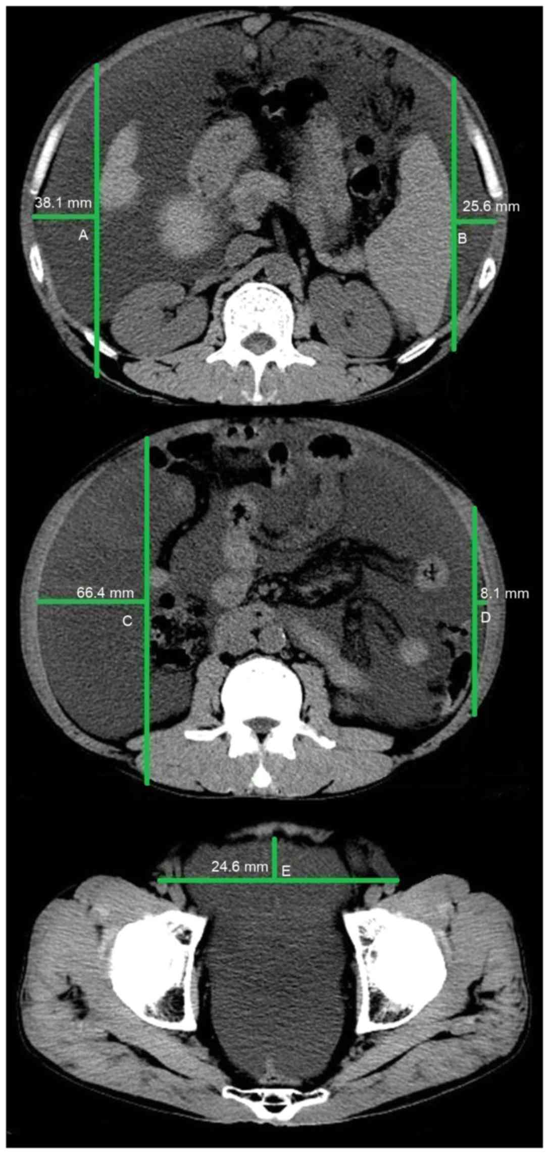
Oriuchi N, Nakajima T, Mochiki E,
Takeyoshi I, Kanuma T, Endo K and Sakamoto J: A new, accurate and
conventional five-point method for quantitative evaluation of
ascites using plain computed tomography in cancer patients. Jpn J
Clin Oncol. 35:386–390. 2005. View Article : Google Scholar : PubMed/NCBI
|
|
10
|
Imamoto H, Oba K, Sakamoto J, Iishi H,
Narahara H, Yumiba T, Morimoto T, Nakamura M, Oriuchi N, Kakutani
C, et al: Assessing clinical benefit response in the treatment of
gastric malignant ascites with non-measurable lesions: A
multicenter phase II trial of paclitaxel for malignant ascites
secondary to advanced/recurrent gastric cancer. Gastric Cancer.
14:81–90. 2011. View Article : Google Scholar : PubMed/NCBI
|
|
11
|
Ishiguro T, Kumagai Y, Baba H, Tajima Y,
Imaizumi H, Suzuki O, Kuwabara K, Matsuzawa T, Sobajima J, Fukuchi
M, et al: Predicting the amount of intraperitoneal fluid
accumulation by computed tomography and its clinical use in
patients with perforated peptic ulcer. Int Surg. 99:824–829. 2014.
View Article : Google Scholar : PubMed/NCBI
|
|
12
|
Pugh RN, Murray-Lyon IM, Dawson JL,
Pietroni MC and Williams R: Transection of the oesophagus for
bleeding oesophageal varices. Br J Surg. 60:646–649. 1973.
View Article : Google Scholar : PubMed/NCBI
|
|
13
|
Kamath PS, Wiesner RH, Malinchoc M,
Kremers W, Therneau TM, Kosberg CL, D'Amico G, Dickson ER and Kim
WR: A model to predict survival in patients with end-stage liver
disease. Hepatology. 33:464–470. 2001. View Article : Google Scholar : PubMed/NCBI
|
|
14
|
Kamath PS and Kim WR: Advanced Liver
Disease Study Group: The model for end-stage liver disease (MELD).
Hepatology. 45:797–805. 2007. View Article : Google Scholar : PubMed/NCBI
|
|
15
|
Peng Y, Qi X, Dai J, Li H and Guo X:
Child-Pugh versus MELD score for predicting the in-hospital
mortality of acute upper gastrointestinal bleeding in liver
cirrhosis. Int J Clin Exp Med. 8:751–757. 2015.PubMed/NCBI
|
|
16
|
Barakat AA, Metwaly AA, Nasr FM,
El-Ghannam M, El-Talkawy MD and Taleb HA: Impact of hyponatremia on
frequency of complications in patients with decompensated liver
cirrhosis. Electron Physician. 7:1349–1358. 2015.PubMed/NCBI
|
|
17
|
Kim WR, Biggins SW, Kremers WK, Wiesner
RH, Kamath PS, Benson JT, Edwards E and Therneau TM: Hyponatremia
and mortality among patients on the liver-transplant waiting list.
N Engl J Med. 359:1018–1026. 2008. View Article : Google Scholar : PubMed/NCBI
|
|
18
|
Maeda H, Kobayashi M and Sakamoto J:
Evaluation and treatment of malignant ascites secondary to gastric
cancer. World J Gastroenterol. 21:10936–10947. 2015. View Article : Google Scholar : PubMed/NCBI
|
|
19
|
Italian Association for the Study of the
Liver (AISF); Italian Society of Transfusion Medicine and
Immunohaematology (SIMTI): AISF-SIMTI position paper: The
appropriate use of albumin in patients with liver cirrhosis. Dig
Liver Dis. 48:4–15. 2016. View Article : Google Scholar : PubMed/NCBI
|
|
20
|
Campillo B, Richardet JP, Scherman E and
Bories PN: Evaluation of nutritional practice in hospitalized
cirrhotic patients: Results of a prospective study. Nutrition.
19:515–521. 2003. View Article : Google Scholar : PubMed/NCBI
|
|
21
|
Garcia-Martinez R, Caraceni P, Bernardi M,
Gines P, Arroyo V and Jalan R: Albumin: Pathophysiologic basis of
its role in the treatment of cirrhosis and its complications.
Hepatology. 58:1836–1846. 2013. View Article : Google Scholar : PubMed/NCBI
|