Observation of corneal transplantation in peripheral corneal disease postoperatively
- Authors:
- Published online on: April 25, 2018 https://doi.org/10.3892/etm.2018.6100
- Pages: 5384-5388
Metrics: Total
Views: 0 (Spandidos Publications: | PMC Statistics: )
Total PDF Downloads: 0 (Spandidos Publications: | PMC Statistics: )
Abstract
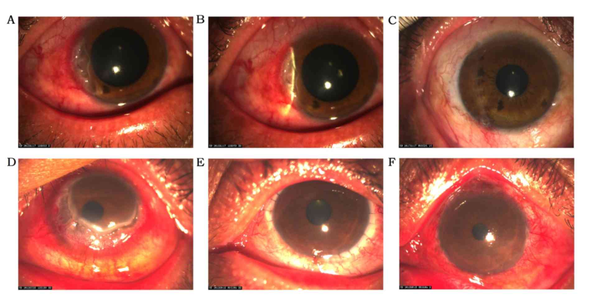
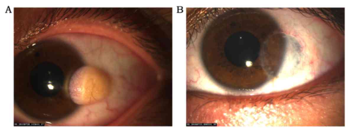
The aim of the present study was to investigate the role of the limbal stem cells in corneal epithelial homeostasis in patients with peripheral corneal disease who received corneal transplantation surgery. This retrospective study enrolled 85 patients (85 eyes) with peripheral corneal lesion who underwent corneal transplantation at Department of Ophthalmology in First Hospital of Jilin University, Jilin, China. All patients during each follow‑up period were examined for best corrected visual acuity, corneal reconstruction (assessed by slit‑lamp biomicroscopy), anterior segment optical coherence tomography, and confocal microscopy. Patients were followed up for 3.5±2.4 years. All patients had improved postoperative best corrected visual acuity. Neither vascularization nor conjunctivalization of the graft occurred. Scanning of the graft was covered by normal corneal epithelium as confirmed on the laser scanning in vivo confocal microscopy. No evidence of normal limbal was detected in any of the operation eyes. The limbal stem cells may not play a critical role during normal corneal epithelial turnover in patients with peripheral corneal disease following corneal transplantation surgery.