Introduction
Cerebral aneurysms are present in an estimated 6% of
the global population (1,2). Cerebral aneurysms rupture in 1% of
these patients, which, depending on their size, ultimately lead to
subarachnoid hemorrhage (3). The
1-year mortality rate was 75% in 6,850 patients with saccular, but
untreated ruptured intracranial aneurysms admitted to hospital for
a week between 1968 and 2007 in Finland (4). Traditional clipping and emergence
coiling are the two necessary treatment measures applied to
eliminate cerebral aneurysms (1).
The results of the International Subarachnoid Aneurysm Trial (ISAT)
(5,6)
and the International Study of Unruptured Intracranial Aneurysms
(ISUIA) (1) indicated that overall
morbidity and mortality of patients after a 1-year follow-up that
received endovascular coiling was lower than those that received
surgical clipping. The associated complications of coiling have
become more common, as it has become a more acceptable method that
is used for the majority of patients.
Coil migration is a lethal intraprocedural
complication of endovascular aneurysm treatment; however, there is
no standard method for managing coil migration (7). Although the majority of physicians
administer anticoagulants to address coil migration, certain
physicians will use a stent to localize a dislodged coil into a
large vessel (8), while others will
retrieve coils via surgical or endovascular methods (9). Hopf-Jensen et al (10) demonstrated that the Solitaire™ AB
Neurovascular Remodeling Device exhibited a number of advantages
over other devices for the retrieval of migrated coils, including
large cell retriever properties and electrolyte detachment design.
These results are similar to those from Liu et al (11), who analyzed Trevo Stentriever, which
is another frequently used stent. In addition to the inherent
stiffness and inconvenience of migrated stents, vessel damage can
be caused by stent movement (12).
The present study aimed to develop a novel device
for resolving coil migration that was simple to assemble, easy and
safe to use, and presented a lower risk to fragile arteries in the
brain. The current study designed a novel device, termed magnetic
wires, consisting of two microwires, the tips of which were
attached with small magnetic rings. A prototype device was
constructed from commercially available materials. The feasibility
and safety of this device compared with the Solitaire™ AB stent
retriever was evaluated in vitro and in vivo.
Materials and methods
Prototype production and
operation
A Mirage™ .008′ Hydrophilic Guidewire (Micro
Therapeutics, Inc., Irvine, CA, USA) was attached to a small (0.8
mm outside diameter; 0.25 mm inside diameter; 1 mm thickness)
samarium cobalt magnetic ring (SmCo; Wanxuan Magnet Company,
Shenzheng, China) using ethyl-α-cyanoacrylate glue (Hubei Huitian
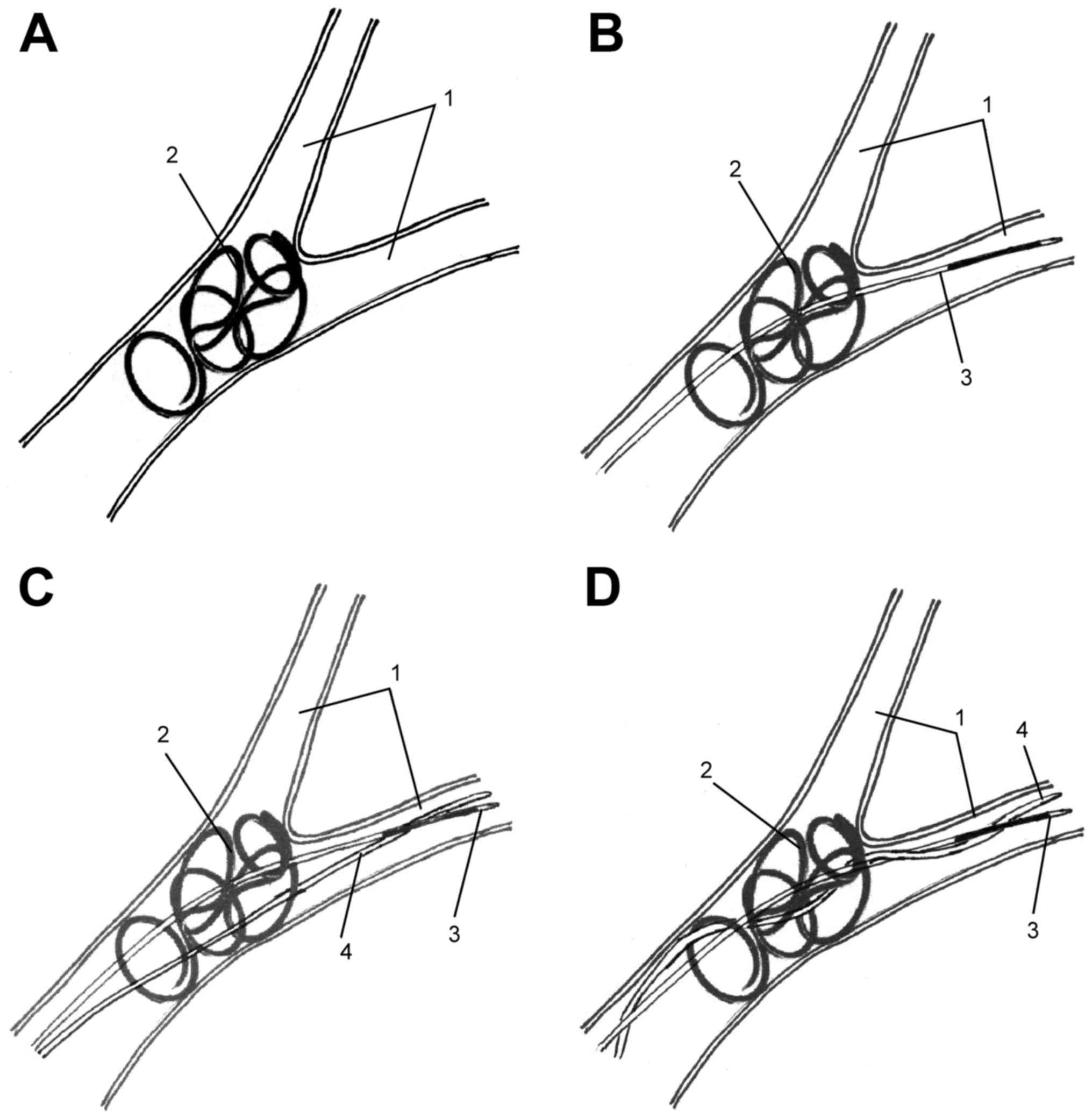
New Materials Co., Ltd., Shanghai, China). The microwires of the
device may be advanced through the dislodged coil one at a time to
retrieve the coil via the attraction of the magnetic tips, by
forming a loop similar to the configuration of chopsticks (Fig. 1).
In vitro assessment
A Portable FlowTek A-215 Pulsating Flow Circulator
(DialAct Corporation, Fremont, CA, USA) was used to compare the
prototype with the Solitaire™ AB stent retriever (Medtronic,
Minneapolis, MN, USA). The pulsatile pump pushes saline at a rate
of 50 cm/sec to simulate arterial blood flow. The insect
Axium™ Detachable Coil (4/8 mm; Medtronic) was inserted
into the tube of the circulator through a one-way valve. The
corresponding retrieval device was then pushed through the valve to
catch the coil. The stent microtube was then Sent through,
exceeding the coil and and the tube was then withdrawn to open the
stent (like a net) to catch and retrieve the coil. The coil was
then detached and withdrawn by pulling out the push-pole. The
method, which uses magnetic wires to retrieve the coil, is
described in Fig. 1. For A total of
five retrieval trials were conducted for each device. The time from
the release to the return of the coil to the tube entrance was
recorded.
Animal preparation
All procedures were conducted according to the
National Institutes of Health (Bethesda, MA, USA) guidelines
(13) for the humane handling of
animals and were approved by the Animal Care and Use Committee of
The Second Xiangya Hospital of Central South University (Changsha,
China). A total of 5 male healthy piglets (age, 2 months; weight,
15–17 kg) were obtained from the Experimental Pig Production
Institute (Laboratory Animal Center of PLA General Hospital,
Beijing, China). Pigs were housed at a temperature of 18–25°C and a
humidity of 40–60%, with a 12 h light/dark cycle. Food was provided
twice a day at 2–3% of the pigs' total body weight, with free
access to water. General anesthesia was induced using pentobarbital
sodium (Merck KGaA, Darmstadt, Germany) (25 mg/kg, intravenously)
in 100% oxygen and a 6-F femoral artery sheath was inserted into
each animal's right femoral artery following successful Seldinger
puncture (14).
Coil release and retrieval
An angiogram was performed after a 6-F guiding
catheter (Envoy® Guiding Catheter; Cordis Neurovascular,
Inc., Miami, FL, USA) was placed in the proximal carotid artery.
The DSA machine can estimate the diameter of vessel, using a metal
ball, which may be more accurate. Therefore, the diameter of the
carotid artery was measured via angiography using a 1 cm diameter
iron ball (Shandong Donge Steel Ball Group Co., Ltd., Shangdong,
China) attached to the right side of the head as a reference.
Following the insertion of an Axium™ Detachable Coil into the
carotid artery, the retrieval procedure was initiated. The coil,
which was released into the left and right carotid arteries, was
carefully withdrawn using the magnetic wires or the Solitaire™ AB
stent retriever as aforementioned (Figs.
2 and 3). The time from coil
release to retrieval was recorded for each retrieval system.
All in vivo assessments were performed under
X-Ray guidance. Using a digital subtraction angiography machine
(Innova IGS 530; GE Healthcare, Chicago, IL, USA), the spring coil
could be observed. Furthermore, the magnetic micro-conductance
wires could be manipulated to pass through the gap of the spring
ring under X-ray fluoroscopy, making the magnet end of the micro
guidewire connect closely.
Histopathological evaluation of coil
retrieval method safety
Following sacrifice of all animals with 20 ml
potassium chloride (Jinzhong Development Zone China capital
Chemical Technology Co., Ltd., Shaanxi, China; 100 g/l)
intravenously, specimens were collected via neck dissection and
craniotomy. From the middle portion of the carotid artery, where
the retrieval was conducted, two specimens were obtained from each
animal. Of these specimens, those for light microscopy (LM) were
fixed using 4% formaldehyde solution for 48 h at 22°C. The samples
were embedded in paraffin and 4 µm sections were sliced. Standard
histological methods (washing with xylene) were used in order to
remove paraffin and the samples were rehydrates with a graduated
alcohol series. Masson's trichrome staining was performed using a
Masson's trichrome kit (cat. no. ab150686; Abcam, Cambridge, UK)
according to the manufacturer's protocol to identify collagen
fibers within vessels. Specimens for scanning electron microscopy
(SEM) were immersed in 4% paraformaldehyde for 4 h at 22°C, fixed
using 1% glutaraldehyde for 2 h at 22°C. They were then dehydrated
in a series of graded alcohol, coated with platinum-palladium using
an ion coater (cat. no. IB-5; Eiko Engineering Co., Ltd.,
Hitachinaka, Japan). The tissues were observed using a scanning
electron microscope (cat. no. S-2380N; Hitachi, Ltd., Tokyo,
Japan). Endothelial lesions presented as defects in the internal
elastic lamina via LM and as denudation of the wavy endothelial
surface via SEM.
Results
In vitro assessment
The magnetic wires and Solitaire™ AB stent retriever
were able to successfully retrieve the coils released in
vitro (100%; Table I). However,
the magnetic wires had a shorter retrieval time compared with the
stent retrieval system (Table
I).
 | Table I.Coil retrieval time using the magnetic
wires and the Solitaire™ AB stent retriever in vitro and
in vivo. |
Table I.
Coil retrieval time using the magnetic
wires and the Solitaire™ AB stent retriever in vitro and
in vivo.
|
|
| Retrieval trial,
sec |
|---|
|
|
|
|
|---|
| Condition | Device used | 1 | 2 | 3 | 4 | 5 |
|---|
| In vitro | Magnetic wires | 228 | 204 | 210 | 189 | 193 |
|
| Solitaire stent | 273 | 235 | 264 | 203 | 221 |
| In vivo | Magnetic wires | Failed | 453 | 436 | Failed | 415 |
|
| Solitaire stent | 427 | Failed | 392 | 418 | 387 |
In vivo assessment
The magnetic wires were successful in 3/5 (60%) of
the in vivo retrieval trials (Table I). However, the Solitaire™ AB stent
retriever successfully captured the migrated coil in 4/5 (80%) of
the trials (Table I). There were no
incidences of vasospasm or vessel perforation in either group when
comparing angiograms prior to and following surgery.
Histopathological safety
Specimens from the right and left carotid arteries,
in which the Solitaire™ AB stent retriever and magnetic wires were
employed, respectively, were collected from each animal, and
processed for LM and SEM. Endothelial lesions were observed in 3
(60%) right carotid arteries (Solitaire™ AB stent retriever) and in
1 (20%) left carotid artery (magnetic wires) (Fig. 3; Table
II). There was no gross intimal dissection or any abnormalities
beneath the internal elastic lamina in any of the 4 cases in which
endothelial lesions were observed via LM. No other specimens
indicated any evidence of endothelial injury via either LM or
SEM.
| Table II.Detection of EI following the in
vitro and in vivo application of the magnetic wires or
the Solitaire™ AB stent retriever. |
Table II.
Detection of EI following the in
vitro and in vivo application of the magnetic wires or
the Solitaire™ AB stent retriever.
|
|
| Retrieval device,
presence of EI |
|---|
|
|
|
|
|---|
| Animal | Detection method | Magnetic wires | Solitaire stent |
|---|
| 1 | LM | No | Yes |
|
| SEM | No | Yes |
| 2 | LM | No | Yes |
|
| SEM | No | Yes |
| 3 | LM | No | No |
|
| SEM | No | No |
| 4 | LM | Yes | Yes |
|
| SEM | Yes | Yes |
| 5 | LM | No | No |
|
| SEM | No | No |
Discussion
There have been limited reports of the successful
use of various devices for retrieving a coil that has migrated or
become misplaced, including the Microsnare (15), a microcatheter retriever
(Retriever-10 and Retriever-18), the Alligator Retrieval Device
(9,15), Merci retriever (16), the Solitaire™ AB stent retriever
(10) and the Trevo Stentriever
device (11,17). Several experts have shaped microwires
to capture migrated coils (18,19);
however, the majority of clinicians use one of the following four
types of coil retrieval devices: Snares, clamps, stents or spiral
devices.
Existing coil retrieval devices are mechanically
complex, rendering them difficult to manufacture and maneuver.
Furthermore, the materials of these devices are too stiff and large
for use in the fragile cerebral arteries. Watanabe et al
(20) described the first use of a
loop snare to retrieve a migrated coil during the treatment of a
superior cerebellar artery aneurysm. However, since then, there
have only been a few reported cases of snares being used to capture
dislodged coils (18,21,22); the
lack of use of these snares may be due to difficulties in opening a
snare in small vessels in an appropriate position for coil
retrieval. The use of clamps, including the Alligator Retrieval
Device, and spiral devices, including Merci, has been reported even
less frequently (20,23).
The latest and the most popular type of retrieval
device, the stent retrieval system, has a number of marked flaws.
The recommended vessel diameter of Solitaire™ AB stent retriever is
from 2.2 mm to 6 mm as according to the manufacturer's protocol.
Thus, it was considered that if the small artery diameter is
<2.2 mm, the stent remains too large; and may consequently push
the dislodged coil further into unreachable arterial branches. The
movement of the net-like stent within the artery may damage the
vessel wall, specifically endothelial cells and the internal
elastic lamina, increasing the risk of inflammation and thrombosis
(12). The present study also
considered that when withdrawing the stent attached to the
dislodged coil along the parent artery near the neck of an
aneurysm, the coils in the aneurysm may be pulled out by the stent.
These potential complications demonstrate that a stent-like device
is not the optimal choice for coil retrieval.
The present study aimed to design an alternative
type of retrieval device. The original concept of this device
originated from a report describing how a number of physicians use
two microwires wrapped together to capture dislodged material in a
vessel (24). However, this
procedure is difficult and is therefore affected by the skills of
the clinician. There are a number of similarities between the
two-microwire device used in the present study and chopsticks, with
magnets being the key feature uniting the two microwires. The
magnets used in the present study to produce the prototype device
were composed of SmCo; these magnets were produced to power the
stepping motor of an electronic watch and their size fits the
0.008′ microwires.
In vitro, the prototype device demonstrated
the convenience of the wire shape for advancing through the
migrated coil and that the attractive force between the two magnets
was sufficient for coil retrieval. These findings establish the
feasibility of the magnetic wire design. However, the in
vivo success rate of the novel device, in 3/5 trials, was
insufficient. The present study concluded that the lack of
interaction with the microwires due to the enlargement of the
magnetic ring at the tips resulted in difficulties controlling the
device. This impedes the retrieval of a dislodged coil from an area
with rapid blood flow. Furthermore, the microwire used in the
current study was composed of nitinol, which contains ~51% nickel
and thus may be attracted to magnets; this feature of the microwire
may therefore interfere with its intended function. In the present
study, definite influence was observed when the second magnetic
microwire tip reached and caught the former remote end. Future
magnetic microwire should therefore not contain ferromagnetic
material.
The LM and SEM results demonstrated that the extent
of endothelial injury was smaller with the magnetic wire group
compared with the Solitaire™ AB stent retriever.
In conclusion, the present study designed a simple
novel device that has great potential for the retrieval of
dislodged coils during interventional surgery. However, this device
requires further development and validation in order to produce an
optimal tool for resolving this complication of endovascular
aneurysm treatment.
Acknowledgements
The present study was supported by Dr. Gary
Duckwiler of the Department of Neuroradiology of David Geffen
School of Medicine at UCLA (Los Angeles, CA, USA), who provided the
sample coils.
Glossary
Abbreviations
Abbreviations:
|
LM
|
light microscopy
|
|
SEM
|
scanning electron microscopy
|
|
SmCo
|
samarium cobalt
|
References
|
1
|
Wiebers DO, Whisnant JP, Huston J III,
Meissner I, Brown RD Jr, Piepgras DG, Forbes GS, Thielen K, Nichols
D, O'Fallon WM, et al: Unruptured intracranial aneurysms: Natural
history, clinical outcome, and risks of surgical and endovascular
treatment. Lancet. 362:103–110. 2003. View Article : Google Scholar : PubMed/NCBI
|
|
2
|
Winn HR and Britz GW: Unruptured
aneurysms. J Neurosurg. 104:179–180. 2006. View Article : Google Scholar : PubMed/NCBI
|
|
3
|
Broderick JP, Brott TG, Duldner JE,
Tomsick T and Huster G: Volume of intracerebral hemorrhage. A
powerful and easy-to-use predictor of 30-day mortality. Stroke.
24:987–993. 1993. View Article : Google Scholar : PubMed/NCBI
|
|
4
|
Korja M, Kivisaari R, Jahromi Rezai B and
Lehto H: Natural history of ruptured but untreated intracranial
aneurysms. Stroke. 48:1081–1084. 2017. View Article : Google Scholar : PubMed/NCBI
|
|
5
|
Molyneux A, Kerr R, Stratton I, Sandercock
P, Clarke M, Shrimpton J and Holman R: International Subarachnoid
Aneurysm Trial (ISAT) Collaborative Group: International
subarachnoid aneurysm trial (ISAT) of neurosurgical clipping versus
endovascular coiling in 2143 patients with ruptured intracranial
aneurysms: A randomised trial. Lancet. 360:1267–1274. 2002.
View Article : Google Scholar : PubMed/NCBI
|
|
6
|
Molyneux AJ, Kerr RS, Yu LM, Clarke M,
Sneade M, Yarnold JA and Sandercock P: International Subarachnoid
Aneurysm Trial (ISAT) Collaborative Group: International
subarachnoid aneurysm trial (ISAT) of neurosurgical clipping versus
endovascular coiling in 2143 patients with ruptured intracranial
aneurysms: A randomised comparison of effects on survival,
dependency, seizures, rebleeding, subgroups, and aneurysm
occlusion. Lancet. 366:809–817. 2005. View Article : Google Scholar : PubMed/NCBI
|
|
7
|
Leslie-Mazwi TM, Heddier M, Nordmeyer H,
Stauder M, Velasco A, Mosimann PJ and Chapot R: Stent retriever use
for retrieval of displaced microcoils: A consecutive case series.
AJAR Am J Neuroradiol. 34:1996–1999. 2013. View Article : Google Scholar
|
|
8
|
Schütz A, Solymosi L, Vince GH and
Bendszus M: Proximal stent fixation of fractured coils: Technical
note. Neuroradiology. 47:874–878. 2005. View Article : Google Scholar : PubMed/NCBI
|
|
9
|
Oh J, Kim J, Hong S, Hu C, Pyen J, Whang
K, Cho S and You DS: Retrieval of unintended migrated detached
coil: Case report. J Cerebrovasc Endovasc Neurosurg. 16:268–274.
2014. View Article : Google Scholar : PubMed/NCBI
|
|
10
|
Hopf-Jensen S, Hensler HM, Preiss M and
Muller-Hulsbeck S: Solitaire® stent for endovascular
coil retrieval. J Clin Neurosci. 20:884–886. 2013. View Article : Google Scholar : PubMed/NCBI
|
|
11
|
Liu KC, Ding D, Starke RM, Geraghty SR and
Jensen ME: Intraprocedural retrieval of migrated coils during
endovascular aneurysm treatment with the trevo stentriever device.
J Clin Neurosci. 21:503–506. 2014. View Article : Google Scholar : PubMed/NCBI
|
|
12
|
Park S, Hwang SM, Song JS, Suh DC and Lee
DH: Evaluation of the solitaire system in a canine arterial
thromboembolic occlusion model: Is it safe for the endothelium?
Interv Neuroradiol. 19:417–424. 2013. View Article : Google Scholar : PubMed/NCBI
|
|
13
|
Guide for the Care and Use of Laboratory
Animals, . NIH Publication no. 85-23. Revised 1985.
|
|
14
|
Seldinger SI: Catheter replacement of the
needle in percutaneous arteriography; A new technique. Acta Radiol.
39:368–376. 1953. View Article : Google Scholar : PubMed/NCBI
|
|
15
|
Dinc H, Kuzeyli K, Kosucu P, Sari A and
Cekirge S: Retrieval of prolapsed coils during endovascular
treatment of cerebral aneurysms. Neuroradiology. 48:269–272. 2006.
View Article : Google Scholar : PubMed/NCBI
|
|
16
|
Vora N, Thomas A, Germanwala A, Jovin T
and Horowitz M: Retrieval of a displaced detachable coil and
intracranial stent with an L5 merci retriever during endovascular
embolization of an intracranial aneurysm. J Neuroimaging. 18:81–84.
2008. View Article : Google Scholar : PubMed/NCBI
|
|
17
|
Kabbani MR, Smith A and Leider M:
Endovascular coil retrieval using a trevoprovue stentriever. J
Neurointerv Surg. 7:e192015. View Article : Google Scholar : PubMed/NCBI
|
|
18
|
Lee CY: Use of wire as a snare for
endovascular retrieval of displaced or stretched coils: Rescue from
a technical complication. Neuroradiology. 53:31–35. 2011.
View Article : Google Scholar : PubMed/NCBI
|
|
19
|
Standard SC, Chavis TD, Wakhloo AK, Ahuja
A, Guterman LR and Hopkins LN: Retrieval of a guglielmi detachable
coil after unraveling and fracture: Case report and experimental
results. Neurosurgery. 35:994–998. 1994. View Article : Google Scholar : PubMed/NCBI
|
|
20
|
Watanabe A, Hirano K, Mizukawa K, Kamada
M, Okamura H, Suzuki Y and Ishii R: Retrieval of a migrated
detachable coil–case report. Neurol Med Chir (Tokyo). 35:247–250.
1995. View Article : Google Scholar : PubMed/NCBI
|
|
21
|
Koseoglu K, Parildar M, Oran I and Memis
A: Retrieval of intravascular foreign bodies with goose neck snare.
Eur J Radiol. 49:281–285. 2004. View Article : Google Scholar : PubMed/NCBI
|
|
22
|
Prestigiacomo CJ, Fidlow K and
Pile-Spellman J: Retrieval of a fractured Guglielmi detachable coil
with use of the goose neck snare ‘twist’ technique. J Vasc Interv
Radiol. 10:1243–1247. 1999. View Article : Google Scholar : PubMed/NCBI
|
|
23
|
Ding D and Liu KC: Management strategies
for intraprocedural coil migration during endovascular treatment of
intracranial aneurysms. J Neurointerv Surg. 6:428–431. 2014.
View Article : Google Scholar : PubMed/NCBI
|
|
24
|
Gurley JC, Booth DC, Hixson C and Smith
MD: Removal of retained intracoronary percutaneous transluminal
coronary angioplasty equipment by a percutaneous twin guidewire
method. Cathet Cardiovasc Diagn. 19:251–256. 1990. View Article : Google Scholar : PubMed/NCBI
|