Clinical and pathological studies of eight cases of lipidized fibrous histiocytoma
- Authors:
- Fangyun Liu
- Hongjun Wang
- Guoying Wu
- Congyang Li
View Affiliations
Affiliations: Fenlan Laboratory, Hangzhou, Zhejiang 310056, P.R. China, Department of Pathology, Yexian First People's Hospital, Pingdingshan, Henan 467200, P.R. China, Department of Pathology, People's Liberation Army 989 Hospital, Pingdingshan, Henan 467000, P.R. China
- Published online on: January 9, 2023 https://doi.org/10.3892/etm.2023.11792
-
Article Number:
93
-
Copyright: © Liu
et al. This is an open access article distributed under the
terms of Creative
Commons Attribution License.
Metrics: Total
Views: 0 (Spandidos Publications: | PMC Statistics: )
Metrics: Total PDF Downloads: 0 (Spandidos Publications: | PMC Statistics: )
This article is mentioned in:
Abstract
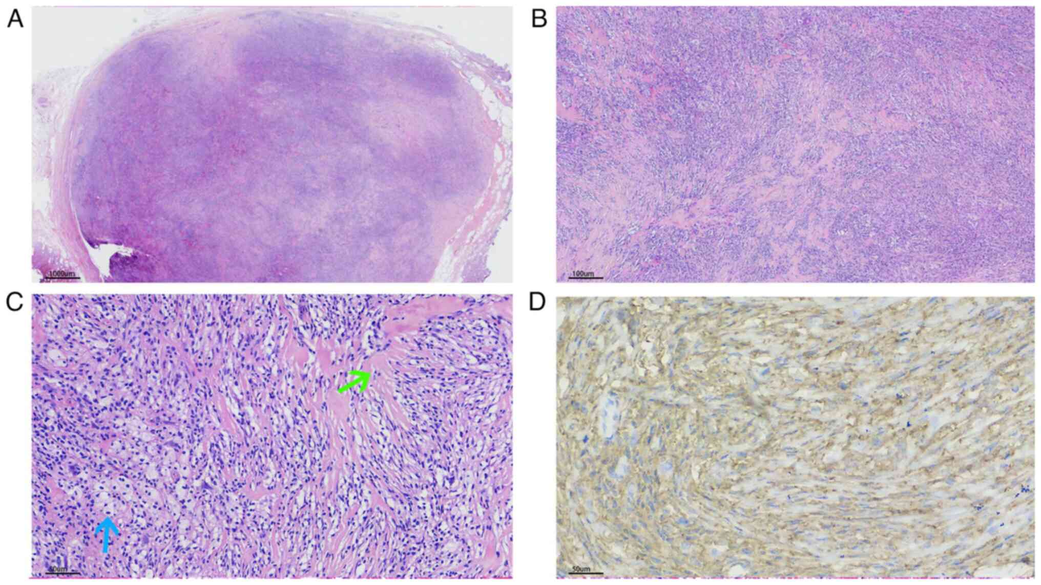
Lipidized fibrous histiocytoma (FH) is a rare type of FH. The present study aimed to describe the clinical and pathological features of lipidized FH. A total of eight patients diagnosed with lipidized FH were retrospectively reviewed in the present study. The cohort included three male and five female patients (male to female ratio, 1.7:1) with a mean age of 48 years (range, 38‑62 years). In total, four tumors were located on the buttock, three on the lower leg and one on the forearm. Histological, lipidized FH showed a wide spectrum. Some cases included prominent stromal hyalinization and hyalinized vessels with scant lipid‑laden histiocytes. Other cases exhibited the prominent lipid‑laden histiocytes and scant stromal hyalinization. Overall, lipidized FH must be differentiated from other benign and malignant tumors, taking into account the therapeutic and prognostic differences between these different entities.
View References
1
|
Meister P, Konrad E and Krauss F: Fibrous
histiocytoma: A histological and statistical analysis of 155 cases.
Pathol Res Pract. 162:361–379. 1978.PubMed/NCBI View Article : Google Scholar
|
2
|
Gonzalez S and Duarte I: Benign fibrous
histiocytoma of the skin. A morphologic study of 290 cases. Pathol
Res Pract. 174:379–391. 1982.PubMed/NCBI View Article : Google Scholar
|
3
|
Seo JK, Shin EJ, Jeong KH and Shin MK:
Lipidized fibrous histiocytoma: Differential diagnosis from
juvenile xanthogranuloma. Ann Dermatol. 31:254–256. 2019.PubMed/NCBI View Article : Google Scholar
|
4
|
Iwata J and Fletcher CD: Lipidized fibrous
histiocytoma: Clinicopathologic analysis of 22 cases. Am J
Dermatopathol. 22:126–134. 2000.PubMed/NCBI View Article : Google Scholar
|
5
|
Alves JV, Matos DM, Barreiros HF and
Bártolo EA: Variants of dermatofibroma-a histopathological study.
An Bras Dermatol. 89:472–477. 2014.PubMed/NCBI View Article : Google Scholar
|
6
|
Sommer W, Knöfel AK, Izykowski N, Oldhafer
F, Avsar M, Jonigk D, Warnecke G and Haverich A: Physical exercise
reduces transplant arteriosclerosis in a mouse aorta
transplantation model. J Thorac Cardiovasc Surg. 149:330–337.
2015.PubMed/NCBI View Article : Google Scholar
|
7
|
Zaballos P, Mir-Bonafé JF, Avilés JA and
Bañuls J: Dermoscopy of lipidised dermatofibroma: A morphological
study of 13 cases. Aus J Dermatopathol. 60:e127–e131.
2019.PubMed/NCBI View Article : Google Scholar
|
8
|
Wagamon K, Somach SC, Bass J, Sigel JE,
Xue W, Schluchter M and Jaworsky C: Lipidized dermatofibromas and
their relationship to serum lipids. J Am Acad Dermatol. 54:494–498.
2006.PubMed/NCBI View Article : Google Scholar
|
9
|
Kosemehmetoglu K, Ardic F, Kilpatrick SE,
Aydingoz U, Sumathi VP and Michal M: Sclerosing epithelioid
fibrosarcoma of bone: morphological, immunophenotypical, and
molecular findings of 9 cases. Virchows Arch. 478:767–777.
2021.PubMed/NCBI View Article : Google Scholar
|
10
|
Wilkinson PE, Merkourea S, Gopalakrishnan
R and Argyris PP: Primary intraosseous xanthomas of the jaws: A
series of six cases including an example with formation of
apoptosis-related hyaline globules, so-called ‘thanatosomes’. Head
Neck Pathol. 14:859–868. 2020.PubMed/NCBI View Article : Google Scholar
|