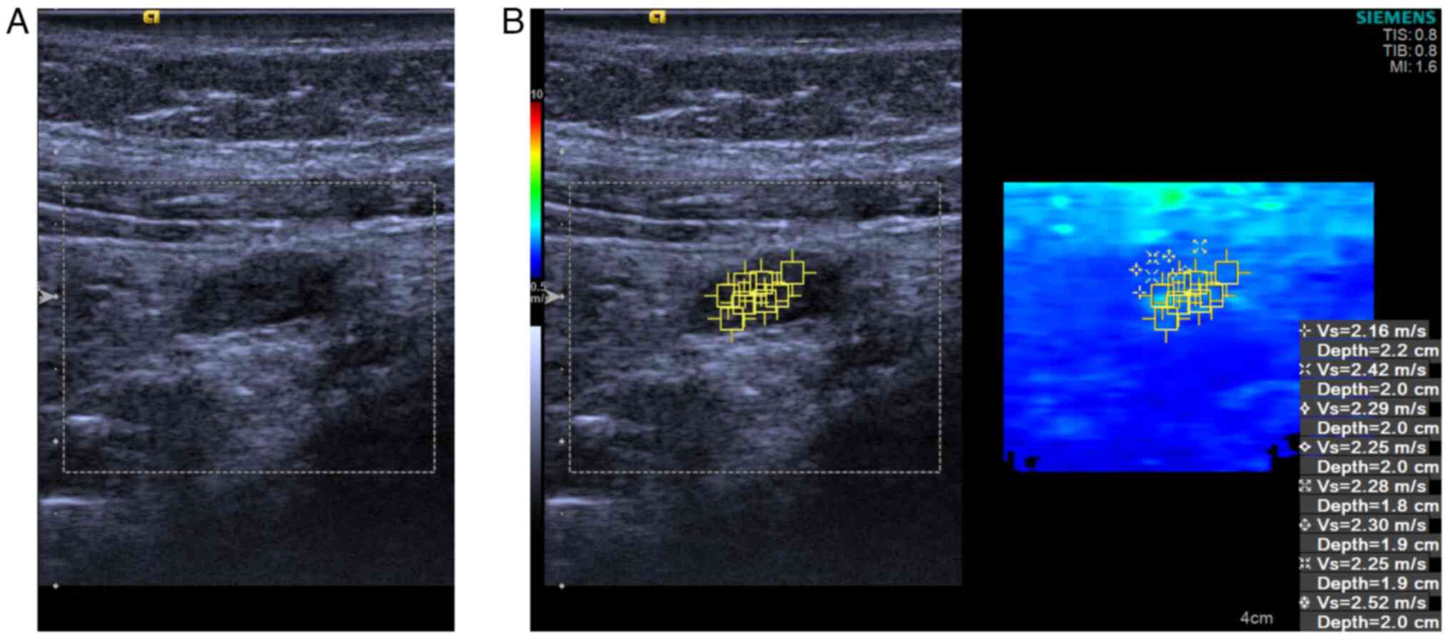
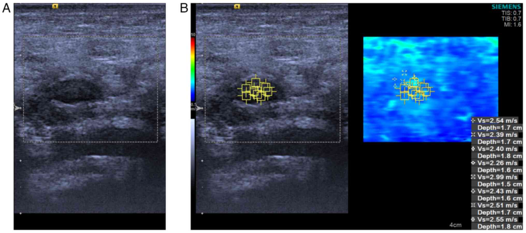
|
1
|
Devine M and Coffey JC: Mesenteric
Adenopathy and Adenitis. Prog Inflammation Res. 90:127–148.
2023.
|
|
2
|
Simanovsky N and Hiller N: Importance of
sonographic detection of enlarged abdominal lymph nodes in
children. J Ultrasound Med. 26:581–584. 2007.PubMed/NCBI View Article : Google Scholar
|
|
3
|
Toorenvliet B, Vellekoop A, Bakker R,
Wiersma F, Mertens B, Merkus J, Breslau P and Hamming J: Clinical
differentiation between acute appendicitis and acute mesenteric
lymphadenitis in children. Eur J Pediatr Surg. 21:120–123.
2011.PubMed/NCBI View Article : Google Scholar
|
|
4
|
Ozdamar MY and Karavas E: Acute mesenteric
lymphadenitis in children: Findings related to differential
diagnosis and hospitalization. Arch Med Sci. 16:313–320.
2018.PubMed/NCBI View Article : Google Scholar
|
|
5
|
Evans A, Whelehan P, Thomson K, McLean D,
Brauer K, Purdie C, Baker L, Jordan L, Rauchhaus P and Thompson A:
Invasive breast cancer: Relationship between shear-wave
elastographic findings and histologic prognostic factors.
Radiology. 263:673–677. 2012.PubMed/NCBI View Article : Google Scholar
|
|
6
|
Wong VW, Vergniol J, Wong GL, Foucher J,
Chan HL, Le Bail B, Choi PC, Kowo M, Chan AW, Merrouche W, et al:
Diagnosis of fibrosis and cirrhosis using liver stiffness
measurement in nonalcoholic fatty liver disease. Hepatology.
51:454–462. 2010.PubMed/NCBI View Article : Google Scholar
|
|
7
|
Sun CY, Lei KR, Liu BJ, Bo XW, Li XL, He
YP, Wang D, Ren WW, Zhao CK and Xu HX: Virtual touch tissue imaging
and quantification (VTIQ) in the evaluation of thyroid nodules: The
associated factors leading to misdiagnosis. Sci Rep.
7(41958)2017.PubMed/NCBI View Article : Google Scholar
|
|
8
|
Glišić TM, Perišić MD, Dimitrijevic S and
Jurišić V: Doppler assessment of splanchnic arterial flow in
patients with liver cirrhosis: Correlation with ammonia plasma
levels and MELD score. J Clin Ultrasound. 42:264–269.
2014.PubMed/NCBI View Article : Google Scholar
|
|
9
|
Zhou H, Zhou XL, Xu HX, Li DD, Liu BJ,
Zhang YF, Xu JM, Bo XW, Li XL, Guo LH and Qu S: Virtual touch
tissue imaging and quantification in the evaluation of thyroid
nodules. J Ultrasound Med. 36:251–260. 2017.PubMed/NCBI View Article : Google Scholar
|
|
10
|
Kong WT, Zhou WJ, Wang Y, Zhuang XM and Wu
M: The value of virtual touch tissue imaging quantification in the
differential diagnosis between benign and malignant breast lesions.
J Med Ultrason (2001). 46:459–466. 2019.PubMed/NCBI View Article : Google Scholar
|
|
11
|
Ruger H, Psychogios G, Jering M and Zenk
J: Multimodal ultrasound including virtual touch imaging
quantification for differentiating cervical lymph nodes. Ultrasound
Med Biol. 46:2677–2682. 2020.PubMed/NCBI View Article : Google Scholar
|
|
12
|
Bayramoglu Z, Caliskan E, Karakas Z,
Karaman S, Tugcu D, Somer A, Acar M, Akıcı F and Adaletli I:
Diagnostic performances of superb microvascular imaging, shear wave
elastography and shape index in pediatric lymph nodes
categorization: A comparative study. Br J Radiol.
91(20180129)2018.PubMed/NCBI View Article : Google Scholar
|
|
13
|
Jurisic V, Terzic T, Colic S and Jurisic
M: The concentration of TNF-alpha correlate with number of
inflammatory cells and degree of vascularization in radicular
cysts. Oral Dis. 14:600–605. 2008.PubMed/NCBI View Article : Google Scholar
|
|
14
|
Dzopalić T, Božić-Nedeljković B and
Jurišić V: Function of innate lymphoid cells in the immune-related
disorders. Hum Cell. 32:231–239. 2019.PubMed/NCBI View Article : Google Scholar
|
|
15
|
Handorf AM, Zhou Y, Halanski MA and Li WJ:
Tissue stiffness dictates development, homeostasis, and disease
progression. Organogenesis. 11:1–15. 2015.PubMed/NCBI View Article : Google Scholar
|
|
16
|
Desmots F, Fakhry N, Mancini J, Reyre A,
Vidal V, Jacquier A, Santini L, Moulin G and Varoquaux A: Shear
wave elastography in head and neck lymph node assessment: Image
quality and diagnostic impact compared with B-Mode and doppler
ultrasonography. Ultrasound Med Biol. 42:387–398. 2016.PubMed/NCBI View Article : Google Scholar
|
|
17
|
Yang JR, Song Y, Jia YL and Ruan LT:
Application of multimodal ultrasonography for differentiating
benign and malignant cervical lymphadenopathy. Jpn J Radiol.
39:938–945. 2021.PubMed/NCBI View Article : Google Scholar
|
|
18
|
Dudea SM, Botar-Jid C, Dumitriu D,
Vasilescu D, Manole S and Lenghel ML: Differentiating benign from
malignant superficial lymph nodes with sonoelastography. Med
Ultrason. 15:132–139. 2013.PubMed/NCBI View Article : Google Scholar
|
|
19
|
Grossman M and Shiramizu B: Evaluation of
lymphadenopathy in children. Curr Opin Pediatr. 6:68–76.
1994.PubMed/NCBI View Article : Google Scholar
|
|
20
|
Lau WY, Fan ST, Yiu TF, Chu KW and Wong
SH: Negative findings at appendectomy. Am J Surg. 148:375–378.
1984.PubMed/NCBI View Article : Google Scholar
|
|
21
|
Gross I, Siedner-Weintraub Y, Stibbe S,
Rekhtman D, Weiss D, Simanovsky N, Arbell D and Hashavya S:
Characteristics of mesenteric lymphadenitis in comparison with
those of acute appendicitis in children. Eur J Pediatr.
176:199–205. 2017.PubMed/NCBI View Article : Google Scholar
|
|
22
|
Schulte B, Beyer D, Kaiser C, Horsch S and
Wiater A: Ultrasonography in suspected acute appendicitis in
childhood-report of 1285 cases. Eur J Ultrasound. 8:177–182.
1998.PubMed/NCBI View Article : Google Scholar
|
|
23
|
Sivit CJ, Newman KD and Chandra RS:
Visualization of enlarged mesenteric lymph nodes at US examination.
Clinical significance. Pediatr Radiol. 23:471–475. 1993.PubMed/NCBI View Article : Google Scholar
|


