Introduction
Ankylosing spondylitis (AS), a chronic inflammation
attacking sacroiliac joints and the spine, is a complex genetic
disease (1) with 3‰ incidence
rate in yellow race (2,3). Although the exact cause of AS is
still unclear, there is a close relationship between the genetic
information and the occurrence of AS. Studies show that the major
histocompatibility complex region (MHC) is closely related to AS,
and about 95% AS patients are born with a gene named HLA-B27.
However, not only the HLA-B27 gene, but also other important genes
are believed to participate in the process of AS (4). In addition, numerous AS-related
genes remain to be identified.
Gene expression profiling analysis is a
bioinformatics approaches whose application is rising in recent
years. This approach can perform high throughput analysis of
thousands of genes at one time. The combination of gene expression
profiling and disease genomics results in deep understanding of the
relationship of hereditary disease and genes, which has become an
important research direction.
There are many ethical issues regarding the
performance of experimental assays in humans. Our study utilized
the AS mouse model for the research of the disease (5). First, the gene expression data were
integrated into the public Gene Expression Omnibus (GEO), and
bioinformatics analyses were performed to screen the potential
disease-associated genes in a large scale. Blood samples of normal
and AS model mice before and after treatment of TNF-α were
gathered, and RT-PCR and ELISA assays, were carried out to examine
tehe mRNA and protein levels of the genes, respectively. The
association of the potential gene with the disease was assesssed by
examining the transcriptional and translational levels of the
genes, and by functional analysis of one gene. This study provides
novel insights into the diagnosis, prevention and treatment of the
disease.
Materials and methods
Screening of differentially expressed
AS-related genes
Gene chip data GSE25101 were downloaded from the
United States National Center for Biotechnology Information Sharing
database GEO, and analyzed by the Vizard 1.2 software. After signal
extraction, normalization and chip comparison analysis, the
AS-related genes were screened according to the ratio values.
Through gene ontology (GO) analysis performed on the http://www.gosurfer.org website, the molecular
function, biological pathways and cellular components of
differentially expressed genes were obtained. The target genes
associated with AS were screened by combination of the
pathophysiology process of disease and the related study.
Establishment of the AS mouse model
The AS mouse model was established with a transgenic
method. The plasmid was constructed by our laboratory, and the
Kunming mice were purchased from the Institute of Experimental
Animals, Chinese Academy of Medical Sciences. Development of the
mouse model has been previously described. The animals that were
successfully converted into AS model mice were administered the TNF
inhibitor etanercept (Shanghai CITIC Health Pharmaceutical Co.,
Ltd) for over 6 months (1).
Genomic DNA extraction
Venous blood (1 ml) was taken from normal and AS
model mice, and 0.4 ml anticoagulants was added to extract genomic
DNA with the high salt precipitation method (blood was taken from
model mice before and after treatment). The DNA concentration was
obtained with the NanoDrop 2000 spectrophotometer (Thermo
Scientific). The DNA samples were conserved at −80°C.
Detection of gene transcription
levels
According to the screened genes in step 1, real-time
PCR primers were designed using the online website http://www.idtdna.com. The relative expression
quantity of the amplified target gene and 18sRNA gene fragments
were detected with SYBR-Green I dye method. The PCR conditions were
95°C for 3 sec, 60°C for 5 sec, 72°C for 30 sec, for 40 cycles.
Real-time detection of fluorescence intensity in each cycle during
the PCR reaction process was obtained to achieve equivalent
detection. The standard curve of the respective standard samples
were established, and the relative expression levels of the target
genes in the control group and the AS model group after treatment
were analyzed based on the control gene of 18sRNA. All experimental
date were statistically analyzed by SPSS 15.0 software, and data
were obtained from the software in the CFX-96 real-time
quantitative PCR instrument (Bio-Rad) with One-way ANOVA variance
analysis. P<0.05 represents a significant difference, and
P<0.01 represents a very significant difference.
Examination of protein expression
levels
The protein expression levels of the screened genes
were examined with ELISA kits (Biosource) in the serum according to
the manufacturer’s instructions.
Functional analysis of the target gene
cytotoxic T lymphocyte-associated antigen 4 (CTLA4)
T lymphocytes were isolated from peripheral blood
with immunomagnetic beads, and then cultured in RPMI-1640 medium
supplemented with 10% fetal bovine serum in 24-well plates with
5×10⁵ cells in each well. The cells were divided into 5
groups. Each groups was treated with 30, 60, 90 or 120 ng/ml CTLA4
and the saline group was used as the control. After 48 h, each
group of cells was harvested, and the levels of interleukin 5
(IL-5) in the supernatant were detected with the ELISA kit
(Biosource).
Results
Screening of differentially expressed
genes
Through screening differentially expressed genes, a
total of 83 AS-associated genes were obtained, of which 74 genes
were upregulated and 9 genes were downregulated. Differential genes
related to AS are involved in immunity, cell cycle, apoptosis,
differentiation, signal transduction, cell activity, cell
structure, development, stress response, transcription regulation,
metabolism and other processes. The results of the present study
showed that the molecular function and biological pathway of 13
genes participated in the inflammatory and immune responses,
accounting for 15.7% (Table
I).
 | Table I.Differentially expressed genes
associated with AS. |
Table I.
Differentially expressed genes
associated with AS.
| Gene | Fold-difference |
|---|
| ITGA6 | 1.5 |
| PSMB9 | 0.8 |
| TRAT1 | 1.6 |
| PHACTR1 | 1.6 |
| CTLA4 | 1.5 |
| BIRC3 | 1.3 |
| THBD | 1.4 |
| IL-15 | 1.8 |
| BCL11A | 0.9 |
| NKG7 | 1.5 |
| VNN1 | 1.2 |
| MRPL43 | 1.4 |
| PGGT1B | 1.4 |
Based on the microarray results, disease
pathophysiology and former research, we further embarked on a
clinical study, and screened target genes associated with AS. A
total of 3 target genes were identified: cell receptor related to
membrane ligands (TRAT1), CTLA4, IL-15.
Establishment of the AS mouse model
Three hundred Kunming mice were injected with
hormones for superovulation, and fertilized eggs were acquired.
Genomic DNA fragments containing the HLA-B2704 gene were
microinjected into the zygote pronuclear, and two-cell stage
fertilized eggs that survived were further transferred to the
fallopian tube in pseudopregnant animals to produce the offspring.
The expression level of the HLA-B2704 gene in the blood was
detected by reverse transcription PCR (Fig. 1). Ten positive mice were
successfully obtained, and the AS mouse model was established.
Transcription level of differentially
expressed gene
An online primer design website were used to design
primers of the 3 screened genes, namely TRAT1, CTLA4 and IL-15,
with the housekeeping gene 18S as a reference. The primer sequences
are shown in Table II.
| Table II.Primer sequence. |
Table II.
Primer sequence.
| Gene | Forward primer F | Reverse primer |
|---|
| TRAT1 |
5′-TGTAAGCTCGAGTTGTGGGCATCT-3′ |
5′-TTCAGGAAGATTCGGGCACCTGAT-3′ |
| CTLA4 |
5′-AGACCTGAACACCGCTCCCATAAAG-3′ |
5′GCATTGCTTTGCAGAAGACAGGGA-3′ |
| IL-15 |
5′-CCATGTGGCTCTTTGGAGCAATGT-3′ |
5′-TCTTGTATGGGCTGGCTATCTGCT-3′ |
| 18sRNA |
5′-CCTGGATACCGCAGCTAGGA-3′ |
5′-GCGGCGCAATACGAATGCCCC-3′ |
Ten mice from the AS mouse model pretreatment group,
10 from the AS mouse model post-treatment group, and 10 normal mice
were examined. The expression of the above genes in blood samples
were detected by real-time PCR, and the results are shown in
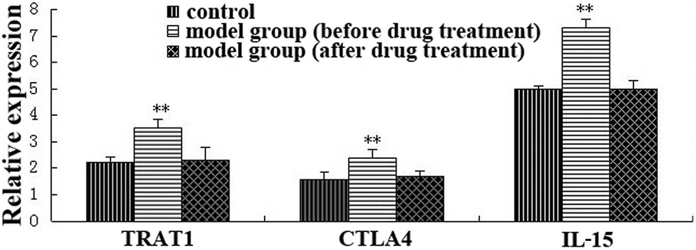
Fig. 2.
As shown in Fig.
2, the expression of TRAT1, CTLA4 and IL-15 in the model group
before drug treatment were 1.6, 1.5, 1.5-fold of the control group,
which was consistent with the screening results of differentially
expressed genes (P<0.05). However, the expression of these genes
were restored to normal levels after drug treatment, confirming
that overexpression of these genes are associated with the
occurrence of AS.
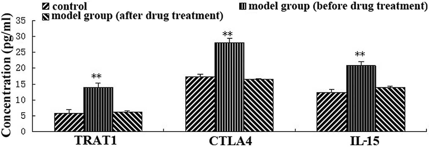
Protein levels of differentially
expressed gene
To further study the relationship between these
three genes and the disease, the protein expression levels of the
genes in peripheral blood from 10 AS model mice before and after
TNF treatment and 10 control group mice were detected by ELISA. The
results are shown in Fig. 3.
As demonstrated in Fig. 3, consistent with the changes in
gene expression, the protein (cytokine) level in the serum samples
of the model group before drug administration was significantly
higher than those of the control group. However, the protein levels
were restored to normal levels after drug administration,
suggesting that AS is not only associated with the mRNA expression
of the three genes, but is also correlated with their protein
expression levels.
Functional analysis of the CTLA4
gene
According to the screening results of differentially
expressed genes, we then studied the role of CTLA4 in disease and
its effect on T lymphocyte activity under different concentrations.
T lymphocytes were treated with different concentrations of CTLA4
protein, and the concentration of IL-5 in culture was examined.
With the increase of CTLA4 concentration, the level of IL-5 was
significantly reduced compared with the control group, and
stabilized after reaching 90 ng/ml (Fig. 4).
Discussion
Ankylosing spondylitis (AS) is a progressive
disease. Its first sign is pain in the unilateral sacroiliac joint
and further leads to pain in the bilateral sacroiliac joint, lower
legs, spines and if finally affects the ribs. In severe cases,
thoracic expansion is impaired and cervical becomes mandatory,
which limits the movement of the head and neck. Although the
pathogenesis is not fully elucidated at present, many studies show
that the occurrence of AS is closely related with hereditary genes,
especially genes associated with the immune system. Studies have
shown that, in patients suffering from AS, the expression of Th1
cytokines, such as TNF-α and IFN-γ is significantly reduced, while
adaptive immunity associated Th2 and Th17 expression is increased
(6). In addition, many studies
have demonstrated that the HLA-B27 gene is closely linked to AS,
but the underlying mechanism remains unclear. Although HLA-B27 can
hijack the CD4+ and CD8+ T cells that appear
in joint fluid in aAS patients, it is possible that CD4+
and CD8+ T cells act at the onset of the disease. Asides
from HLA-B27, HLA-B60, HLA-DR1, CARD15, TFGF-β as well as IL-2 are
implicated in the occurrence of the disease (3,4,7).
We studied the disease at a genome-wide scale through differential
gene expression profiling, obtaining related differentially
expressed genes which underwent confirmation analyses.
Cytotoxic T lymphocyte-associated antigen 4 (CTLA-4)
is a transmembrane receptor on T cells, which shares B7 molecular
ligand with CD28. Since the binding affinity between CTLA4 and B7
is 10–20 times higher than that of CD28 and B7, only a small
quantity of CTLA4 can block the binding of CD28 and B7, inhibit T
cell activation, and cause organic pathology, thus participating in
negative regulation of the immune response (8). Takatsu (9) found that in T cell-conditioned
culture medium, IL-5 can replace the in vitro synergistic
thymus-dependent antigen antibody responses of T cells, indicating
that IL-5 is closely linked to T cell activity. Our study
demonstrates that increasing TCLA4 concentration can significantly
reduce T lymphocyte activity in the blood, suggesting that the
CTLA4 gene may act through inhibition of T cell activity, thus
leading to AS.
T cell receptor-related membrane ligand (TRAT1), a
29 kDa dimer with disulfide bonds, consists of 159 amino acids, of
which 3 tyrosine residues may participate in SH2-mediated
intracellular molecular signaling. When T cells are activated, the
TRAT1 protein can be phosphorylated rapidly and acts coordinately
with the p58 regulatory subunit, presenting intracellular signals
to the plasma membrane (10). At
present, the function of TRAT1 has not been fully elucidated.
Overexpression of TRAT1 has no effect on T cell receptor mediated
activation of NAFT or IL-2 promoter transcriptional activity, which
proves that TRAT1 may not regulate the occurrence of disease via
the IL-2 pathway (11).
Interleukin 15 (IL-15) is mainly produced by
mononuclear macrophage secretion, and exerts its biological
function via the IL-2 receptor β chain and γ chain (IL-2Rβ, γ). In
addition, IL-15 possesses the specific IL-15a receptor chain, which
is a high affinity IL-15 receptor chain. IL-15 can stimulate
phytohemagglutinin-activated peripheral blood T cell proliferation
and B cell secreting antibodies. Besides, IL-15 promotes NK cell
proliferation and cytokine secretion, thus enhancing NK cell
cytotoxic activity. The overexpression of IL-15 in tissues may lead
to disordered immune system, triggering the occurrence of the
disease.
Our study shows that AS pathogenesis is mainly
related to immune-related genes, whose expression at both the mRNA
and protein level is changed. Functional analysis identified that
CTLA4 reduces IL-5 levels in T cell culture medium, suggesting that
CTLA4 may act through inhibition of T cell activity, thus leading
to AS pathogenesis. However, the mechanism of TRAT1 and IL-15
mediated AS pathogenesis remains to be further studied.
References
|
1.
|
S AssassiJD ReveilleFC ArnettWhole-blood
gene expression profiling in ankylosing spondylitis shows
upregulation of Toll-like receptor 4 and 5J
Rheumatol388798201110.3899/jrheum.10046920952467
|
|
2.
|
YY GuoLL YangHD CuiS ZhaoN ZhangCoexisting
ankylosing spondylitis and rheumatoid arthritis: a case report with
literature reviewChin Med J (Engl)12434303432201122088550
|
|
3.
|
HS HowePL CheungKO KongTransforming growth
factor beta-1 and gene polymorphisms in oriental ankylosing
spondylitisRheumatology445154200510.1093/rheumatology/keh42615479754
|
|
4.
|
TH KimWS UhmRD InmanPathogenesis of
ankylosing spondylitis and reactive arthritisCurr Opin
Rheumatol17400405200510.1097/01.bor.0000163447.44037.c415956835
|
|
5.
|
DJV SpencerThe involvement of HLA-B27 in
ankylosing spondylitisMedicine (SCJMM)684892005
|
|
6.
|
B SzalayG MeszarosA CsehAdaptive immunity
in ankylosing spondylitis: phenotype and functional alterations of
T-cells before and during infliximab therapyClin Dev
Immunol2012808724201210.1155/2012/80872421969839
|
|
7.
|
RA GeorgeJY LiuLL FengRJ
Bryson-RichardsonD FatkinMA WoutersAnalysis of protein sequence and
interaction data for candidate disease gene predictionNucleic Acids
Res34e130200610.1093/nar/gkl70717020920
|
|
8.
|
MA OosterwegelRJ GreenwaldDA MandelbrotRB
LorsbachAH SharpeCTLA-4 and T cell activationCurr Opin
Immunol11294300199910.1016/S0952-7915(99)80047-810375557
|
|
9.
|
K TakatsuInterleukin 5 and B cell
differentiationCytokine Growth Factor
Rev92535199810.1016/S1359-6101(97)00034-8
|
|
10.
|
E BruynsA Marie-CardineH KirchgessnerT
cell receptor (TCR) interacting molecule (TRIM), a novel
disulfide-linked dimer associated with the TCR-CD3-zeta complex,
recruits intra-cellular signaling proteins to the plasma membraneJ
Exp Med188561575199810.1084/jem.188.3.561
|
|
11.
|
W ZhangLE SamelsonThe role of
membrane-associated adaptors in T cell receptor signallingSemin
Immunol123541200010.1006/smim.2000.020510723796
|