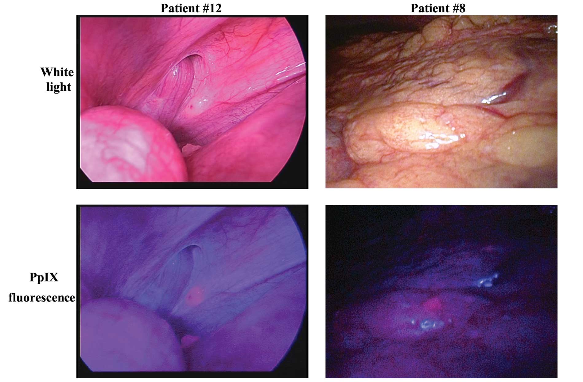
|
1.
|
Sugarbaker PH: Peritonectomy procedures.
Ann Surg. 221:29–42. 1995. View Article : Google Scholar : PubMed/NCBI
|
|
2.
|
Glehen O, Gilly FN, Boutitie F, Bereder
JM, Quenet F, Sideris L, Mansvelt B, Lorimier G, Msika S and Elias
D: Toward curative treatment of peritoneal carcinomatosis from
nonovarian origin by cytoreductive surgery combined with
perioperative intraperitoneal chemotherapy: a multi-institutional
study of 1,290 patients. French Surgical Association Cancer.
116:5608–5618. 2010.
|
|
3.
|
Brücher BL, Piso P, Verwaal V, Esquivel J,
Derraco M, Yonemura Y, Gonzalez-Moreno S, Pelz J, Königsrainer A,
Ströhlein M, Levine EA, Morris D, Bartlett D, Glehen O, Garofalo A
and Nissan A: Peritoneal carcinomatosis: cytoreductive surgery and
HIPEC - overview and basics. Cancer Invest. 30:209–224.
2012.PubMed/NCBI
|
|
4.
|
Koppe MJ, Boerman OC, Oyen WJG and
Bleichrodt RP: Peritoneal carcinomatosis of colorectal origin. Ann
Surg. 243:212–222. 2006. View Article : Google Scholar : PubMed/NCBI
|
|
5.
|
Bamba Y, Itabashi M and Kameoka S:
Clinical use of PET/CT in peritoneal carcinomatosis from colorectal
cancer. Hepatogastroenterology. 59:1408–1411. 2012.PubMed/NCBI
|
|
6.
|
Ohgari Y, Nakayasu Y, Kitajima S, Sawamoto
M, Mori H, Shimokawa O, Matsui H and Taketani S: Mechanisms
involved in delta-aminolevulinic acid (ALA)-induced
photosensitivity of tumor cells: relation of ferrochelatase and
uptake of ALA to the accumulation of protoporphyrin. Biochem
Pharmacol. 71:42–49. 2005. View Article : Google Scholar
|
|
7.
|
Kriegmair M, Baumgartner R, Knuchel R,
Stepp H, Hofstadter F and Hofstetter A: Detection of early bladder
cancer by 5-aminolevulinic acid induced porphyrin fluorescence. J
Urol. 155:105–109. 1996. View Article : Google Scholar : PubMed/NCBI
|
|
8.
|
Jichlinski P, Forrer M, Mizeret J,
Glanzmann T, Braichotte D, Wagnieres G, Zimmer G, Guillou L,
Schmidlin F, Graber P, van den Bergh H and Leisinger HJ: Clinical
evaluation of a method for detecting superficial surgical
transitional cell carcinoma of the bladder by light induced
fluorescence of protoporphyrin IX following the topical application
of 5-aminolevulinic acid: preliminary results. Lasers Surg Med.
20:402–408. 1997. View Article : Google Scholar
|
|
9.
|
Stummer W, Stocker S, Wagner S, Stepp H,
Fritsch C, Goetz C, Goetz AE, Kiefmann R and Reulen HJ:
Intraoperative detection of malignant gliomas by 5-aminolevulinic
acid-induced porphyrin fluorescence. Neurosurgery. 42:518–526.
1998. View Article : Google Scholar : PubMed/NCBI
|
|
10.
|
Friesen SA, Hjortland GO, Madsen SJ,
Hirschberg H, Engebraten O, Nesland JM and Peng Q: 5-Aminolevulinic
acid-based photodynamic detection and therapy of brain tumors
(review). Int J Oncol. 21:577–582. 2002.PubMed/NCBI
|
|
11.
|
Hatakeyama T, Murayama Y, Komatsu S,
Shiozaki A, Kuriu Y, Ikoma H, Nakanishi M, Ichikawa D, Fujiwara H,
Okamoto K, Ochiai T, Kokuba Y, Inoue K, Nakajima M and Otsuji E:
Efficacy of 5-aminolevulinic acid-mediated photodynamic therapy
using light-emitting diodes in human colon cancer cells. Oncol Rep.
29:911–916. 2013.PubMed/NCBI
|
|
12.
|
Hino H, Murayama Y, Nakanishi M, Inoue K,
Nakajima M and Otsuji E: 5-Aminolevulinic acid-mediated
photodynamic therapy using light-emitting diodes of different
wavelengths in a mouse model of peritoneally disseminated gastric
cancer. J Surg Res. 185:119–126. 2013. View Article : Google Scholar
|
|
13.
|
Murayama Y, Harada Y, Imaizumi K, Dai P,
Nakano K, Okamoto K, Otsuji E and Takamatsu T: Precise detection of
lymph node metastases in mouse rectal cancer by using
5-aminolevulinic acid. Int J Cancer. 125:2256–2263. 2009.
View Article : Google Scholar : PubMed/NCBI
|
|
14.
|
Murayama Y, Ichikawa D, Koizumi N, Komatsu
S, Shiozaki A, Kuriu Y, Ikoma H, Kubota T, Nakanishi M, Harada Y,
Fujiwara H, Okamoto K, Ochiai T, Kokuba Y, Takamatsu T and Otsuji
E: Staging fluorescence laparoscopy for gastric cancer by using
5-aminolevulinic acid. Anticancer Res. 2:5421–5427. 2012.PubMed/NCBI
|
|
15.
|
Koizumi N, Harada Y, Murayama Y, Harada K,
Beika M, Yamaoka Y, Dai P, Komatsu S, Kubota T, Ichikawa D, Okamoto
K, Yanagisawa A, Otsuji E and Takamatsu T: Detection of meta-static
lymph nodes using 5-aminolevulinic acid in patients with gastric
cancer. Ann Surg Oncol. 20:3541–3548. 2013. View Article : Google Scholar : PubMed/NCBI
|
|
16.
|
Klaver YLB, Lemmens VEPP, Creemers GJ,
Rutten HJT, Nienhuijs SW and de Hingh IHJT: Population-based
survival of patients with peritoneal carcinomatosis from colorectal
origin in the era of increasing use of palliative chemotherapy. Ann
Oncol. 22:2250–2256. 2011. View Article : Google Scholar : PubMed/NCBI
|
|
17.
|
Verwaal VJ: Randomized trial of
cytoreduction and hyperthermic intraperitoneal chemotherapy versus
systemic chemotherapy and palliative surgery in patients with
peritoneal carcinomatosis of colorectal cancer. J Clin Oncol.
21:3737–3743. 2003. View Article : Google Scholar
|
|
18.
|
Maggiori L and Elias D: Curative treatment
of colorectal peritoneal carcinomatosis: current status and future
trends. Eur J Surg Oncol. 36:599–603. 2010. View Article : Google Scholar : PubMed/NCBI
|
|
19.
|
Cao C, Yan TD, Black D and Morris DL: A
systematic review and meta-analysis of cytoreductive surgery with
perioperative intraperitoneal chemotherapy for peritoneal
carcinomatosis of colorectal origin. Ann Surg Oncol. 16:2152–2165.
2009. View Article : Google Scholar : PubMed/NCBI
|
|
20.
|
Zöpf T, Schneider AR, Weickert U, Riemann
JF and Arnold JC: Improved preoperative tumor staging by
5-aminolevulinic acid induced fluorescence laparoscopy.
Gastrointest Endosc. 62:763–767. 2005.PubMed/NCBI
|


