Open Access

Value of radial probe endobronchial ultrasound-guided localization of solitary pulmonary nodules with the combination of ultrathin bronchoscopy and methylene blue prior to video-assisted thoracoscopic surgery

  • Authors:
    • Chun-Hua Xu
    • Li-Ke Yu
    • Lan Cao
    • Rusong Yang
    • Jun Yan
    • Zheng-Cheng Liu
    • Yan Wang
  • View Affiliations

  • Published online on: May 26, 2016     https://doi.org/10.3892/mco.2016.913
  • Pages: 279-282
  • Copyright: © Xu et al. This is an open access article distributed under the terms of Creative Commons Attribution License.

Metrics: Total Views: 0 (Spandidos Publications: | PMC Statistics: )
Total PDF Downloads: 0 (Spandidos Publications: | PMC Statistics: )


Abstract

The aim of this study was to assess the clinical value of radial probe endobronchial ultrasound (RP-EBUS)-guided localization of solitary pulmonary nodules (SPNs) with the combination of ultrathin bronchoscopy and methylene blue prior to video-assisted thoracoscopic surgery (VATS). An ultrathin bronchoscope was used to localize the lesions under RP-EBUS guidance in 48 patients (18 men and 30 women; age range, 41‑72 years; mean age, 54 years), who subsequently underwent VATS resection. The lesion size, distance from the parietal pleura, localization time and complications were evaluated. The RP-EBUS-guided localization success rate was 72.9%. The lesion size ± standard deviation was 12.8±4.2 mm and the mean distance from the parietal pleura was 11.2±9.7 mm. The mean localization time was 15.7±8.3 min. The major complication of RP-EBUS‑guided localization was asymptomatic hemorrhage in 4 patients (8.3%). The VATS resection success rate was 95.8%. In terms of pathological type, the 48 lesions included atypical adenomatous hyperplasia (n=4), adenocarcinoma in situ (n=5), minimally invasive adenocarcinoma (n=7), adenocarcinoma (n=18), squamous cell carcinoma (n=1), inflammation (n=6), hamartoma (n=4) and tuberculosis (n=3). Therefore, RP-EBUS-guided localization with the combination of an ultrathin bronchoscope and methylene blue prior to VATS resection is a promising technique for SPNs, it plays an important role in the accurate localization of SPNs and it is an effective and safe technique to assist VATS resection of such nodules.
View Figures
View References

Related Articles

Journal Cover

August-2016
Volume 5 Issue 2

Print ISSN: 2049-9450
Online ISSN:2049-9469

Sign up for eToc alerts

Recommend to Library

Copy and paste a formatted citation
x
Spandidos Publications style
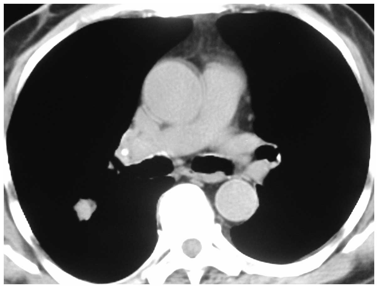
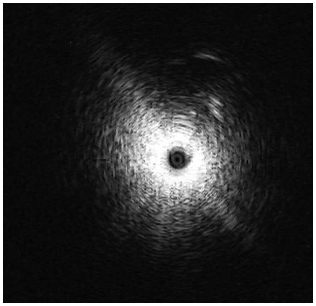
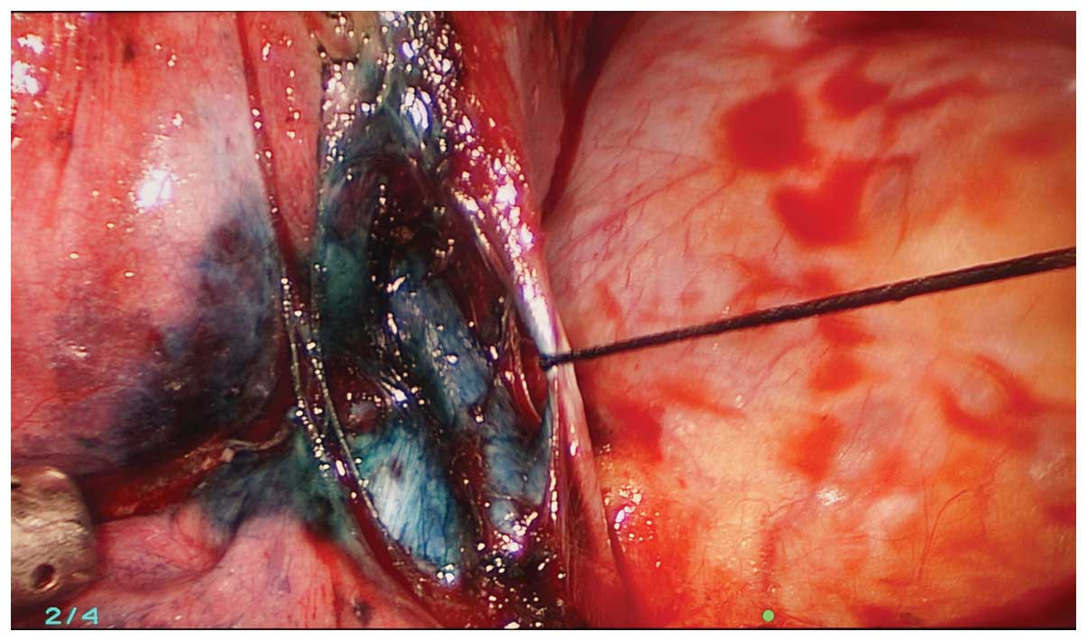
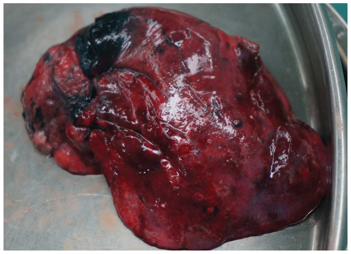
Xu C, Yu L, Cao L, Yang R, Yan J, Liu Z and Wang Y: Value of radial probe endobronchial ultrasound-guided localization of solitary pulmonary nodules with the combination of ultrathin bronchoscopy and methylene blue prior to video-assisted thoracoscopic surgery. Mol Clin Oncol 5: 279-282, 2016
APA
Xu, C., Yu, L., Cao, L., Yang, R., Yan, J., Liu, Z., & Wang, Y. (2016). Value of radial probe endobronchial ultrasound-guided localization of solitary pulmonary nodules with the combination of ultrathin bronchoscopy and methylene blue prior to video-assisted thoracoscopic surgery. Molecular and Clinical Oncology, 5, 279-282. https://doi.org/10.3892/mco.2016.913
MLA
Xu, C., Yu, L., Cao, L., Yang, R., Yan, J., Liu, Z., Wang, Y."Value of radial probe endobronchial ultrasound-guided localization of solitary pulmonary nodules with the combination of ultrathin bronchoscopy and methylene blue prior to video-assisted thoracoscopic surgery". Molecular and Clinical Oncology 5.2 (2016): 279-282.
Chicago
Xu, C., Yu, L., Cao, L., Yang, R., Yan, J., Liu, Z., Wang, Y."Value of radial probe endobronchial ultrasound-guided localization of solitary pulmonary nodules with the combination of ultrathin bronchoscopy and methylene blue prior to video-assisted thoracoscopic surgery". Molecular and Clinical Oncology 5, no. 2 (2016): 279-282. https://doi.org/10.3892/mco.2016.913