Morphology as a risk factor for the malignant potential of T2 colorectal cancer

  • Authors:
    • Yuichi Mori
    • Shin‑Ei Kudo
    • Shungo Endo
    • Chiyo Maeda
    • Shumpei Mukai
    • Yasuharu Maeda
    • Shinichi Kataoka
    • Kenichi Takeda
    • Katsuro Ichimasa
    • Hideyuki Miyachi
    • Naruhiko Sawada
    • Eiji Hidaka
    • Fumio Ishida
  • View Affiliations

  • Published online on: July 7, 2016     https://doi.org/10.3892/mco.2016.951
  • Pages: 223-226
Metrics: Total Views: 0 (Spandidos Publications: | PMC Statistics: )
Total PDF Downloads: 0 (Spandidos Publications: | PMC Statistics: )


Abstract

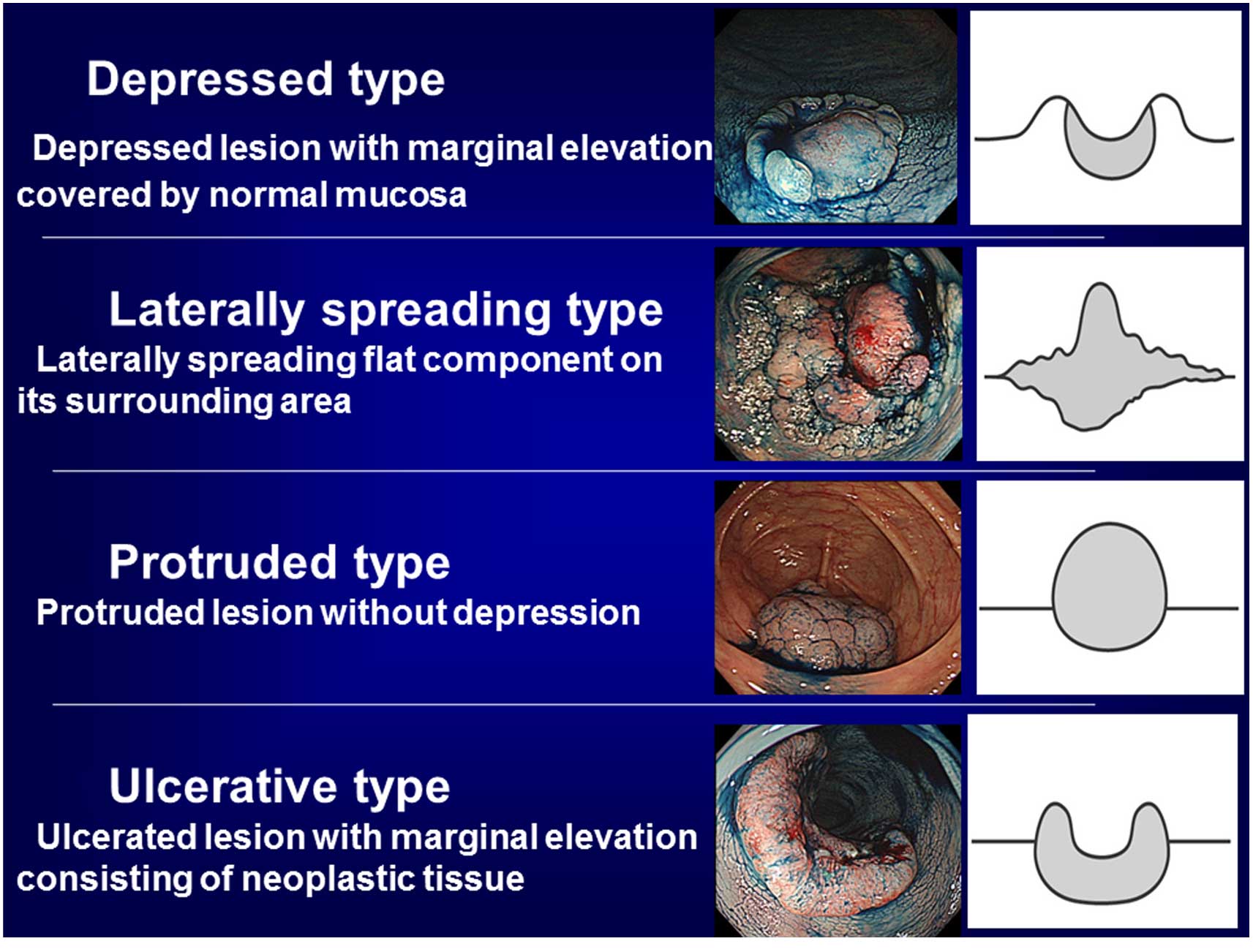
Currently, depressed type T1 colorectal cancer is often detected and its malignant potential is being revealed. However, few reports have focused on the morphology of T2 colorectal cancer types. The aim of the present retrospective study was to clarify the characteristics of colorectal T2 cancer types derived from depressed type T1 cancer. The present study included 195 patients with colorectal T2 cancer surgically resected at a referral hospital between April 2001 and April 2009. A total of 48 lesions (25%) with lymph node metastasis and 4 (2%) with distant metastasis were found. The lesions were classified by their form during endoscopy into four categories: Group A, depressed type (the periphery consists of normal mucosa); group B, laterally spreading type; group C, protruded type; group D, ulcerative type (the periphery consists of neoplastic mucosa). Tumor size, lymphatic and venous involvement, lymph node metastasis, and distant metastasis were compared between the four categories. The distribution of patients were as follows: Group A, 73 (37%), group B, 26 (13%), group C, 24 (12%) and group D, 72 (37%). The average tumor size for each category was 23, 51, 30 and 36 mm for groups A, B, C and D, respectively. The number of positive findings for lymphatic involvement, venous involvement, lymph node metastasis and distant metastasis for each category were: Group A, 50 (69%), 54 (74%), 19 (26%) and 1 (1%); group B, 14 (54%), 12 (46%), 8 (31%) and 0 (0%); group C, 14 (58%), 8 (33%), 6 (25%) and 0 (0%); group D 35 (49%), 41 (57%), 16 (22%) and 3 (4%). Group A demonstrated a significantly smaller tumor size (P<0.01), and higher rates of lymphatic and venous involvement (P=0.0333 and P=0.0019, respectively). No significant differences were observed between categories with regards to nodal and distant metastases. Of the T2 cancer types, ~40% were the depressed type. Compared with the other forms, depressed type lesions exhibited a smaller tumor size, and higher rates of lymphatic and venous involvement, which suggested these lesions were of a more malignant nature.
View Figures
View References

Related Articles

Journal Cover

September-2016
Volume 5 Issue 3

Print ISSN: 2049-9450
Online ISSN:2049-9469

Sign up for eToc alerts

Recommend to Library

Copy and paste a formatted citation
x
Spandidos Publications style
Mori Y, Kudo SE, Endo S, Maeda C, Mukai S, Maeda Y, Kataoka S, Takeda K, Ichimasa K, Miyachi H, Miyachi H, et al: Morphology as a risk factor for the malignant potential of T2 colorectal cancer. Mol Clin Oncol 5: 223-226, 2016
APA
Mori, Y., Kudo, S., Endo, S., Maeda, C., Mukai, S., Maeda, Y. ... Ishida, F. (2016). Morphology as a risk factor for the malignant potential of T2 colorectal cancer. Molecular and Clinical Oncology, 5, 223-226. https://doi.org/10.3892/mco.2016.951
MLA
Mori, Y., Kudo, S., Endo, S., Maeda, C., Mukai, S., Maeda, Y., Kataoka, S., Takeda, K., Ichimasa, K., Miyachi, H., Sawada, N., Hidaka, E., Ishida, F."Morphology as a risk factor for the malignant potential of T2 colorectal cancer". Molecular and Clinical Oncology 5.3 (2016): 223-226.
Chicago
Mori, Y., Kudo, S., Endo, S., Maeda, C., Mukai, S., Maeda, Y., Kataoka, S., Takeda, K., Ichimasa, K., Miyachi, H., Sawada, N., Hidaka, E., Ishida, F."Morphology as a risk factor for the malignant potential of T2 colorectal cancer". Molecular and Clinical Oncology 5, no. 3 (2016): 223-226. https://doi.org/10.3892/mco.2016.951