Chondroblastic osteosarcoma arising in the maxilla mimicking the radiographic and histological characteristics of cemento-osseous lesions: A case report
- Authors:
- Bin‑Bin Li
- Jian‑Yun Zhang
- Yan Gao
View Affiliations
Affiliations: Department of Oral Pathology, Peking University School and Hospital of Stomatology, Beijing 100081, P.R. China
- Published online on: April 10, 2017 https://doi.org/10.3892/mco.2017.1219
-
Pages:
733-736
Metrics: Total
Views: 0 (Spandidos Publications: | PMC Statistics: )
Metrics: Total PDF Downloads: 0 (Spandidos Publications: | PMC Statistics: )
This article is mentioned in:
Abstract
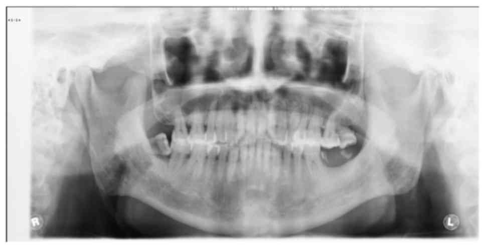
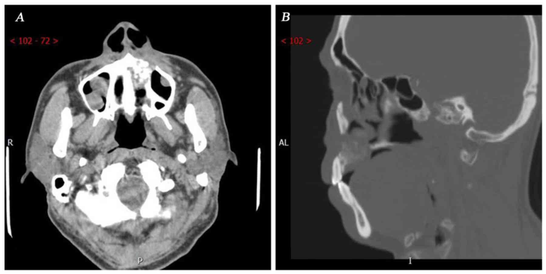
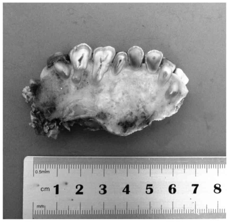
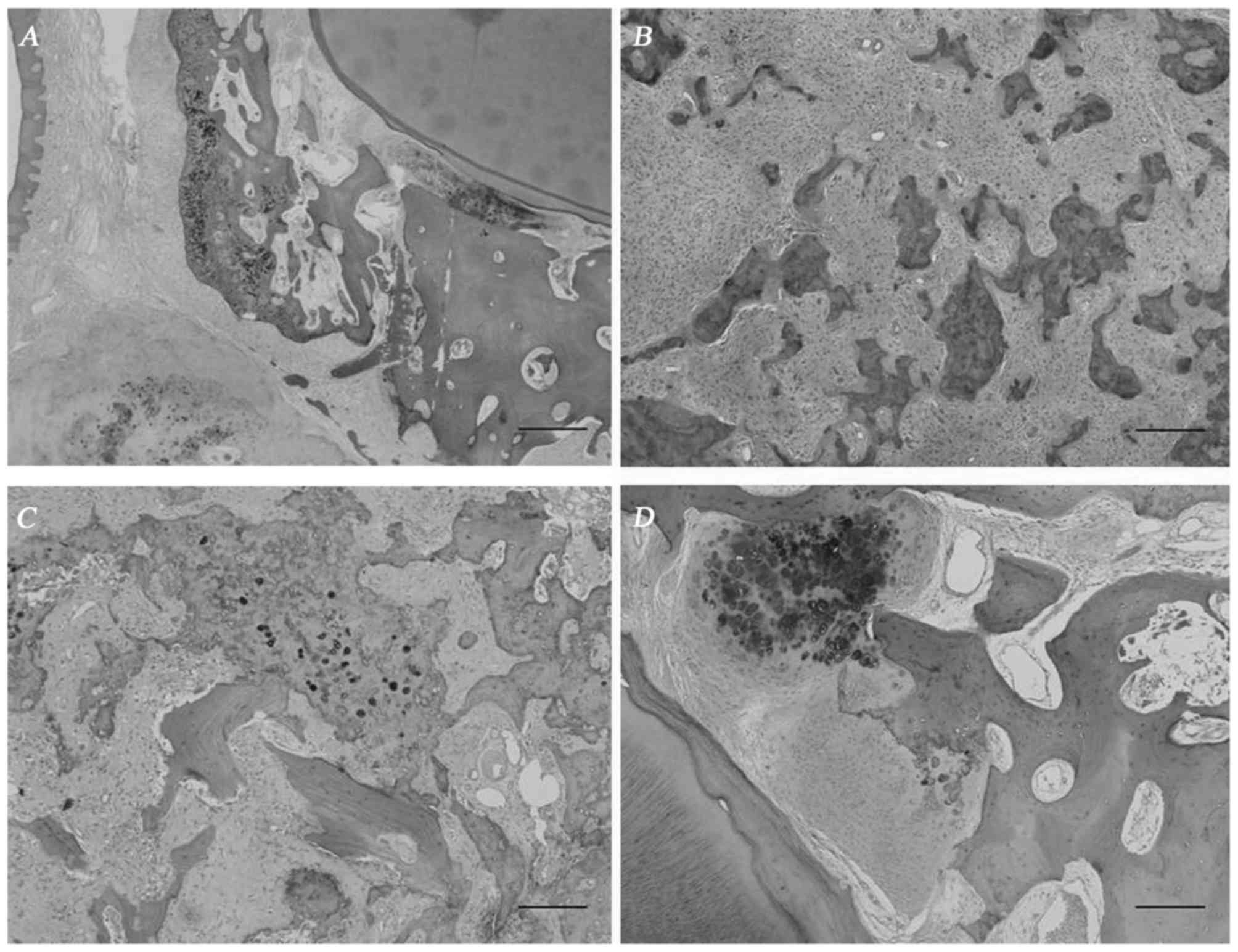
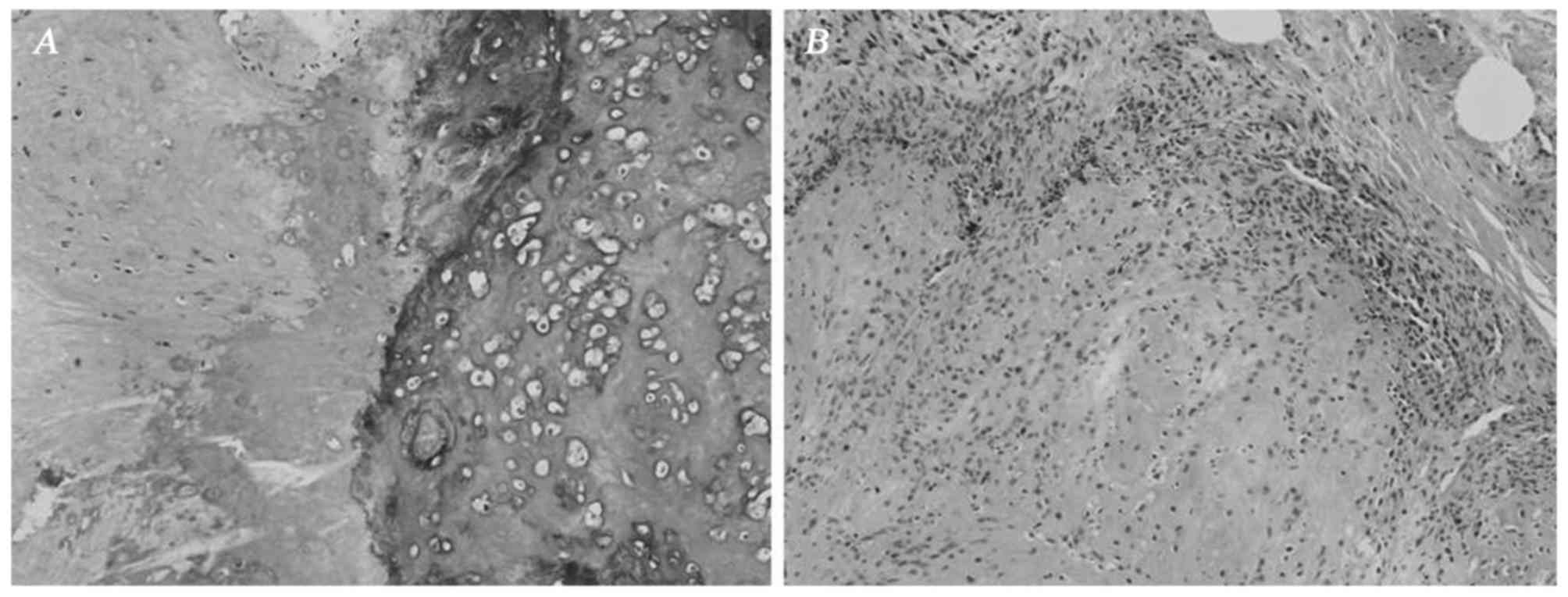
Osteosarcomas of the jaw are comparatively rare and represent only 2-10% of all osteosarcomas. We herein present a rare case of an osteosarcoma exhibiting the radiographic and histological characteristics of cemento‑osseous lesions in the alveolar ridge of the maxilla. A 53-year-old male patient presented with the complaint of gradual swelling of the left maxilla over 4 years. Radiography revealed an ill‑defined radioopaque mass, intimately associated with the apices of the involved teeth, without a periosteal reaction. Microscopically, a cementicle‑like structure was identified in the alveolar bone. In addition, the lesion exhibited typical characteristics of chondroblastic osteosarcoma in the body of the maxilla. The tumor contained abundant osteoid and cartilage intimately associated with anaplastic tumor cells. The cartilage displayed malignant‑appearing cells in lacunae, and there was crowding at the periphery of the lobule where the spindle cells formed sheets. The differential diagnosis included primary osteosarcoma, concurrent cemento‑osseous dysplasia and osteosarcoma, or a secondary osteosarcoma based on a pre‑existing cemento‑osseous lesion. The presence of the cementicle‑like structure in the alveolar bone and the involvement of the periodontal ligament and alveolar bone proper were unique in our case. The general invasive growth pattern and the abundance of the irregular tumor bone helped establish the diagnosis of primary osteosarcoma. This case may represent evidence of the pathogenesis of primary osteosarcoma in the jaw.
View References
1
|
Sato K and Unni KK: Malignant tumors of
bone and cartilagePathology and Genetics of Head and Neck Tumors:
World Health Organization Classification of Tumors. Barnes L,
Eveson JW, Reichart P and Sidransky D: IARC Press; Lyon: pp. 7–52.
2005
|
2
|
Nthumba PM: Osteosarcoma of the jaws: A
review of literature and a case report on synchronous multicentric
osteosarcomas. World J Surg Oncol. 10:2402012. View Article : Google Scholar : PubMed/NCBI
|
3
|
Forteza G, Colmenero B and López-Barea F:
Osteogenic sarcoma of maxilla and mandible. Oral Surg Oral Med Oral
Pathol. 62:179–184. 1986. View Article : Google Scholar : PubMed/NCBI
|
4
|
Paparella ML, Olvi LG, Brandizzi D,
Keszler A, Santini-Araujo E and Cabrini RL: Osteosarcoma of the
jaw: An analysis of a series of 74 cases. Histopathology.
63:551–557. 2013.PubMed/NCBI
|
5
|
Wang S, Shi H and Yu Q: Osteosarcoma of
the jaws: Demographic and CT imaging features. Dentomaxillofac
Radiol. 41:37–42. 2012. View Article : Google Scholar : PubMed/NCBI
|
6
|
Jia S and Li B: Osteosarcoma of the Jaws:
Case report on synchronous multicentric osteosarcomas. J Clin Diagn
Res. 8:ZD01–ZD03. 2014.PubMed/NCBI
|
7
|
Carnelio S, Pai K, Rao N, Solomon M and
Ahasan A: Metastatic osteosarcoma to the maxilla: A case report and
a review of the literature. Quintessence Int. 33:397–399.
2002.PubMed/NCBI
|
8
|
Olusanya AA, Adeyemi BF and Adisa AO:
Concurrent cemento-osseous dysplasia and osteogenic sarcoma: Report
of two cases. Case Rep Med. 2012:1805612012.PubMed/NCBI
|
9
|
Koury ME, Regezi JA, Perrott DH and Kaban
LB: ‘Atypical’ fibro-osseous lesions: Diagnostic challenges and
treatment concepts. Int J Oral Maxillofac Surg. 24:162–169. 1995.
View Article : Google Scholar : PubMed/NCBI
|
10
|
Matsuzaka K, Shimono M, Uchiyama T, Noma H
and Inoue T: Lesions related to the formation of bone, cartilage or
cementum arising in the oral area: A statistical study and review
of the literature. Bull Tokyo Dent Coll. 43:173–180. 2002.
View Article : Google Scholar : PubMed/NCBI
|
11
|
Roguljic H, Matthews BG, Yang W, Cvija H,
Mina M and Kalajzic I: In vivo identification of periodontal
progenitor cells. J Dent Res. 92:709–715. 2013. View Article : Google Scholar : PubMed/NCBI
|