Non‑neural granular cell tumor of the uterine corpus mimicking uterine leiomyoma: A case report
- Authors:
- Published online on: July 13, 2017 https://doi.org/10.3892/mco.2017.1324
- Pages: 351-354
-
Copyright: © Deguchi et al. This is an open access article distributed under the terms of Creative Commons Attribution License.
Metrics: Total
Views: 0 (Spandidos Publications: | PMC Statistics: )
Total PDF Downloads: 0 (Spandidos Publications: | PMC Statistics: )
Abstract
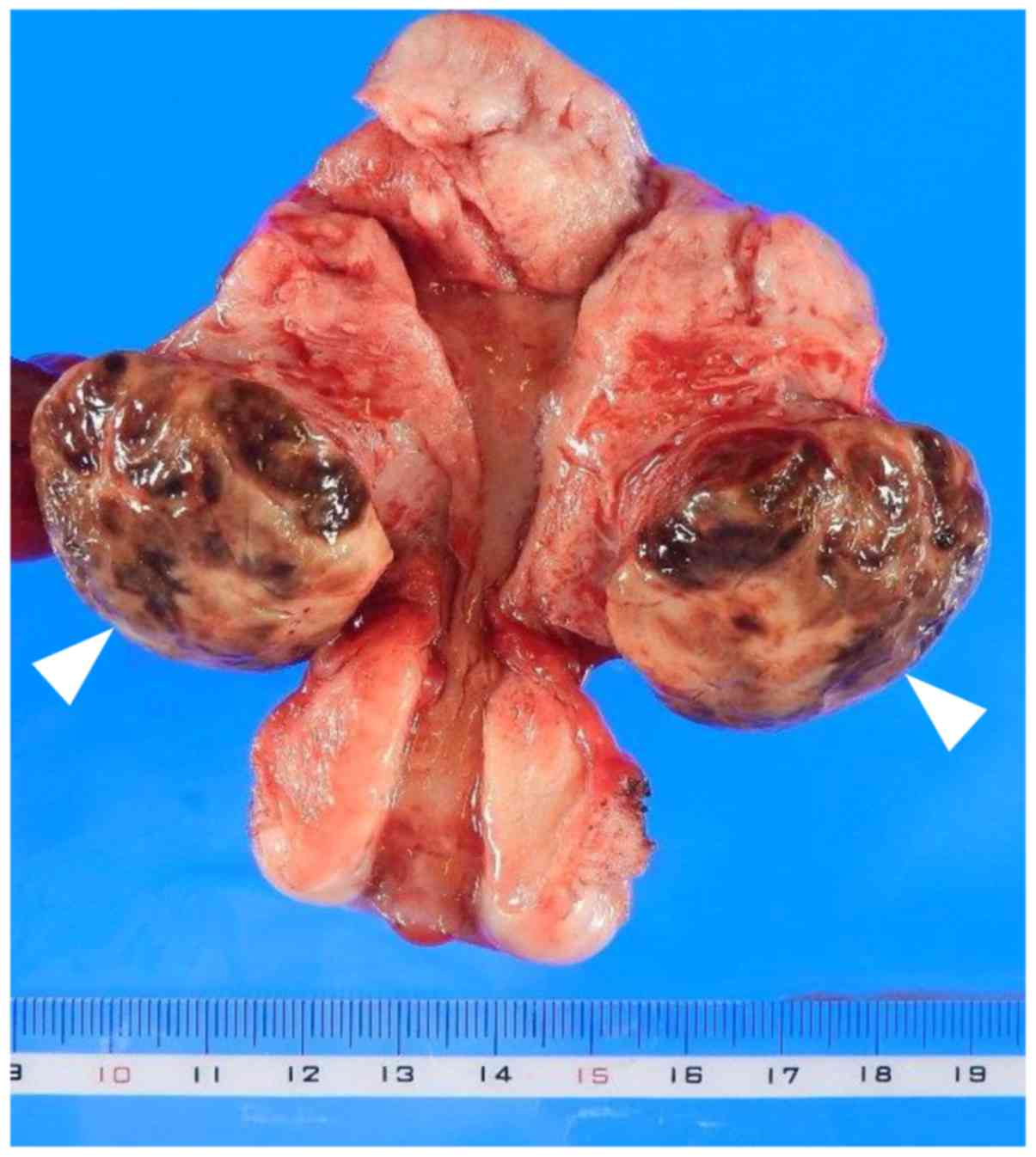
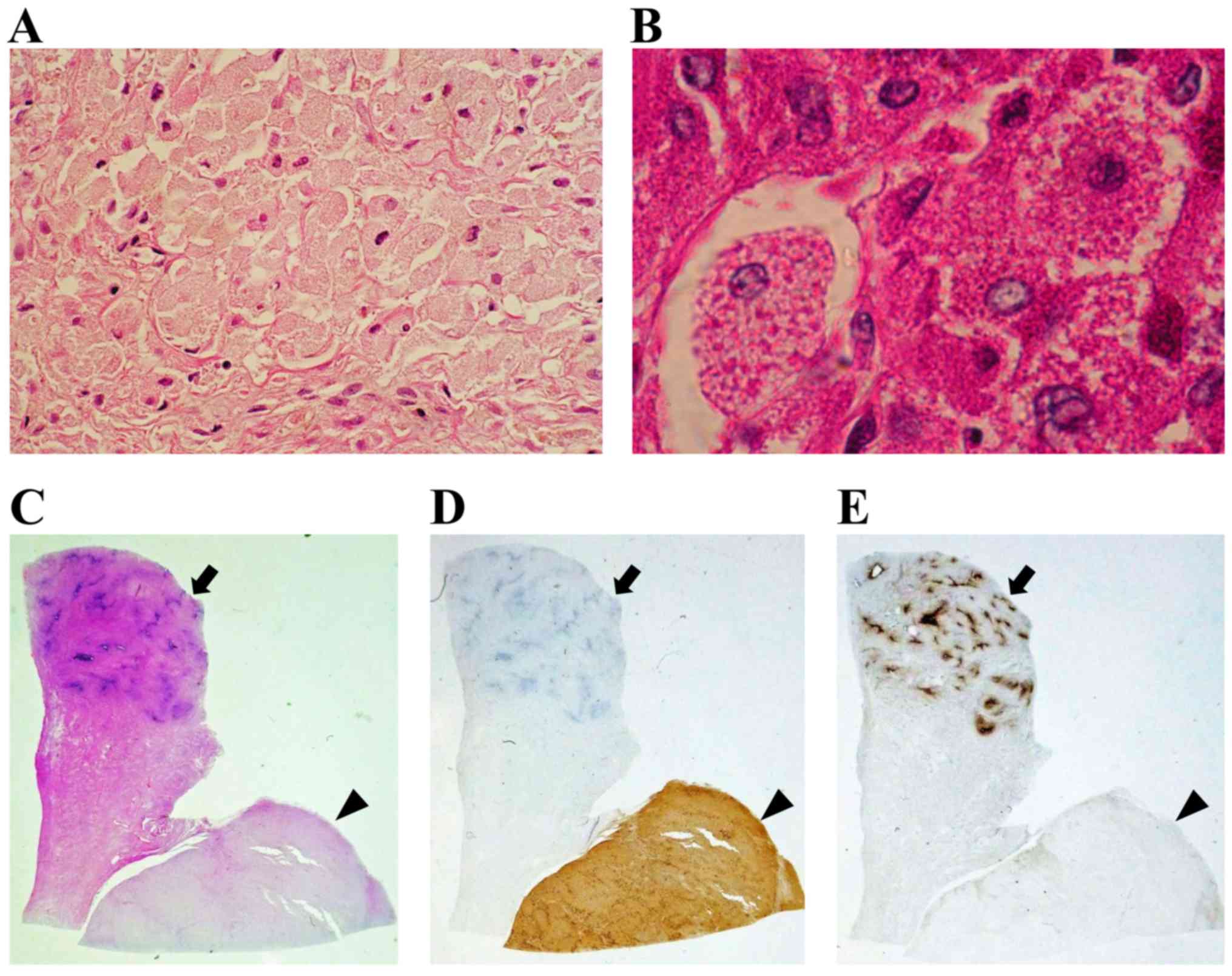
Non‑neural granular cell tumors (GCTs) are clinically rare, whereas cases arising in the uterine corpus are exceedingly rare. Only three uterine cervical cases of GCTs have been reported to date and, to the best of our knowledge, there are no reports of GCT of the uterine corpus in the literature. We herein describe the first case of non‑neural GCT arising from the uterine corpus reported to date. A 55‑year‑old premenopausal woman was referred to the Department of Obstetrics and Gynecology of Hashimoto Municipal Hospital (Wakayama, Japan) with a suspected uterine tumor. The tumor presented as a uterine leiomyoma‑like mass on radiological examinations, but the diagnosis of non‑neural GCT was established based on pathological and immunohistochemical examinations. Microscopically, histological examination of the entire surgical specimen revealed large polygonal cells with abundant eosinophilic granular cytoplasm and round to oval nuclei. Immunohistochemistry revealed positive periodic acid‑Schiff staining of the cytoplasmic granules, which was resistant to diastase. In addition, the tumor cells stained positive for CD68, but negative for S‑100, neuron‑specific enolase, cytokeratin, CD34, α‑smooth muscle actin, desmin, estrogen receptor and progesterone receptor. It is important for gynecologists to be aware of the possibility of non‑neural GCT of the uterine corpus, for which accurate diagnosis, complete resection and long‑term follow‑up are crucial, as it may be easily misdiagnosed as uterine leiomyoma.