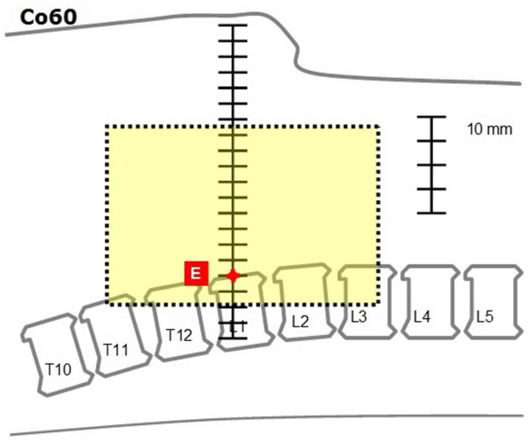
Definition of fields margins for palliative radiotherapy of pancreatic carcinoma
- Authors:
- Published online on: April 12, 2018 https://doi.org/10.3892/mco.2018.1605
- Pages: 715-718
-
Copyright: © Buwenge et al. This is an open access article distributed under the terms of Creative Commons Attribution License.
Metrics: Total
Views: 0 (Spandidos Publications: | PMC Statistics: )
Total PDF Downloads: 0 (Spandidos Publications: | PMC Statistics: )
Abstract
The present study aimed to provide practical guidelines for palliative treatment of advanced carcinoma of the pancreas (CAP) with the 2D technique. Fifteen patients with locally advanced CAP consecutively treated with radiation therapy at the Radiation Oncology Center, Research and Care Foundation ‘Giovanni Paolo II’ (Campobasso, Italy) underwent computed tomography simulation in supine position. Definition of the clinical target volume (CTV) included the head and body of the pancreas, and the retropancreatic space. The planning target volume was defined by adding a margin of 14 mm to the CTV in the cranio‑caudal direction and of 11 mm in radial direction. For each patient, 3 treatment plans were calculated using a cobalt source, 6 MV photons and 15 MV photons (box technique). Beams were drawn using the primary collimators without using multileaf collimators, and progressively optimized in order to respect the minimum dose (Dmin>90%) constraint. Once the final plan was achieved, distances of the fields edges from a set of reference points (bony or duodenal landmarks) were measured. Using this technique, 15 anterior‑posterior and postero‑anterior (AP‑PA) beams and 15 pairs of lateral‑lateral (LL) beams were defined for the different patients. Finally, the single minimal AP‑PA and LL beams able to include the 15 sets of AP‑PA and LL beams were defined. The results of this analysis are reported in tabular form. Guidelines are provided for treatment based on cobalt unit or Linear accelerator (both 6 and 15 MV photons). This study provides information regarding field size and position. A dosimetric study has been planned to identify the dose to be administered with this technique taking into account current dose‑volume constraints.